. 2012;7(10):e47713.
doi: 10.1371/journal.pone.0047713. Epub 2012 Oct 16.

High fat diet-induced gut microbiota exacerbates inflammation and obesity in mice via the TLR4 signaling pathway

Affiliations

High fat diet-induced gut microbiota exacerbates inflammation and obesity in mice via the TLR4 signaling pathway

Kyung-Ah Kim et al. PLoS One. 2012.

Abstract

Background & aims: While it is widely accepted that obesity is associated with low-grade systemic inflammation, the molecular origin of the inflammation remains unknown. Here, we investigated the effect of endotoxin-induced inflammation via TLR4 signaling pathway at both systemic and intestinal levels in response to a high-fat diet.

Methods: C57BL/6J and TLR4-deficient C57BL/10ScNJ mice were maintained on a low-fat (10 kcal % fat) diet (LFD) or a high-fat (60 kcal % fat) diet (HFD) for 8 weeks.

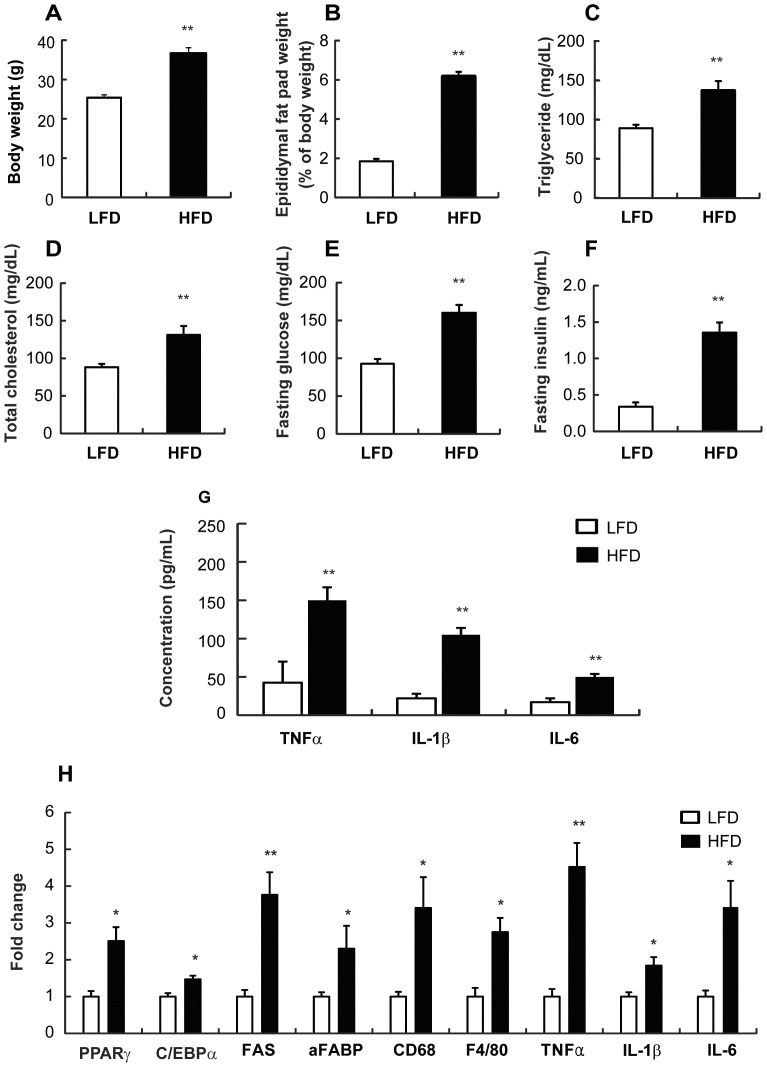
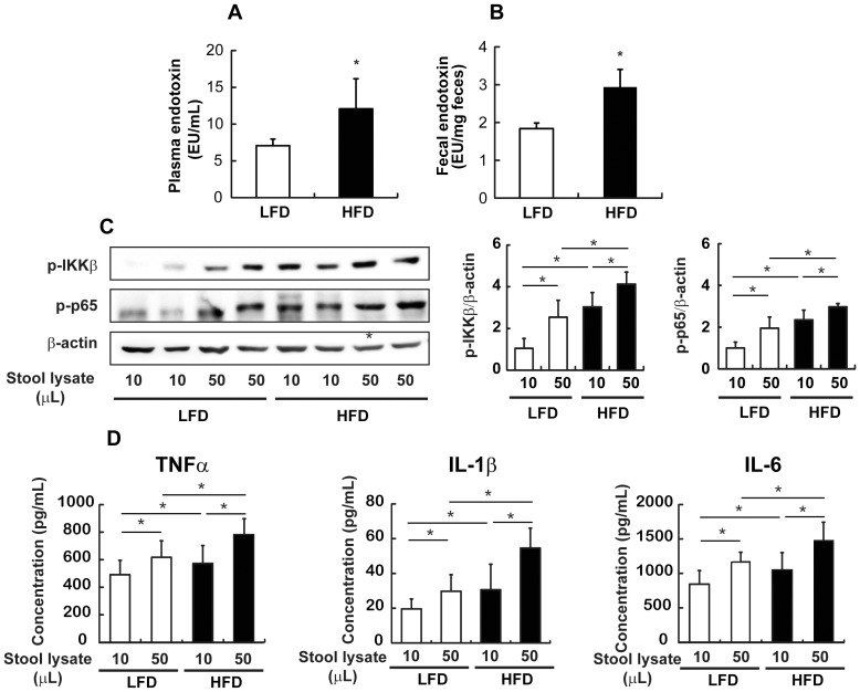
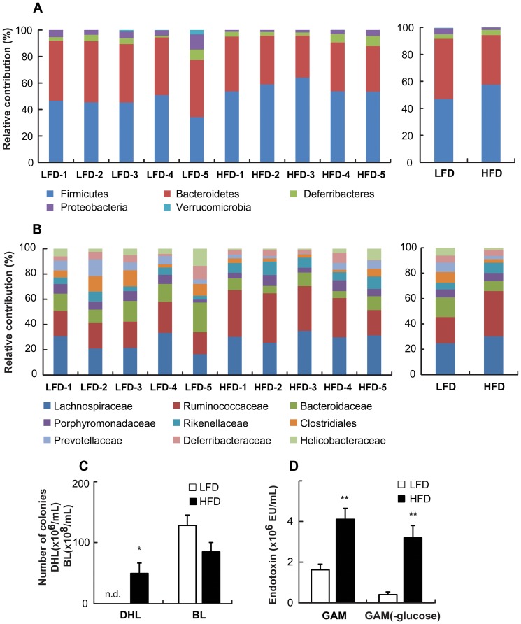
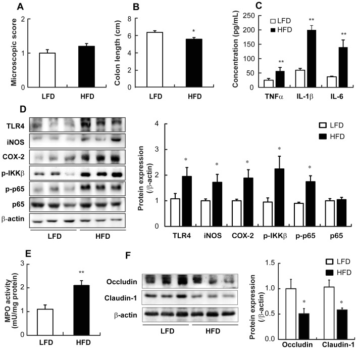
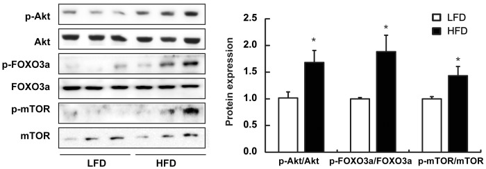
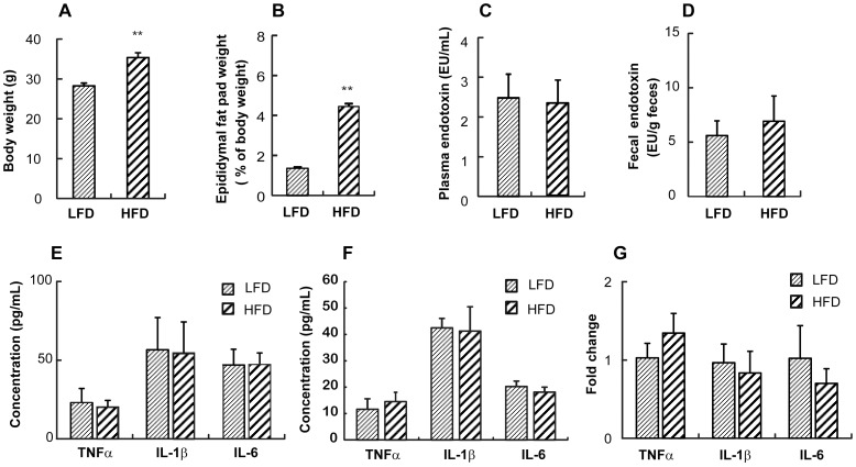
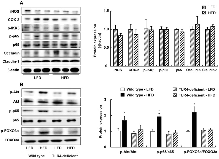
Results: HFD induced macrophage infiltration and inflammation in the adipose tissue, as well as an increase in the circulating proinflammatory cytokines. HFD increased both plasma and fecal endotoxin levels and resulted in dysregulation of the gut microbiota by increasing the Firmicutes to Bacteriodetes ratio. HFD induced the growth of Enterobecteriaceae and the production of endotoxin in vitro. Furthermore, HFD induced colonic inflammation, including the increased expression of proinflammatory cytokines, the induction of Toll-like receptor 4 (TLR4), iNOS, COX-2, and the activation of NF-κB in the colon. HFD reduced the expression of tight junction-associated proteins claudin-1 and occludin in the colon. HFD mice demonstrated higher levels of Akt and FOXO3 phosphorylation in the colon compared to the LFD mice. While the body weight of HFD-fed mice was significantly increased in both TLR4-deficient and wild type mice, the epididymal fat weight and plasma endotoxin level of HFD-fed TLR4-deficient mice were 69% and 18% of HFD-fed wild type mice, respectively. Furthermore, HFD did not increase the proinflammatory cytokine levels in TLR4-deficient mice.

Conclusions: HFD induces inflammation by increasing endotoxin levels in the intestinal lumen as well as in the plasma by altering the gut microbiota composition and increasing its intestinal permeability through the induction of TLR4, thereby accelerating obesity.


Conflict of interest statement

Competing Interests: The authors have declared that no competing interests exist.

Figures

Figure 1
Figure 1. Effect of HFD on macrophage infiltration and inflammation in the adipose tissue and the circulating proinflammatory cytokines in mice.
Male C57 BL/6J mice (4 weeks old) were fed LFD or HFD for 8 weeks. (A) Body weight (g) was measured. (B) Epididymal fat pad (% of body weight) was calculated. Plasmatic concentrations for (C) triglyceride (mg/dL), (D) total cholesterol (mg/dL), (E) fasting glucose (mg/dL), and (F) fasting insulin (mg/dL) were measured. (E) Concentration of circulating proinflammatory cytokines (pg/mL) were measured by ELISA. (H) mRNA expression levels in the adipose tissue were measured using real-time PCR. All values are indicated as the mean ± SEM (n = 10). *, p<0.05 and **, p<0.01 compared with LFD.
Figure 2
Figure 2. Effect of HFD on the LPS levels in mice.
(A) LAL assay was used to measure the plasma endotoxin concentration (EU/mL) and (B) fecal endotoxin concentration (EU/g feces). (C) Western blot analysis (left) and quantification of western blot (right) from isolated peritoneal macrophages which were incubated with 10 or 50 µl of stool solution were performed to determine IKKβ and p65 activation levels. (D) Concentrations of proinflammatory cytokines (pg/mL) in the culture medium of isolated peritoneal macrophages incubated with 10 or 50 µl of stool lysate were measured by ELISA. All values were indicated as the mean ± SEM (n = 10). *, p<0.05 compared with LFD.
Figure 3
Figure 3. Effect of HFD on changes in the gut microbiota system of mice.
Taxonomy compositions: (A) phylum and (B) family levels are shown (individual samples are on the left panels and pooled samples are on the right panels). Genomic DNA was extracted from the cecal samples taken from mice maintained for 8 weeks on a LFD and HFD. Samples were analyzed for the bacterial composition by pyrosequencing of the bacterial 16S rRNA fragments (n = 5). (C) Number of Enterobacteriaceae in DHL and Bifidobacteria in BL agar plates and (D) endotoxin content of fecal microflora cultured in GAM or GAM (-glucose) containing LFD or HFD were measured. n.d., not detected, All values were indicated as the mean ± SEM (n = 7). *, p<0.05
Figure 4
Figure 4. Effect of HFD on colitis in mice.
(A) Plotted is the macroscopic score assigned to the colitis. (B) Colon length (cm) was measured and data plotted. (C) Colonic concentrations of proinflammatory cytokines (pg/mL) were measured by ELISA. (D) Western blot analysis for colonic TLR4, iNOS, COX-2, p-IKKβ, and p-p65 protein levels (left) and quantification of western blot (right) are shown. (E) Intestinal MPO activity was measured. (F) Western blot analysis for occludin and claudin-1 levels (left) and quantification of western blot (right) in the colon are shown. All values were indicated as the mean ± SEM (n = 10). *, p<0.05 and **, p<0.01 compared with LFD.
Figure 5
Figure 5. Effect of HFD on the activation of Akt, FOXO3, and mTOR in the colon of mice.
Western blot analysis (left) and quantification of western blot (right) was performed on colon lysates from mice maintained on LFD or HFD.
Figure 6
Figure 6. Effect of HFD on the adiposity, the plasma and fecal LPS contents, and the proinflammatory cytokines in TLR4 deficient mice.
(A) Body weight (g) was measured. (B) Epididymal fat pad (% of body weight) was calculated. LAL assay was used to measure (C) fecal endotoxin concentration (EU/g feces) and (D) plasma endotoxin concentration (EU/mL). (E) Concentrations of proinflammatory cytokines (pg/mL) in the plasma (left) and colon (right) were measured by ELISA. (F) mRNA expression levels in the adipose tissue were measured using real-time PCR. All values were indicated as the mean ± SEM (n = 5). **, p<0.01 compared with LFD.
Figure 7
Figure 7. Effect of HFD on NF-κB activation, Akt and FOXO3a phosphorylation, and tight junction–associated protein levels in the colon of TLR4-deficient mice.
(A) Western blot analysis for iNOS, COX-2, p-IKKβ, p-p65 occludin, and claudin-1 protein levels (left) and quantification of western blot (right) in the colon lysates from TLR4-deficient mice are shown. (B) Western blot analysis for p-p65,p-65, p-Akt, Akt, p-FOXO3a, and FOXO3a protein levels (left) and quantification of western blot (right) in the colon lysates from wild-type or TLR4-deficient mice are shown. All values were indicated as the mean ± SEM (n = 5).

References

    1. Faulds MH, Dahlman-Wright K (2012) Metabolic diseases and cancer risk. Curr Opin Oncol 24: 58–61. - PubMed
    1. Wellen KE, Hotamisligil GS (2005) Inflammation, stress, and diabetes. J Clin Invest 115: 1111–1119. - PMC - PubMed
    1. Hotamisligil GS (2006) Inflammation and metabolic disorders. Nature 444: 860–867. - PubMed
    1. Chakraborty S, Zawieja S, Wang W, Zawieja DC, Muthuchamy M (2010) Lymphatic system: a vital link between metabolic syndrome and inflammation. Ann N Y Acad Sci 1207 Suppl 1: E94–102. - PMC - PubMed
    1. Monteiro R, Azevedo I (2010) Chronic inflammation in obesity and the metabolic syndrome. Mediators Inflamm 2010. - PMC - PubMed

Publication types

MeSH terms