

Case Reports
doi: 10.3340/jkns.2012.52.2.144.
Epub 2012 Aug 31.
Intraventricular cavernous hemangiomas located at the foramen of monro
Affiliations
- PMID: 23091674
- PMCID: PMC3467373
- DOI: 10.3340/jkns.2012.52.2.144
Item in Clipboard
Case Reports
Intraventricular cavernous hemangiomas located at the foramen of monro
J Korean Neurosurg Soc.
2012 Aug.
Erratum in
- J Korean Neurosurg Soc. 2012 Nov;52(5):505
Abstract
Intraventricular cavernous hemangiomas are uncommon. Among them, those occurred at the foramen of Monro in the third ventricle may be of particular interest because of its rarity, development of hydrocephalus, being differentiated from other brain lesions. We present a rare case of intraventricular cavernous hemangioma at foramen of Monro which was resected through microsurgery and also review the relevant literatures.
Keywords: Arteriovenous malformations; Cavernous hemangioma; Foramen of Monro; Hydrocephalus; Intraventricular; Third ventricle.
Figures
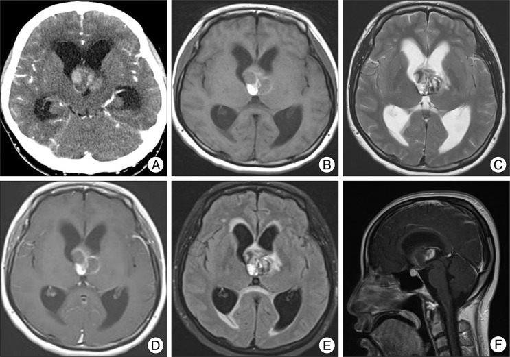
Computed tomogram (CT) shows enlargement of both lateral ventricles which is caused by 3 cm sized multi-lobular calcified mass with slightly peripheral rim enhancement. It is located at the foramen of Monro in the third ventricle and seems to be close to the left thalamus parenchyme partially (A). Magnetic resonance image (MRI) reveals a relatively well-delineated third ventricular mass with heterogeneous signal intensities in the center of lesion on T1-weighted image (B). Typical peripheral hemosiderin rim of low signal intensity is not delineated on T2-weighted image (C). Minimal peripheral enhancement at the right posterior portion of the mass is seen on enhanced T1-weighted image (D). There is a minimal perilesional edema in the left thalamus on the FLAIR image (E). The mass around foramen of Monro is seen on enhanced sagittal T1-weighted image (F).
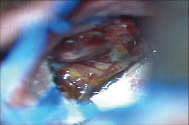
Operative finding shows that the mass with xanthochromic and lobular surface obstructing the foramen of Monro is located in the third ventricle. The left posterior margin of the mass is close to the thalamus.
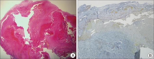
The histological examinations reveal a large hyalinized vessels filled with organizing thrombus (A). Immunostaining for smooth muscle actin (SMA) is positive in the vessel wall (B).
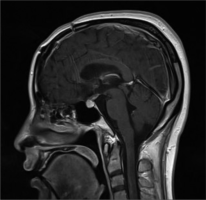
Post operative sagittal MRI shows no evidence of residual mass and improved hydrocephalus.
References
-
- Chen CL, Leu CH, Jan YJ, Shen CC. Intraventricular cavernous hemangioma at the foramen of Monro : case report and literature review. Clin Neurol Neurosurg. 2006;108:604–609. - PubMed
-
- Katayama Y, Tsubokawa T, Maeda T, Yamamoto T. Surgical management of cavernous malformations of the third ventricle. J Neurosurg. 1994;80:64–72. - PubMed
-
- Kivelev J, Niemelä M, Kivisaari R, Hernesniemi J. Intraventricular cerebral cavernomas : a series of 12 patients and review of the literature. J Neurosurg. 2010;112:140–149. - PubMed
-
- Prat R, Galeano I. Endoscopic resection of cavernoma of foramen of Monro in a patient with familial multiple cavernomatosis. Clin Neurol Neurosurg. 2008;110:834–837. - PubMed
Publication types
LinkOut - more resources
Full Text Sources
