

P-cadherin expression in feline mammary tissues
- PMID: 23091776
- PMCID: PMC3469258
- DOI: 10.1155/2012/687424
P-cadherin expression in feline mammary tissues
Abstract
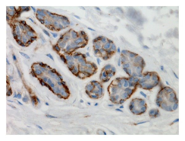
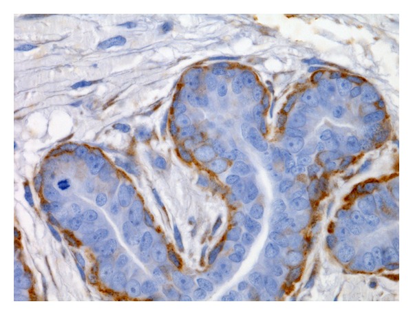
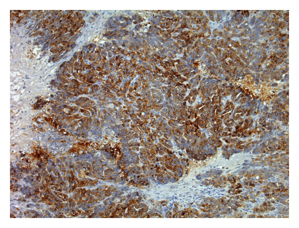
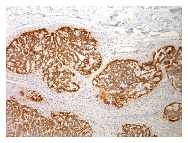
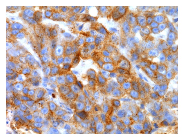
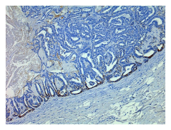
The search for molecular markers in the feline mammary gland, namely, the adhesion molecules belonging to the cadherin family, is useful in the understanding of the development of mammary carcinomas in felines and humans. To study P-cadherin expression in the feline mammary gland, 61 samples of normal (n = 4), hyperplastic (n = 12), and neoplastic (n = 45) feline mammary tissues were examined. In both normal and hyperplastic mammary tissues as well as in benign tumours, P-cadherin immunolabelling was restricted to myoepithelial cells. In malignant tumours, however, there was an aberrant epithelial P-cadherin immunoexpression in 64.1% (n = 25) of cases, with a membranous and/or cytoplasmic pattern of distribution. A statistically significant relationship was seen between epithelial P-cadherin expression and malignant mammary lesions (P = 0.0001). In malignant mammary tumours, there was likewise a statistically significant relationship between aberrant P-cadherin immunoexpression and histological grade (P = 0.0132). Aberrant epithelial P-cadherin expression seems to be related to malignancy in the feline mammary gland. To confirm the results of this investigation, further studies with larger samples and follow-up studies are warranted.
Figures
Similar articles
-
Aberrant P-cadherin expression is associated to aggressive feline mammary carcinomas.BMC Vet Res. 2014 Nov 26;10:270. doi: 10.1186/s12917-014-0270-z. BMC Vet Res. 2014. PMID: 25424750 Free PMC article.
-
P-cadherin expression in canine mammary tissues.J Comp Pathol. 2004 Jan;130(1):13-20. doi: 10.1016/s0021-9975(03)00064-1. J Comp Pathol. 2004. PMID: 14693120
-
CXCR4 expression in feline mammary carcinoma cells: evidence of a proliferative role for the SDF-1/CXCR4 axis.BMC Vet Res. 2012 Mar 14;8:27. doi: 10.1186/1746-6148-8-27. BMC Vet Res. 2012. PMID: 22417013 Free PMC article.
-
P-cadherin role in normal breast development and cancer.Int J Dev Biol. 2011;55(7-9):811-22. doi: 10.1387/ijdb.113382aa. Int J Dev Biol. 2011. PMID: 22161837 Review.
-
Prognostic histopathological and molecular markers in feline mammary neoplasia.Vet J. 2012 Oct;194(1):19-26. doi: 10.1016/j.tvjl.2012.05.008. Epub 2012 Jul 28. Vet J. 2012. PMID: 22841451 Review.
Cited by
-
Aberrant P-cadherin expression is associated to aggressive feline mammary carcinomas.BMC Vet Res. 2014 Nov 26;10:270. doi: 10.1186/s12917-014-0270-z. BMC Vet Res. 2014. PMID: 25424750 Free PMC article.
-
Role of Cadherins in Cancer-A Review.Int J Mol Sci. 2020 Oct 15;21(20):7624. doi: 10.3390/ijms21207624. Int J Mol Sci. 2020. PMID: 33076339 Free PMC article. Review.
References
-
- Angst BD, Marcozzi C, Magee AI. The cadherin superfamily: diversity in form and function. Journal of Cell Science. 2001;114(4):629–641. - PubMed
-
- Takeichi M. Cadherin cell adhesion receptors as a morphogenetic regulator. Science. 1991;251(5000):1451–1455. - PubMed
-
- Takeichi M. Morphogenetic roles of classic cadherins. Current Opinion in Cell Biology. 1995;7(5):619–627. - PubMed
-
- Wheelock MJ, Soler AP, Knudsen KA. Cadherin junctions in mammary tumors. Journal of Mammary Gland Biology and Neoplasia. 2001;6(3):275–285. - PubMed
-
- Nollet F, Kools P, Van Roy F. Phylogenetic analysis of the cadherin superfamily allows identification of six major subfamilies besides several solitary members. Journal of Molecular Biology. 2000;299(3):551–572. - PubMed
LinkOut - more resources
Full Text Sources
