. 2012 Oct;103(4):383-99.
doi: 10.1097/hp.0b013e318266ee13.

Acute gastrointestinal syndrome in high-dose irradiated mice

Affiliations

Acute gastrointestinal syndrome in high-dose irradiated mice

Catherine Booth et al. Health Phys. 2012 Oct.

Abstract

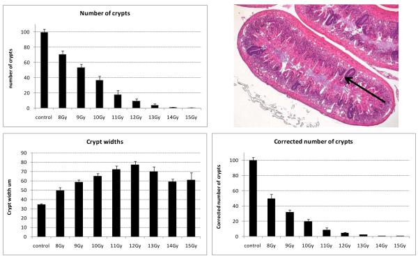
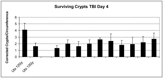
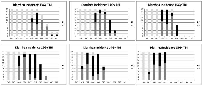
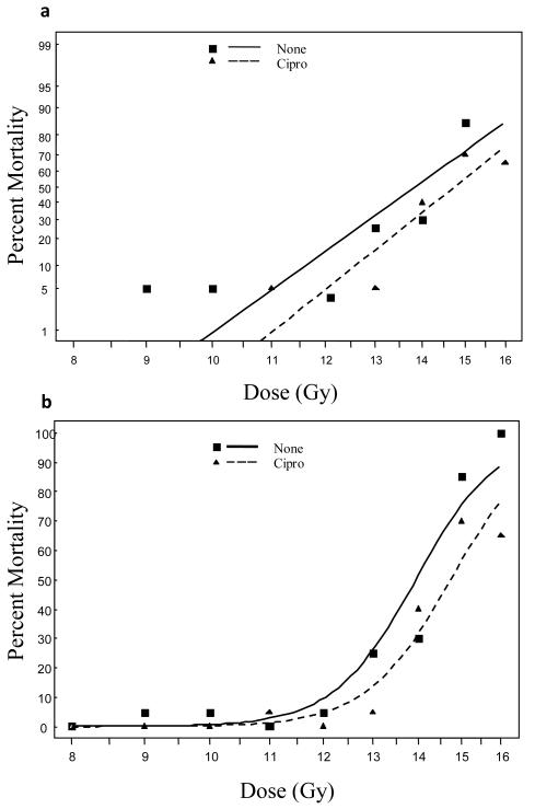
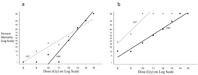
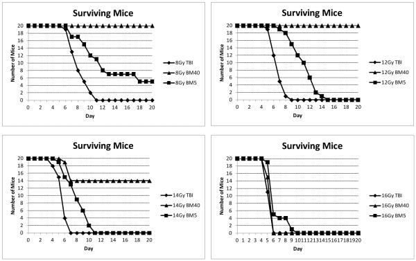
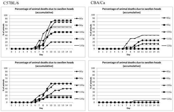
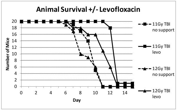
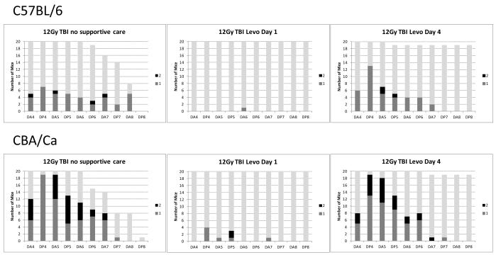
The most detailed reports of the response of the gastrointestinal system to high dose acute radiation have focused mainly on understanding the histopathology. However, to enable medical countermeasure assessment under the animal rule criteria, it is necessary to have a robust model in which the relationship between radiation dose and intestinal radiation syndrome incidence, timing, and severity are established and correlated with histopathology. Although many mortality studies have been published, they have used a variety of mouse strains, ages, radiation sources, and husbandry conditions, all of which influence the dose response. Further, it is clear that the level of bone marrow irradiation and supportive care can influence endpoints. In order to create robust baseline data, the authors have generated dose response data in adult male mice maintained under identical conditions and exposed to either total or partial-body irradiation. Partial-body irradiation includes both extensive (40%) and minimal (5%) bone marrow sparing models, the latter designed to correlate with an established primate model and allow assessment of effects of any medical countermeasure on all three major radiation syndromes (intestinal, bone marrow, and lung) in the surviving mice. Lethal dose (LD(30), LD(50), and LD(70)) data are described in the various models, along with the impact of enteric flora and response to supportive care. Correlation with diarrhea severity and histopathology are also described. These data can be used to aid the design of good laboratory practice (GLP)-compliant Animal Rule studies that are reflective of the conditions following accidental radiation exposure.


Figures

Fig. 1
Fig. 1
Radiation kills the crypt clonogenic cells. As successive clonogenic cells are killed, or reproductively sterilized, the cellular output onto the villi is reduced. Impaired cell production with continued upward cell migration leads to gradual villus and crypt shrinkage (blunting) resulting in reduced nutrient absorption and impaired barrier function. If no clonogenic cells remain in a crypt it will die and if sufficient crypts are killed ulcers will form. However, if one or more clonogenic cells remain the crypt will regenerate (B) and restore the blunted villi, ultimately regenerating the villus, with a slight ‘overshoot’ in cell production (C) before homeostasis and normal levels resume. A control crypt and villus is shown in (A). Adapted from Booth and Potten 2001.
Fig. 2
Fig. 2
Response of the small intestine to total body irradiation (H&E stained cross sections) A: x5 objective; B: x20 objective. Loss of crypts is visible fours days after 10 Gy, with increasing crypt loss with radiation dose. Two days later the effects of the rapid regeneration can be seen, with crypt numbers being restored after 10 Gy to 12 Gy. At higher doses the progeny of the few surviving crypts are migrating to try and restore the mucosal barrier, although the crypt-villus architecture is still lacking.
Fig. 2
Fig. 2
Response of the small intestine to total body irradiation (H&E stained cross sections) A: x5 objective; B: x20 objective. Loss of crypts is visible fours days after 10 Gy, with increasing crypt loss with radiation dose. Two days later the effects of the rapid regeneration can be seen, with crypt numbers being restored after 10 Gy to 12 Gy. At higher doses the progeny of the few surviving crypts are migrating to try and restore the mucosal barrier, although the crypt-villus architecture is still lacking.
Fig.3
Fig.3
Response of the small intestine to total body irradiation (H&E stained cross section; x20 objective). Illustration of the change in histopathology from days 4 to 10 following 14 Gy irradiation (H&E stained cross section; x10 objective). In order to allow survival to the latter time points and illustrate the regenerative capacity of the small and large intestine the bone marrow has been shielded in these studies.
Fig. 4
Fig. 4
There is a radiation dose-dependent crypt kill. The plots illustrate the number of surviving regenerating crypts present in a small intestinal circumference 4 days following total body irradiation of 10 to 12 week old male C57BL/6 mice. Top: The actual number of surviving regenerating crypts present. The arrow in the photograph illustrates an example of a darkly stained surviving, regenerating crypt, derived from at least one clonogenic cell, that is scored in this assay (H&E stained cross section; x5 objective). Bottom: The change in crypt width with radiation dose on day 4 post irradiation and the corrected number of crypts per intestinal circumference once the impact of width is accounted for in the scoring process (see Materials and Methods).
Fig. 5
Fig. 5
Illustration of the consistency of the mean number of surviving crypts following 13 Gy TBI per cross section in saline placebo control groups from a number of different studies over 3 years. In each case the placebo was administered 24h following irradiation, either once or daily. For comparison the number of surviving crypts in untreated irradiated mice following 12 and 13 Gy are also shown.
Fig. 6
Fig. 6
Loss of crypts is coincident with the onset of diarrhea, dehydration and mortality. The top plots illustrate the number of C57BL/6 mice per radiation dose (y axis) exhibiting diarrhea of a score 1 (mild) or 2 (severe) from days 3 to 7 (am and pm observations indicated by DA3, DP3, DA4, DP4 etc). The lower plots illustrate the same data in CBA/Ca mice, where the diarrhea occurs slightly earlier than the C57BL/6 mice. Grey bars indicate the number of animals remaining in the study.
Fig. 7
Fig. 7
a) Dose response curves using a Probit model b) Logit of C57BL/6 survival following PBI BM40. Ciprofloxacin significantly reduced the mortality rate of mice at 20 days (odds ratio = 0.389, p-value = 0.013).
Fig. 8
Fig. 8
Dose response curves using Probit models C57BL/6 and CBA/Ca survival following PBI BM5 a) day 8 and b) day 20. There was a large difference in the incidence of the swollen muzzle syndrome in the two strains of mice, with associated mortality being higher in the C57BL/6 mice.
Fig. 9
Fig. 9
Examples of animal survival in three independent studies in C57BL/6 mice receiving post-irradiation ciprofloxacin. The plots directly compare the survival time course in TBI, PBI BM5 and PBI BM40. These study data contributed towards the much larger data set used to generate the lethal dose tables.
Fig. 10
Fig. 10
The incidence of notable swollen muzzles in a single study. Two strains of mice were exposed to PBI BM5 irradiation (doses that cause H-ARS but minimal GI-ARS). Top: Unsupported animals, Bottom: Animals receiving ciprofloxacin from day 4 post irradiation. The incidence was clearly lower in the CBA/Ca mice and dramatically reduced by ciprofloxacin.
Fig. 11
Fig. 11
Effect of levofloxacin on C57BL/6 animal survival. Groups of twenty C57BL/6 adult male mice per group were irradiated with TBI. All mice received acid water but the addition of levofloxacin in the water/wetted chow was added from day 4 post irradiation. Mice were checked twice daily and euthanized when moribund or on day 15 post irradiation. The number of surviving mice per time point is plotted.
Fig. 12
Fig. 12
Effect of levofloxacin administered from either day 1 or day 4 post irradiation. levofloxacin prevented mortalities during the first 8 days post irradiation and also prevented diarrhea if administered from day 1 (+24h), but not day 4 post irradiation. This observation was consistent in two strains of adult (10-12 week) male mice. Many animals in the group receiving no antibiotic developed swollen muzzles and had faecal associated enterobacteriacae (E.coli and K.pneumoniae) faecal streptococci (S.faecalis and S.faecium) or P.aeruginosa in more than one organ. These samples also had a positive blood culture containing the same organisms, confirming the leaking of gut flora in to the blood. The infection and incidence of swollen muzzles was eliminated by levofloxacin.

References

    1. Boggs DR. The total marrow mass of the mouse: a simplified method of measurement. Am J Hematol. 1984;16(3):277–286. - PubMed
    1. Booth C, Potten CS. The Intestine as a Model for Studying Stem Cell Behaviour. In: Teicher BA, editor. Tumor Models in Cancer Research. Humana Press; Totawa, New Jersey: 2001a. pp. 337–357.
    1. Booth D, Potten CS. Protection against mucosal injury by growth factors and cytokines. J Natl Cancer Inst Monogr. 2001b;29:16–20. - PubMed
    1. Booth D, Haley JD, Bruskin AM, Potten CS. Transforming growth factor beta 3 protects murine small intestinal crypt stem cells and animal survival after irradiation, possibly by reducing stem cell cycling. Int J Cancer. 2000;86(1):53–59. - PubMed
    1. Cai WB, Roberts SA, Potten CS. The number of clonogenic cells in crypts in three regions of murine large intestine. Int J Radiat Biol. 1997a;71(5):573–579. - PubMed

Publication types