. 2012 Oct;103(4):400-10.
doi: 10.1097/hp.0b013e31826530e2.

Evidence of delayed gastrointestinal syndrome in high-dose irradiated mice

Affiliations

Evidence of delayed gastrointestinal syndrome in high-dose irradiated mice

Catherine Booth et al. Health Phys. 2012 Oct.

Abstract

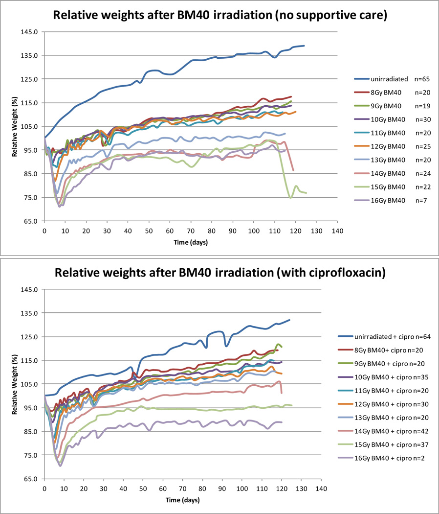
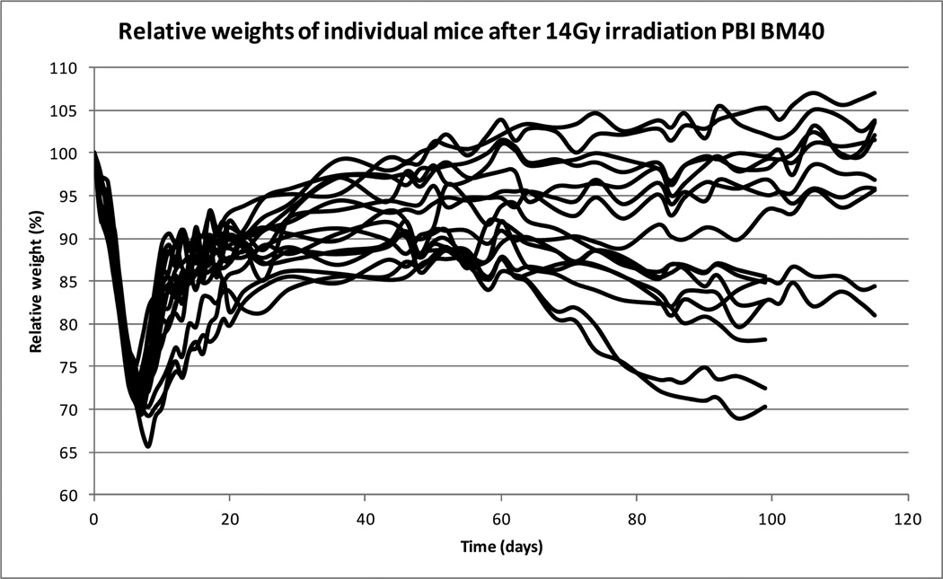
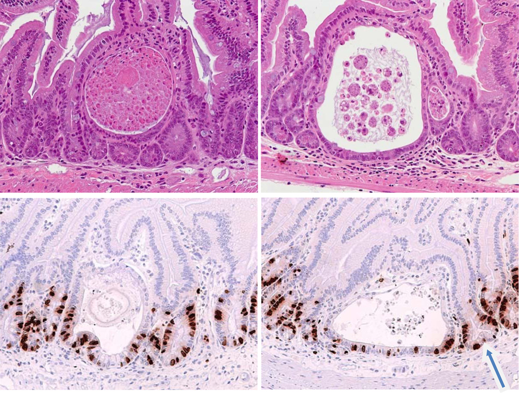
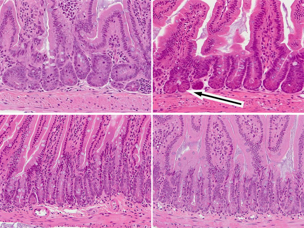
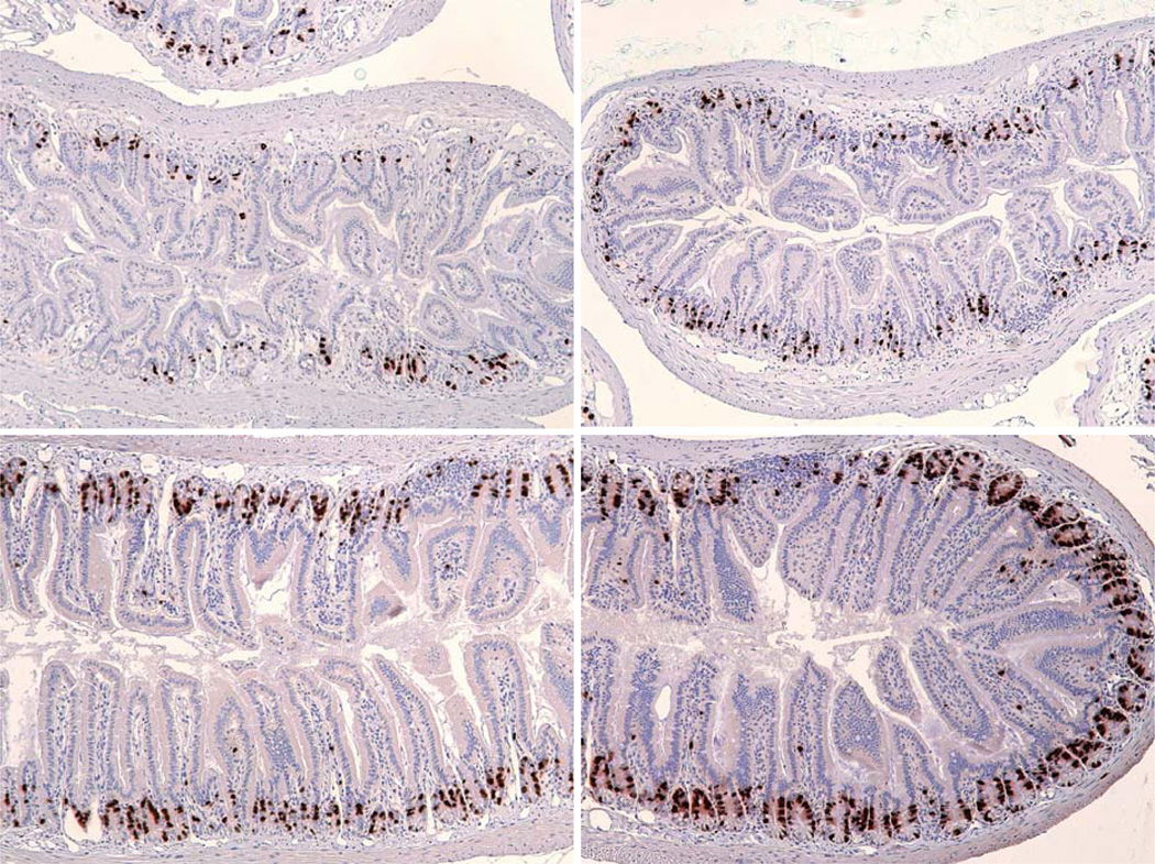
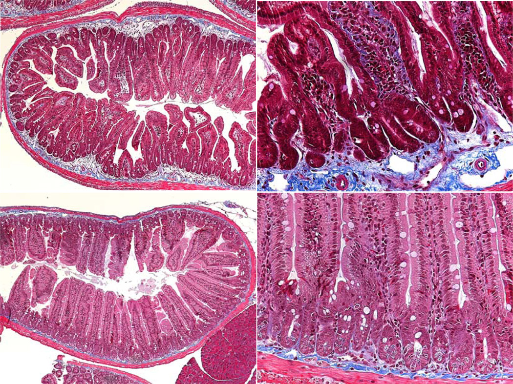
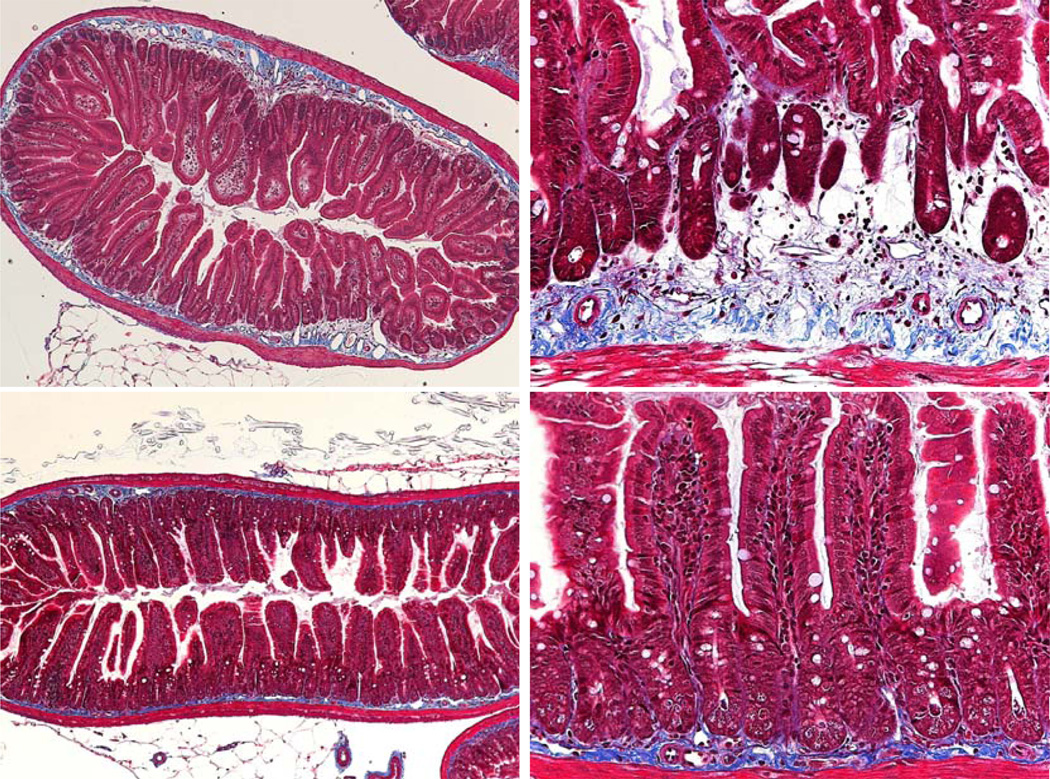
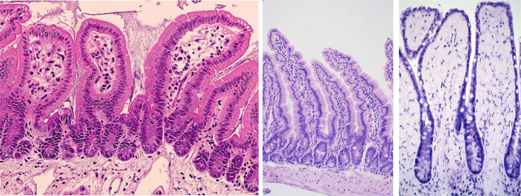
The acute effects of irradiation on the gastrointestinal (GI) system are well documented, but the longer-term effects are less well known. Increased incidence of adenocarcinoma has been noted, but apart from descriptions of fibrosis, the development of other pathologies specific to survivors of acute radiation is poorly understood. Samples were taken from C57BL/6 mice irradiated with partial-body irradiation where the thorax, head, and forelimbs were shielded (i.e., sparing 40% of the bone marrow). Tissue from age-matched controls was also collected. There were clear pathological changes in the intestine associated with DEARE (Delayed Effects of Acute Radiation Exposure) at doses greater than 12 Gy, with a dose-related increase in observed pathologies. Mice maintained on the synthetic antibiotic ciprofloxacin during the acute phase (days 4 to 20), however, had a lower or delayed incidence of symptoms. After 20 d, mice developed structures similar to early adenomas. Abnormally high levels of apoptotic and mitotic cells were present in some crypts, along with the early adenomas, suggesting tissue regeneration and areas of deregulated cell turnover. Over time, there was inhibited crypt cell proliferation in animals with advanced symptoms, a blunting of the crypts and villi, and an enlargement of villus girth, with an increasingly acellular and fibrotic extracellular matrix (a characteristic that has been demonstrated previously in aging mice). Together these changes may lead to a reduced functional surface area and less motile intestine. These observations are similar to those seen in geriatric animals, suggesting a premature aging of the GI tract.


Figures

Figure 1
Figure 1
Weight loss changes post irradiation. The number of animals shown is the number surviving after day 20 from all DEARE studies. Animals were euthanized as scheduled on day 75 or 120, or due to sickness. Animals were euthanized when weight loss exceeded 20% and the animals also exhibited moribund symptoms. Weight gain was reduced in control mice by the presence of ciprofloxacin. After an initial radiation dose dependent weight loss, animals regained weight. All radiation exposures impeded the subsequent weight gain, with the greatest impact at 13 Gy and higher. Antibiotic supportive care may be beneficial at these higher doses.
Figure 2
Figure 2
Weight loss changes post irradiation. An example of the variation within the same treatment group. Some animals appeared healthy and gained weight, whereas others lost weight and in extreme cases became moribund.
Figure 3
Figure 3
Examples of lesions similar to microadenomas in animals exposed to irradiation (typical of lesions seen from 20–200 days post irradiation at doses above 12Gy irrespective of ciprofloxacin treatment). Top: H & E sections of small intestine show examples of the abnormal pathologies seen in DEARE mice four months following 14Gy. The cyst-like structures are typical of microadenomas. Bottom: Similar structures from mice four months following 16Gy, labeled to reveal BrdU labelled cycling cells. The arrow indicates a bifurcating crypt adjacent to a microadenoma.
Figure 4
Figure 4
Examples of changed crypt cell turnover in DEARE mice at a range of doses. Top: Animals four months post irradiation. Bottom: Age matched controls. Post irradiation crypts and villi were both smaller than the controls. Several crypts contained multiple mitotic cells and high levels of crypt fission were seen (arrow). Increased levels of apoptosis (as judged by the stereotypical condensed chromatin and eosinophilic staining were often observed, often at high cell positions.
Figure 5
Figure 5
BrdU labeling in moribund DEARE mice. Mice were injected with BrdU 40 minutes prior to euthanasia and the incorporation revealed by immunohistochemistry. Top: Day 94 following 14 Gy (left), Day 192 following 15 Gy (right). Note the thicker submucosa and the shorter crypts and villi. The diameter of the intestine as a whole is also smaller. Bottom: Unirradiated controls, day 125 (left), day 200 (right).
Figure 6
Figure 6
Masson Trichrome staining in moribund DEARE mice. Top: Day 112 following 16 Gy (left) and day 119 following 14 Gy (right). Bottom: Unirradiated controls, day 125. The increased collagen (blue stain) in the irradiated mice is clearly visible.
Figure 7
Figure 7
Masson Trichrome staining in older moribund DEARE mice. Top: Day 192 following 15 Gy. Bottom: Unirradiated controls, day 200.
Figure 8
Figure 8
Example of the similarity between aged and DEARE mice. Left: H & E section of a 15 Gy day 192 sample. The villi are short and wide with fewer cells in the lamina propria. Mid: A control 5 month old mouse with longer villi and normal looking lamina propria. Right: A 32 month old mouse. The broad spade-like villi result in a reduced absorptive surface area and a less flexible structure, both of which contribute to a failure to thrive.

Similar articles

Cited by

References

    1. Andreyev J. Gastrointestinal complications of pelvic radiotherapy: Are they of importance. Gut. 2005;54:1051–1054. - PMC - PubMed
    1. Boggs DR. The total marrow mass of the mouse: a simplified method of measurement. Am J Hematol. 1984;16(3):277–286. - PubMed
    1. Coia LR, Myerson RJ, Tepper JE. Late effects of radiation therapy on the gastrointestinal tract. Intl J Radiat Oncol Biol Phys. 1995;31(5):1213–1236. - PubMed
    1. Dubios A, Walker RI. Prospects for management of gastrointestinal injury associated with the acute radiation syndrome. Gastroenterology. 1988;95:500–507. - PubMed
    1. Gaugler MH, Vereycken-Holler V, Squiban C, Vandamme M, Vozenin-Brotons MC, Benderitter M. Pravastatin limits endothelial activation after irradiation and decreases the resulting inflammatory and thrombotic responses. Radiat Res. 2005;163(5):479–487. - PubMed

Publication types

MeSH terms