. 2012 Nov 14;134(45):18499-502.
doi: 10.1021/ja307689w. Epub 2012 Nov 2.

Photocleavable DNA barcode-antibody conjugates allow sensitive and multiplexed protein analysis in single cells

Affiliations

Photocleavable DNA barcode-antibody conjugates allow sensitive and multiplexed protein analysis in single cells

Sarit S Agasti et al. J Am Chem Soc. .

Abstract

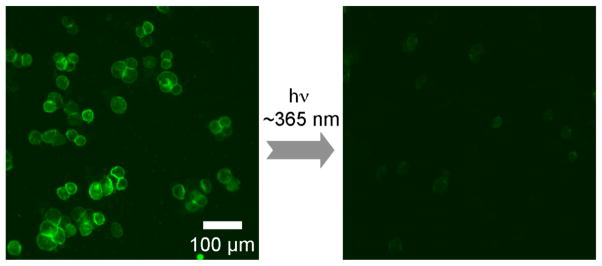
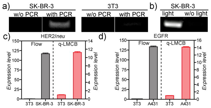
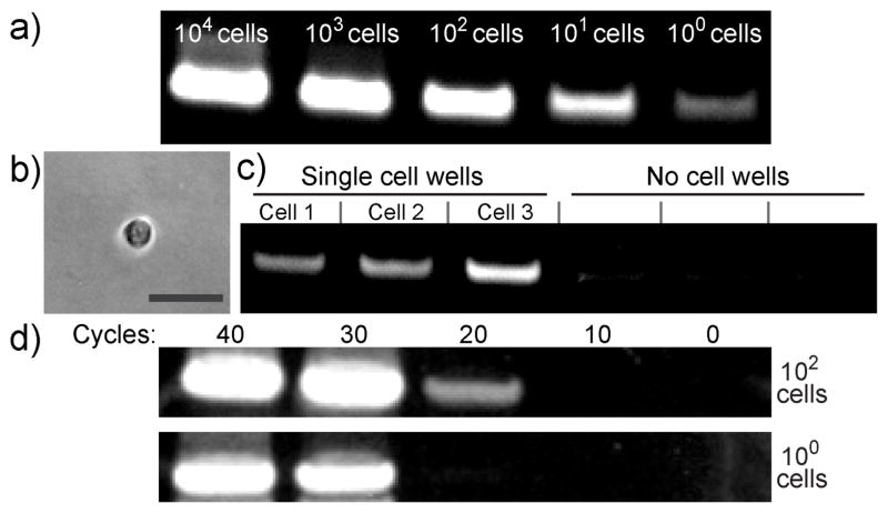
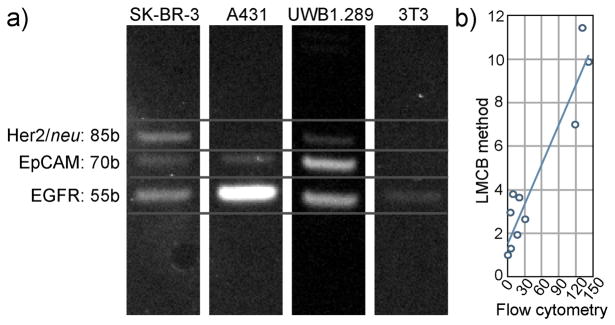
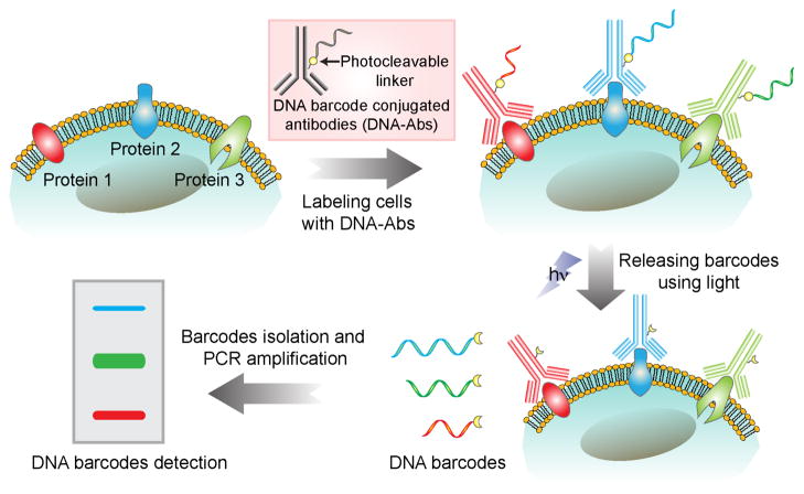
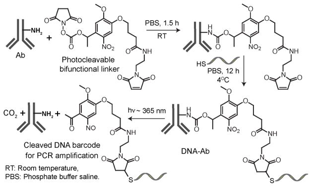
DNA barcoding is an attractive technology, as it allows sensitive and multiplexed target analysis. However, DNA barcoding of cellular proteins remains challenging, primarily because barcode amplification and readout techniques are often incompatible with the cellular microenvironment. Here we describe the development and validation of a photocleavable DNA barcode-antibody conjugate method for rapid, quantitative, and multiplexed detection of proteins in single live cells. Following target binding, this method allows DNA barcodes to be photoreleased in solution, enabling easy isolation, amplification, and readout. As a proof of principle, we demonstrate sensitive and multiplexed detection of protein biomarkers in a variety of cancer cells.


Figures

Figure 1
Figure 1
Fluorescently labeled DNA barcodes conjugated to anti-HER2 antibodies were used to stain SK-BR-3 cells. A decreased fluorescence signal after light irradiation demonstrated that barcodes were released from the labeled cells. See Figure S4 for additional images including bright field images.
Figure 2
Figure 2
a) Detection of HER2/neu in SK-BR-3 cells. After 25 cycles of PCR, a DNA band corresponding to the 85-base bar-code was visible. Control 3T3 cells, consistent with their low expression of HER2/neu, had a minimal 85-base DNA band even after PCR amplification. b) No DNA band was detected in the absence of light irradiation, which demonstrated the critical role of light in the assay method. c) HER2/neu expression and d) EGFR expression (both relative to the control 3T3 cells) from qLMCB correlated well with results from standard flow cytometry-based detection. Error bars represent variation between duplicate measurements.
Figure 3
Figure 3
Detection sensitivity of the LMCB method. a) After 25 cycles of PCR, the DNA barcodes from samples containing varying numbers of SK-BR-3 cells were detected b) Image showing a single SK-BR-3 cell inside a microplate well for digital analysis (scale bar 50 μm). c) Analysis of a single SK-BR-3 cell using the LMCB method in digital format. Following PCR amplification, an 85-base barcode could be detected in individual wells containing single cells (‘single cell wells’). In contrast, wells in which cells were absent (‘no cell wells’) failed to produce a significant band following amplification. d) Gel electrophoresis results showing the detection sensitivity of the LMCB method as a function of PCR cycle number.
Figure 4
Figure 4
a) Multiplexed protein detection using LMCB method. Individual biomarker signals (corresponding to their expression level) can be clearly distinguished from one other based on their barcode size. b) Comparison of LMCB measurements (from Figure 4a) and measurements taken by flow cytometry. For each biomarker, band intensities (normalized to control 3T3 cells) from the gel were plotted against fluorescent intensities (normalized to control 3T3 cells) from flow cytometry (R2 = 0.90).
Scheme 1
Scheme 1
Schematic showing the light-mediated cellular bar-coding strategy. Protein targets were labeled with DNA-Abs and then photocleaved to release DNA barcodes. Amplified bar-codes were analyzed using gel electrophoresis for multiplexed detection of protein biomarkers from single cells.
Scheme 2
Scheme 2
Synthetic scheme of the antibody DNA conjugation and the photocleavage reaction leading to the barcode release.

References

    1. Shackleton M, Quintana E, Fearon ER, Morrison SJ. Cell. 2009;138:822–829. - PubMed
    2. Tay S, Hughey JJ, Lee TK, Lipniacki T, Quake SR, Covert MW. Nature. 2010;466:267–271. - PMC - PubMed
    3. Abbas HA, Pant V, Lozano G. Cell Cycle. 2011;10:3257–3262. - PMC - PubMed
    1. Yu M, Stott S, Toner M, Maheswaran S, Haber DA. J Cell Biol. 2011;192:373–382. - PMC - PubMed
    2. Issadore D, Chung J, Shao H, Liong M, Ghazani AA, Castro CM, Weissleder R, Lee H. Sci Transl Med. 2012;4:141ra92. - PMC - PubMed
    1. Wu M, Singh AK. Curr Opin Biotechnol. 2012;23:83–88. - PMC - PubMed
    2. Altelaar AF, Heck AJ. Curr Opin Chem Biol. 2012;16:206–213. - PubMed
    1. Koob AO, Bruns L, Prassler C, Masliah E, Klopstock T, Bender A. Anal Biochem. 2012;425:120–124. - PMC - PubMed
    2. Homola J. Chem Rev. 2008;108:462–493. - PubMed
    3. Thurer R, Vigassy T, Hirayama M, Wang J, Bakker E, Pretsch E. Anal Chem. 2007;79:5107–5110. - PubMed
    4. Ellerbee AK, Phillips ST, Siegel AC, Mirica KA, Martinez AW, Striehl P, Jain N, Prentiss M, Whitesides GM. Anal Chem. 2009;81:8447–8452. - PubMed
    5. Haun JB, Castro CM, Wang R, Peterson VM, Marinelli BS, Lee H, Weissleder R. Sci Transl Med. 2011;3:71ra16. - PMC - PubMed
    6. Rosi NL, Mirkin CA. Chem Rev. 2005;105:1547–1562. - PubMed
    7. Rotem D, Jayasinghe L, Salichou M, Bayley H. J Am Chem Soc. 2012;134:2781–2787. - PMC - PubMed
    1. Bendall SC, Simonds EF, Qiu P, Amir e-AD, Krutzik PO, Finck R, Bruggner RV, Melamed R, Trejo A, Ornatsky OI, Balderas RS, Plevritis SK, Sachs K, Pe’er D, Tanner SD, Nolan GP. Science. 2011;332:687–696. - PMC - PubMed
    2. Shi Q, Qin L, Wei W, Geng F, Fan R, Shin YS, Guo D, Hood L, Mischel PS, Heath JR. Proc Natl Acad Sci U S A. 2012;109:419–424. - PMC - PubMed

Publication types