

Human embryonic stem cell-derived mesenchymal stem cell seeding on calcium phosphate cement-chitosan-RGD scaffold for bone repair
- PMID: 23092172
- PMCID: PMC3589899
- DOI: 10.1089/ten.TEA.2012.0172
Human embryonic stem cell-derived mesenchymal stem cell seeding on calcium phosphate cement-chitosan-RGD scaffold for bone repair
Abstract
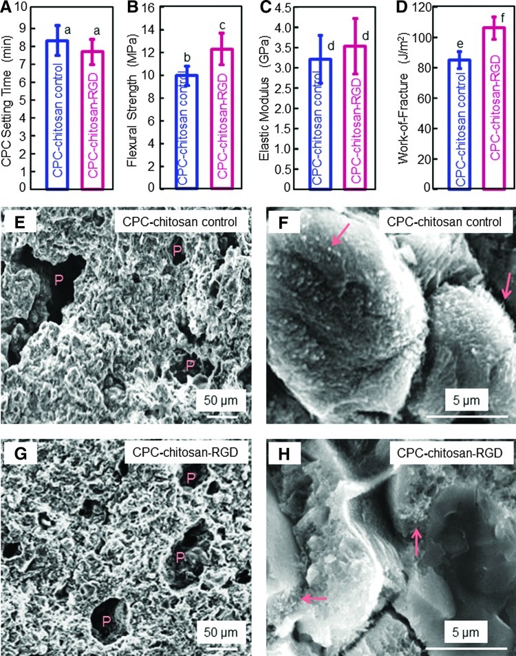
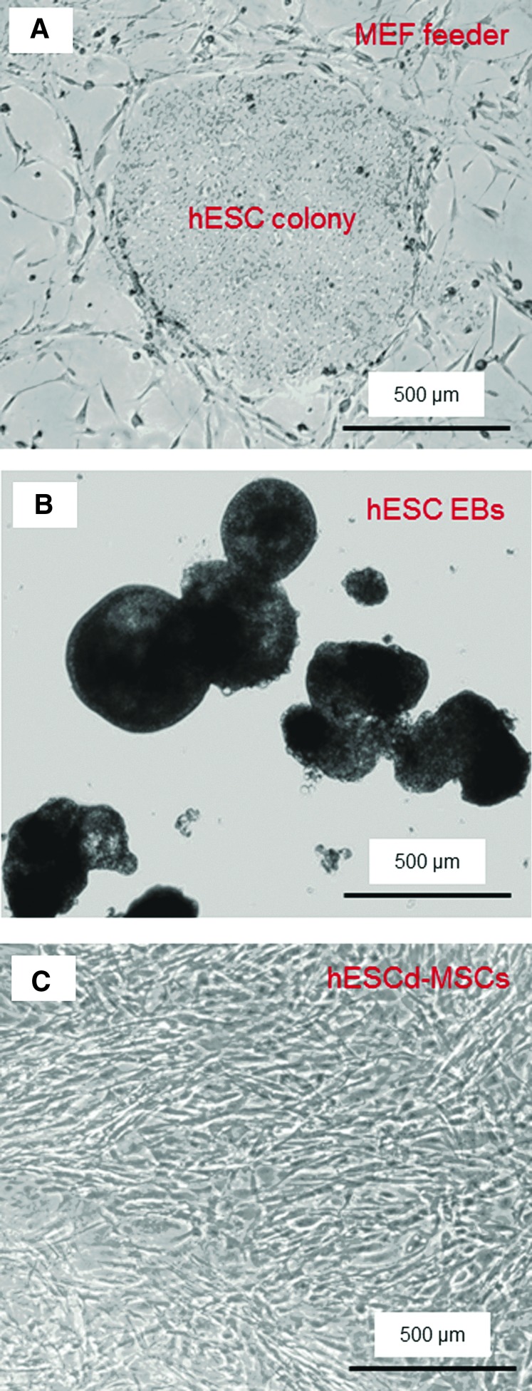
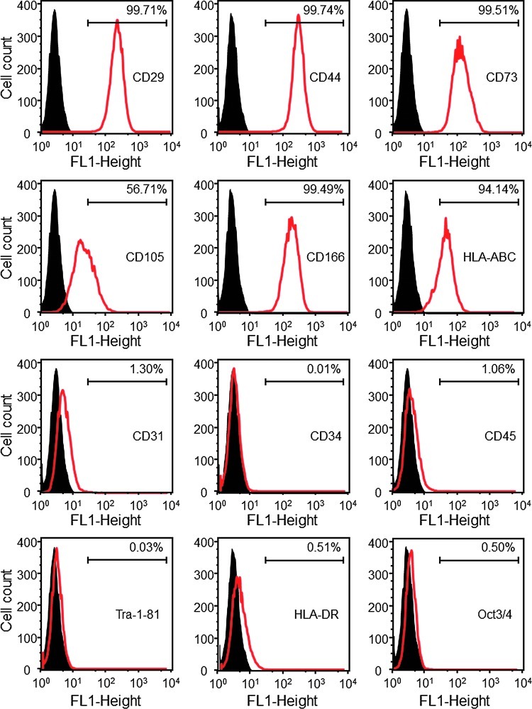
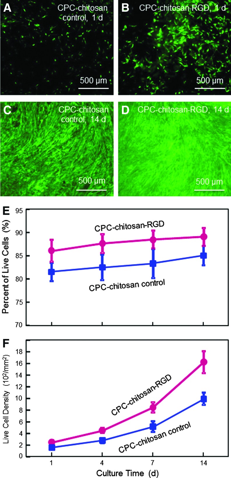
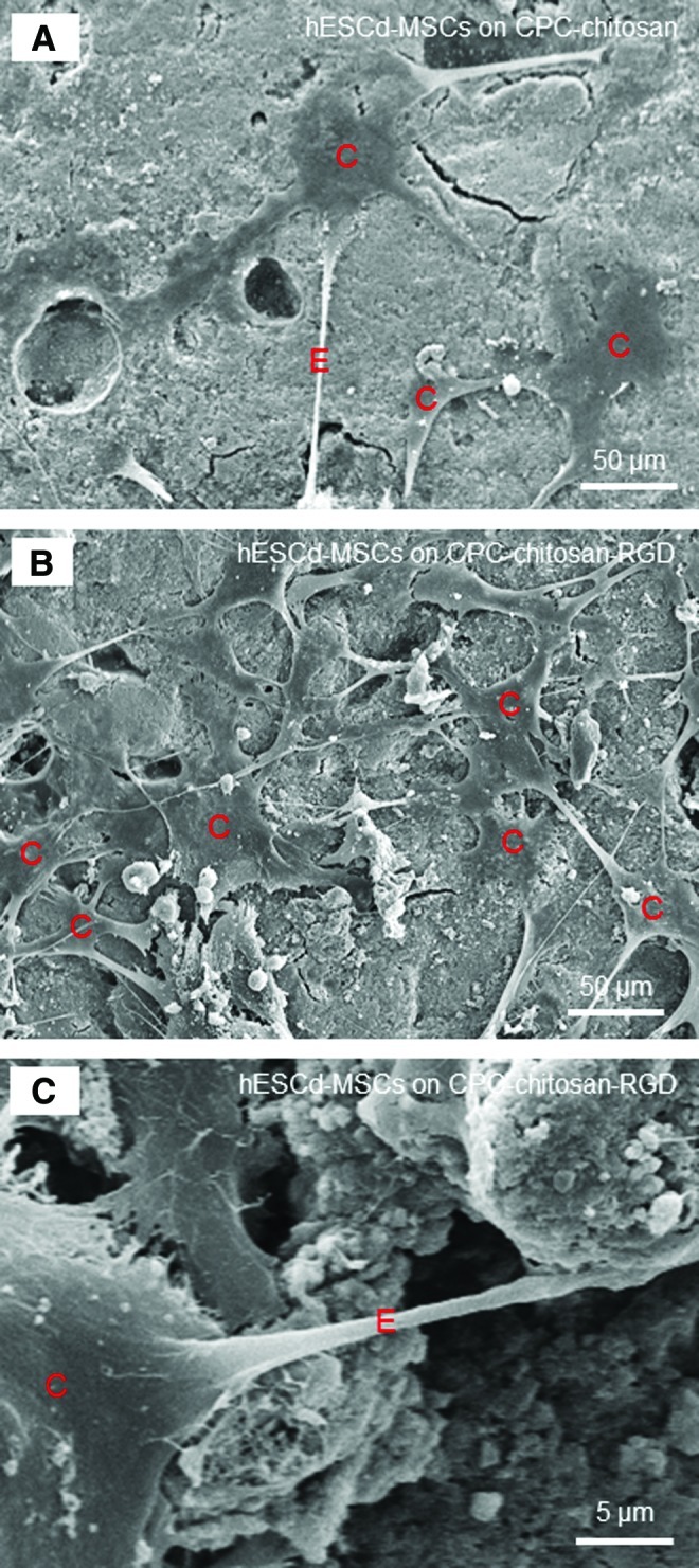
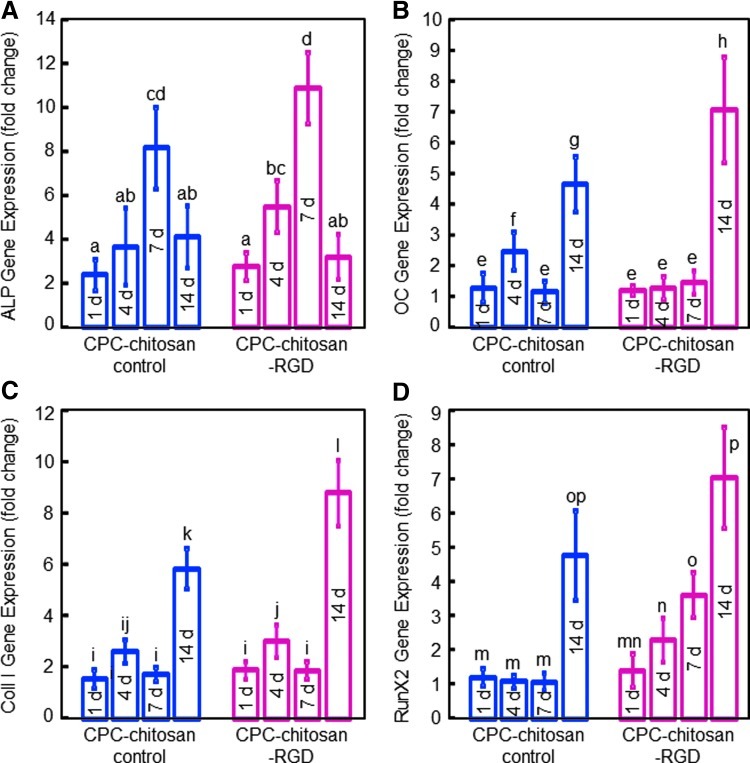
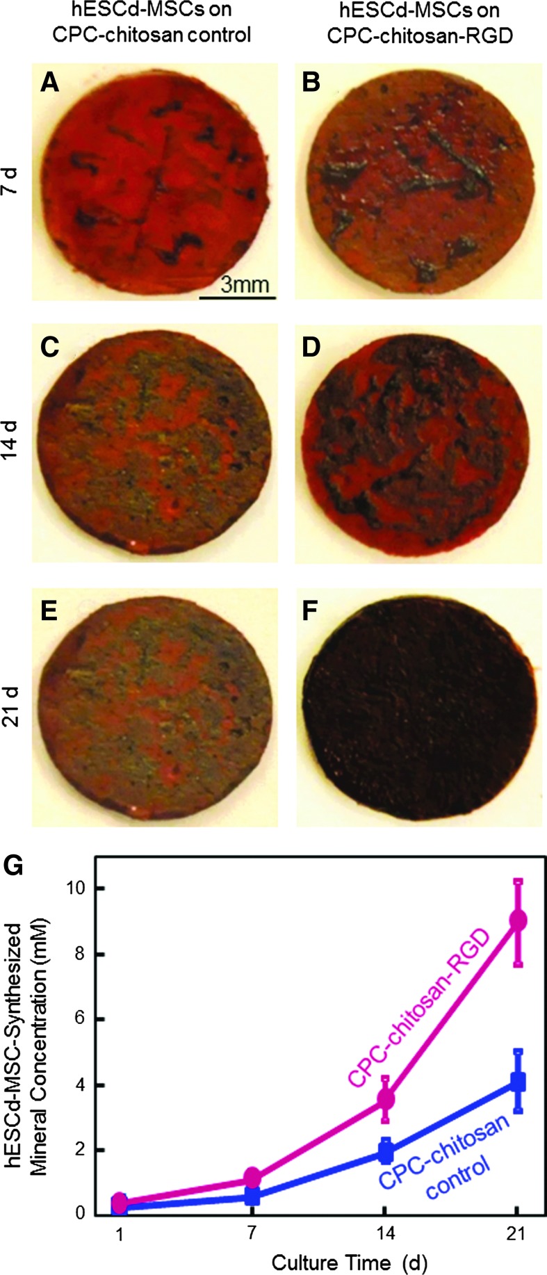
Calcium phosphate cement (CPC) has in situ-setting ability and excellent osteoconductivity. Human embryonic stem cells (hESCs) are exciting for regenerative medicine due to their strong proliferative ability and multilineage differentiation capability. However, there has been no report on hESC seeding with CPC. The objectives of this study were to obtain hESC-derived mesenchymal stem cells (hESCd-MSCs), and to investigate hESCd-MSC proliferation and osteogenic differentiation on novel CPC with chitosan immobilized with RGD (CPC-chitosan-RGD). RGD was covalently bonded with chitosan, which was then incorporated into CPC. The CPC-chitosan-RGD scaffold had higher strength and toughness than CPC-chitosan control without RGD (p<0.05). hESCs were cultured to form embryoid bodies (EBs), and the MSCs were then migrated out of the EBs. Flow cytometry indicated that the hESCd-MSCs expressed typical surface antigen profile of MSCs. hESCd-MSCs had good viability when seeded on CPC scaffolds. The percentage of live cells and the cell density were significantly higher on CPC-chitosan-RGD than CPC-chitosan control. Scanning electron microscope examination showed hESCd-MSCs with a healthy spreading morphology adherent to CPC. hESCd-MSCs expressed high levels of osteogenic markers, including alkaline phosphatase, osteocalcin, collagen I, and Runx2. The mineral synthesis by the hESCd-MSCs on the CPC-chitosan-RGD scaffold was twice that for CPC-chitosan control. In conclusion, hESCs were successfully seeded on CPC scaffolds for bone tissue engineering. The hESCd-MSCs had good viability and osteogenic differentiation on the novel CPC-chitosan-RGD scaffold. RGD incorporation improved the strength and toughness of CPC, and greatly enhanced the hESCd-MSC attachment, proliferation, and bone mineral synthesis. Therefore, the hESCd-MSC-seeded CPC-chitosan-RGD construct is promising to improve bone regeneration in orthopedic and craniofacial applications.
Figures
Similar articles
-
Synergistic effects of 3D chitosan-based hybrid scaffolds and mesenchymal stem cells in orthopaedic tissue engineering.IET Nanobiotechnol. 2023 Apr;17(2):41-48. doi: 10.1049/nbt2.12103. Epub 2023 Jan 28. IET Nanobiotechnol. 2023. PMID: 36708277 Free PMC article. Review.
-
Human induced pluripotent stem cell-derived mesenchymal stem cell seeding on calcium phosphate scaffold for bone regeneration.Tissue Eng Part A. 2014 Apr;20(7-8):1295-305. doi: 10.1089/ten.TEA.2013.0211. Epub 2014 Jan 7. Tissue Eng Part A. 2014. PMID: 24279868 Free PMC article.
-
Human embryonic stem cell encapsulation in alginate microbeads in macroporous calcium phosphate cement for bone tissue engineering.Acta Biomater. 2012 Sep;8(9):3436-45. doi: 10.1016/j.actbio.2012.05.016. Epub 2012 May 22. Acta Biomater. 2012. PMID: 22633970 Free PMC article.
-
Collagen-calcium phosphate cement scaffolds seeded with umbilical cord stem cells for bone tissue engineering.Tissue Eng Part A. 2011 Dec;17(23-24):2943-54. doi: 10.1089/ten.tea.2010.0674. Epub 2011 Aug 18. Tissue Eng Part A. 2011. PMID: 21851269 Free PMC article.
-
Stem Cells and Calcium Phosphate Cement Scaffolds for Bone Regeneration.J Dent Res. 2014 Jul;93(7):618-25. doi: 10.1177/0022034514534689. Epub 2014 May 5. J Dent Res. 2014. PMID: 24799422 Free PMC article. Review.
Cited by
-
Stem Cells in Skeletal Tissue Engineering: Technologies and Models.Curr Stem Cell Res Ther. 2016;11(6):453-474. doi: 10.2174/1574888x10666151001115248. Curr Stem Cell Res Ther. 2016. PMID: 26423296 Free PMC article. Review.
-
An on-site preparable, novel bone-grafting complex consisting of human platelet-rich fibrin and porous particles made of a recombinant collagen-like protein.J Biomed Mater Res B Appl Biomater. 2019 Jul;107(5):1420-1430. doi: 10.1002/jbm.b.34234. Epub 2018 Oct 1. J Biomed Mater Res B Appl Biomater. 2019. PMID: 30270545 Free PMC article.
-
Synergistic effects of 3D chitosan-based hybrid scaffolds and mesenchymal stem cells in orthopaedic tissue engineering.IET Nanobiotechnol. 2023 Apr;17(2):41-48. doi: 10.1049/nbt2.12103. Epub 2023 Jan 28. IET Nanobiotechnol. 2023. PMID: 36708277 Free PMC article. Review.
-
Priming approaches to improve the efficacy of mesenchymal stromal cell-based therapies.Stem Cell Res Ther. 2019 May 2;10(1):131. doi: 10.1186/s13287-019-1224-y. Stem Cell Res Ther. 2019. PMID: 31046833 Free PMC article. Review.
-
The Osteogenic Differentiation Effect of the FN Type 10-Peptide Amphiphile on PCL Fiber.Int J Mol Sci. 2018 Jan 4;19(1):153. doi: 10.3390/ijms19010153. Int J Mol Sci. 2018. PMID: 29300346 Free PMC article.
References
-
- Cutter C.S. Mehrara B.J. Bone grafts and substitutes. J Long Term Eff Med Implants. 2006;16:249. - PubMed
-
- Johnson P.C. Mikos A.G. Fisher J.P. Jansen J.A. Strategic directions in tissue engineering. Tissue Eng. 2007;13:2827. - PubMed
-
- Laurencin C.T. Ambrosio A.M. Borden M.D. Cooper J.A., Jr Tissue engineering: orthopedic applications. Annu Rev Biomed Eng. 1999;1:19. - PubMed
-
- Mistry A.S. Mikos A.G. Tissue engineering strategies for bone regeneration. Adv Biochem Eng Biotechnol. 2005;94:1. - PubMed
Publication types
MeSH terms
Substances
Grants and funding
LinkOut - more resources
Full Text Sources
Other Literature Sources
