Review
. 2013 Jan 24:8:241-76.
doi: 10.1146/annurev-pathol-020712-163930. Epub 2012 Oct 22.

Host responses in tissue repair and fibrosis

Affiliations
Review

Host responses in tissue repair and fibrosis

Jeremy S Duffield et al. Annu Rev Pathol. .

Abstract

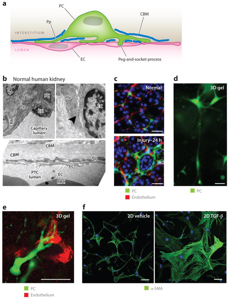
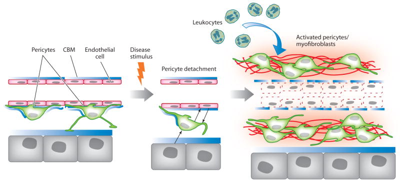
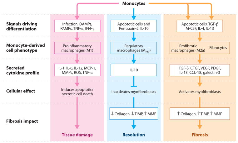
Myofibroblasts accumulate in the spaces between organ structures and produce extracellular matrix (ECM) proteins, including collagen I. They are the primary "effector" cells in tissue remodeling and fibrosis. Previously, leukocyte progenitors termed fibrocytes and myofibroblasts generated from epithelial cells through epithelial-to-mesenchymal transition (EMT) were considered the primary sources of ECM-producing myofibroblasts in injured tissues. However, genetic fate mapping experiments suggest that mesenchyme-derived cells, known as resident fibroblasts, and pericytes are the primary precursors of scar-forming myofibroblasts, whereas epithelial cells, endothelial cells, and myeloid leukocytes contribute to fibrogenesis predominantly by producing key fibrogenic cytokines and by promoting cell-to-cell communication. Numerous cytokines derived from T cells, macrophages, and other myeloid cell populations are important drivers of myofibroblast differentiation. Monocyte-derived cell populations are key regulators of the fibrotic process: They act as a brake on the processes driving fibrogenesis, and they dismantle and degrade established fibrosis. We discuss the origins, modes of activation, and fate of myofibroblasts in various important fibrotic diseases and describe how manipulation of macrophage activation could help ameliorate fibrosis.


Figures

Figure 1
Figure 1
(a) Electron microscope image of myofibroblasts (MFs) in the interstitial space of a kidney from a patient with chronic kidney disease. Note the abundance of rough endoplasmic reticulum in these cells due to high ribosomal activity, and note the markedly expanded interstitial space with collagen fibers. Abbreviations: CBM, capillary basement membrane; EC, endothelial cell; PTC, peritubular capillary; TBM, tubule basement membrane. (b) Confocal image of α–smooth muscle actin–expressing MFs (red ) in adult diseased mouse kidney. Abbreviations: a, arteriole; g, glomerulus.
Figure 2
Figure 2
Mesenchyme cells in normal organs of the mouse. Confocal fluorescence images showing normal adult liver, kidney, lung, and heart with fluorescent labels of the resident mesenchyme cells. All the cells lack markers of leukocytes, endothelial cells, and epithelial cells. The kidney image shows pericytes ( green), endothelium (red ), basement membrane (blue), and nuclei (white). The liver image shows hepatic stellate cells labeled with anti-PDGFR-β (platelet-derived growth factor receptor β) antibodies (red ) and nuclei (blue). The heart image shows normal lung fibroblasts labeled with anti-PDGFR-β antibodies ( green) and SM22 (red ). The lung image shows alveolar spaces with two populations of stromal cells, one expressing collagen Iα1 ( green) and the other PDGFR-β (red ). Scale bars, 25 μm. Images reproduced courtesy of Dr. Michelle Tallquist, University of Texas, San Antonio.
Figure 3
Figure 3
Genetic fate mapping of mesenchymal progenitors in normal adult and injured kidney by use of the Foxd1-Cre;Rosa26-TdTomato-R mouse. (Top) The cross of the Foxd1–Cre recombinase allele with the TdTomato reporter allele, driven by the universal promoters at the Rosa26 locus. Bigenic mice recombine genomic DNA at the Rosa locus only in cells that have activated Foxd1 in nephrogenesis. (Bottom) Confocal images of kidney cortex in normal adult kidney show large numbers of perivascular cells, all of which coexpress platelet-derived growth factor receptor β (PDGFR-β). Vascular smooth muscle cells of the kidney arterioles are also derived from Foxd1 progenitors and coexpress α–smooth muscle actin (α-SMA) intermediate filament in normal kidney, but none of the Foxd1-derived pericytes (arrowheads) or perivascular fibroblasts (arrows) express α-SMA. In kidney injury [shown here is unilateral ureteral obstruction (UUO) day 7], the pericyte and perivascular fibroblast populations expand and continue to express PDGFR-β. However, the entire expanded population of interstitial Foxd1 progenitor–derived cells coexpress α-SMA, the marker that defines these cells as myofibroblasts. Abbreviations: a, arteriole; GFP, green fluorescent protein.
Figure 4
Figure 4
Pericytes (PCs) in the kidney: definitions, functions, and response to cytokines. (a) A PC attached to an endothelial cell (EC) and partially embedded in a duplication of the capillary basement membrane. Note the specific attachment of PC processes (Pp) and the cell body to the EC at several different sites. (b) Electron microscope images of normal human kidney cortex. Shown are peritubular capillaries (PTCs), in which ECs and PCs or Pp are visible. The tubule basement membrane is clearly visible. Note the peg-and-socket process (arrowhead ) at the upper right. (c) PCs attach to ECs in normal mouse kidney, but only 24 h (prior to mitosis) after the onset of obstructive injury, the PCs detach, spread, and migrate from the ECs. (d) PCs in three-dimensional (3D) collagen gel that exhibit long cytoplasmic processes that extend the length of more than 10 cell bodies. (e) PCs in 3D collagen gel home to and bind by attachment specifically to capillary tubes composed of endothelial cells. (f) Kidney PCs cultured on gelatin matrix, stained for α–smooth muscle actin (α-SMA). Note that, in control conditions, PCs show weak α-SMA expression and many long fine processes and elongations, but 24 h after exposure to transforming growth factor (TGF)-β, the PCs change shape, spread, lose their long cytoplasmic processes, upregulate α-SMA, and show distinct cytoplasmic filaments. Scale bars, 25 μm. Abbreviations: 2D, two-dimensional; CBM, capillary basement membrane; RBC, red blood cell.
Figure 5
Figure 5
Schema of pericyte activation by a disease stimulus (based around kidney injury). In response to injury, pericytes become activated and detach from capillaries. This process requires bidirectional signaling between endothelial cells and pericytes. Epithelial cells can also signal to pericytes, and it is unknown whether pericytes signal to epithelial cells. In the presence of persistent injury, activated pericytes proliferate, migrate, and activate genes that give them the myofibroblast phenotype, including upregulated expression of pathological matrix genes, contractile machinery, and immune response genes. This process results in pathological matrix deposition in the virtual interstitial space; recruitment of inflammatory cells; and the loss of pericyte coverage of the endothelial cells, which causes an unstable endothelium that in turn leads to dysangiogenesis and, potentially, rarefaction. Abbreviation: CBM, capillary basement membrane.
Figure 6
Figure 6
Monocyte/macrophage activation pathways involved in fibrosis progression and resolution. Monocytes are recruited to sites of tissue injury and differentiate into distinct specialized effector macrophage populations, depending on the extracellular milieu present at the site of injury. These different effector cell populations can have dramatically different impacts on fibrosis initiation, propagation, and resolution. (Left) Monocytes promote the initiation of fibrosis through differentiation into M1-type macrophages that release cytokines and reactive oxygen species (ROS) that cause additional local tissue injury, and they promote myofibroblast resistance to apoptosis. (Center) Monocytes promote the resolution of fibrotic disease through differentiation into regulatory macrophages (Mreg) that inactivate myofibroblasts and inhibit M1- and M2-type macrophages through local production of interleukin (IL)-10 and/or Arginase-1. (Right) Monocytes promote the progression of fibrotic disease through differentiation into profibrotic (M2a-like) macrophages and fibrocytes that produce various fibroblast stimulatory growth factors and cytokines. Abbreviations: CTGF, connective tissue growth factor; DAMP, damage-associated molecular pattern; IFN, interferon; M-CSF, macrophage colony-stimulating factor; MCP, monocyte chemotactic protein; MMP, matrix metalloproteinase; PAMP, pathogen-associated molecular pattern; PDGF, platelet-derived growth factor; TIMP, tissue inhibitor of metalloproteinase; TGF, transforming growth factor; TNF, tumor necrosis factor; VEGF, vascular endothelial growth factor.

Similar articles

Cited by

References

    1. Levental KR, Yu H, Kass L, Lakins JN, Egeblad M, et al. Matrix crosslinking forces tumor progression by enhancing integrin signaling. Cell. 2009;139:891–906. - PMC - PubMed
    1. Burmolle M, Thomsen TR, Fazli M, Dige I, Christensen L, et al. Biofilms in chronic infections—a matter of opportunity—monospecies biofilms in multispecies infections. FEMS Immunol Med Microbiol. 2010;59:324–36. - PubMed
    1. Wynn TA. Cellular and molecular mechanisms of fibrosis. J Pathol. 2008;214:199–210. - PMC - PubMed
    1. Wynn TA, Barron L. Macrophages: master regulators of inflammation and fibrosis. Semin Liver Dis. 2010;30:245–57. - PMC - PubMed
    1. Hinz B, Phan SH, Thannickal VJ, Prunotto M, Desmouliere A, et al. Recent developments in myofibroblast biology: paradigms for connective tissue remodeling. Am J Pathol. 2012;180:1340–55. - PMC - PubMed

Publication types