Review
. 2013 Feb;61(2):101-15.
doi: 10.1369/0022155412467635. Epub 2012 Oct 23.

Endothelial tip cells in ocular angiogenesis: potential target for anti-angiogenesis therapy

Affiliations
Review

Endothelial tip cells in ocular angiogenesis: potential target for anti-angiogenesis therapy

Martin J Siemerink et al. J Histochem Cytochem. 2013 Feb.

Abstract

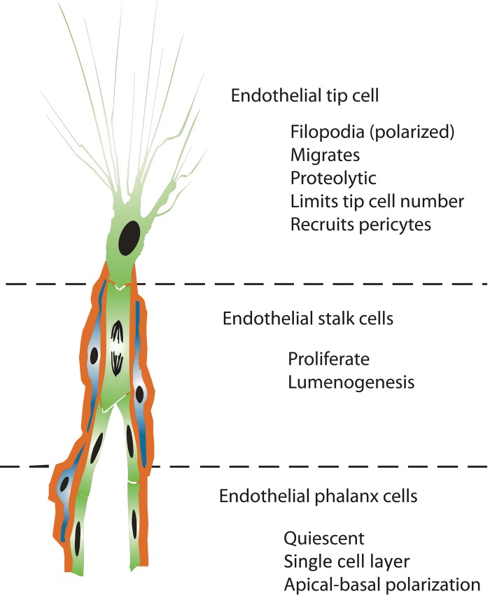
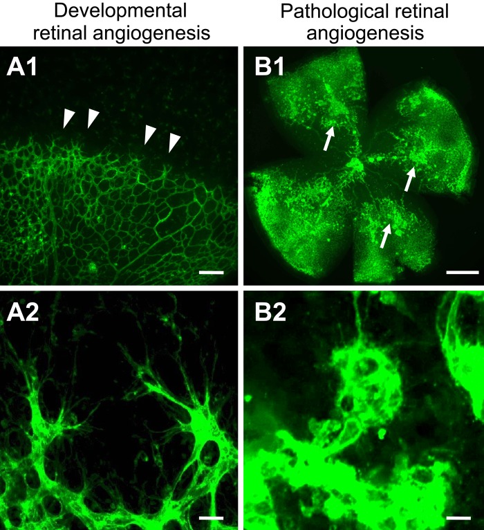
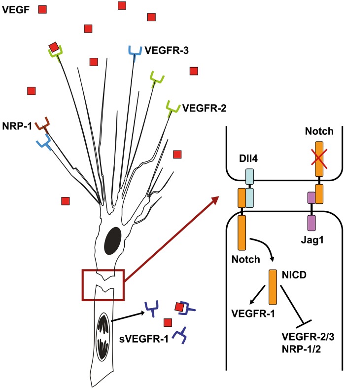
Endothelial tip cells are leading cells at the tips of vascular sprouts coordinating multiple processes during angiogenesis. In the developing retina, tip cells play a tightly controlled, timely role in angiogenesis. In contrast, excessive numbers of tip cells are a characteristic of the chaotic pathological blood vessels in proliferative retinopathies. Tip cells control adjacent endothelial cells in a hierarchical manner to form the stalk of the sprouting vessel, using, among others, the VEGF-DLL-Notch signaling pathway, and recruit pericytes. Tip cells are guided toward avascular areas by signals from the local extracellular matrix that are released by cells from the neuroretina such as astrocytes. Recently, tip cells were identified in endothelial cell cultures, enabling identification of novel molecular markers and mechanisms involved in tip cell biology. These mechanisms are relevant for understanding proliferative retinopathies. Agents that primarily target tip cells can block pathological angiogenesis in the retina efficiently and safely without adverse effects. A striking example is platelet-derived growth factor, which was recently shown to be an efficacious additional target in the treatment of retinal neovascularization. Here we discuss these and other tip cell-based strategies with respect to their potential to treat patients with ocular diseases dominated by neovascularization.


Conflict of interest statement

Declaration of Conflicting Interests: The authors declared no potential conflicts of interest with respect to the research, authorship, and/or publication of this article.

Figures

Figure 1.
Figure 1.
Concept of endothelial cell differentiation during angiogenesis. Angiogenic sprouts are formed by a subset of specialized endothelial cell phenotypes (green), each with a distinct cellular fate. Pericytes (blue) are instantly recruited to ensheathe the sprouting vessel and to produce a basal lamina (red).
Figure 2.
Figure 2.
Tip cells are actively generated in physiological and pathological conditions of the retina. Confocal images of blood vessels from mouse retinas stained with Alexa 488-conjugated isolectin-B4. (A1, A2) Retinal wholemount from postnatal day 5 shows that the superficial vascular plexus is formed by radial outgrowth of vessels from the optic nerve into the periphery. (A1) Tip cells are located at the angiogenic front (arrowheads). Scale bar = 50 µm. (A2) High magnification of tip cells with their typical morphology (highly polarized nature and numerous filopodia probing the environment). Scale bar = 20 µm. (B1, B2) Retinal wholemount from postnatal day 17 in the oxygen-induced retinopathy model when the maximum severity of the pathological neovascularization is reached during relative hypoxia. (B1) Pathological neovessels leave the retina and grow into the vitreous cavity to form unorganized, small-caliber vessels, termed preretinal tufts (arrows). Scale bar = 500 µm. (B2) High magnification of an epiretinal tuft that is formed by activated endothelial cells that extend numerous filopodia in all directions. Scale bar = 20 µm.
Figure 3.
Figure 3.
Schematic representation of the VEGF-DLL4-Notch signaling pathway in angiogenic sprouts. Tip cells express vascular endothelial growth factor receptor 2 (VEGFR2) and vascular endothelial growth factor receptor 3 (VEGFR3) together with several co-receptors, including neuropilin-1 (NRP1), to sense for vascular endothelial growth factor (VEGF) signals. Extracellular gradients of VEGF provide a template for tip cell attraction. Upon activation, tip cells express DLL4, which binds to Notch receptors on follower stalk cells. In stalk cells, Notch is cleaved by gamma-secretase, releasing the Notch intracellular domain (NICD), which dampens the expression of VEGFR2 and VEGFR3, as well as increases VEGFR1 expression. sVEGFR1 is released by stalk cells and binds to VEGF molecules to block local VEGF signaling. Jagged1 is expressed by stalk cells and negatively regulates Notch activity in tip cells.

References

    1. Adams RH, Eichmann A. 2010. Axon Guidance Molecules in Vascular Patterning. Cold Spring Harb Perspect Biol. 2:a001875. - PMC - PubMed
    1. Bai YY, Bai X, Wang ZY, Zhang XF, Ruan CG, Miao JC. 2011. MicroRNA-126 inhibits ischemia-induced retinal neovascularization via regulating angiogenic growth factors. Exp Mol Pathol. 91:471–477 - PubMed
    1. Barnett JM, McCollum GW, Fowler JA, Duan JJW, Kay JD, Liu RQ, Bingaman DP, Penn JS. 2007. Pharmacologic and genetic manipulation of MMP-2 and -9 affects retinal neovascularization in rodent models of OIR. Invest Ophthalmol Vis Sci. 48:907–915 - PMC - PubMed
    1. Benedito R, Roca C, Sorensen I, Adams S, Gossler A, Fruttiger M, Adams RH. 2009. The Notch ligands Dll4 and Jagged1 have opposing effects on angiogenesis. Cell. 137:1124–1135 - PubMed
    1. Benedito R, Rocha SF, Woeste M, Zamykal M, Radtke F, Casanovas O, Duarte A, Pytowski B, Adams RH. 2012. Notch-dependent VEGFR3 upregulation allows angiogenesis without VEGF-VEGFR2 signalling. Nature. 484:110–114 - PubMed

Publication types

MeSH terms

Substances