. 2012 Nov 28;31(23):4441-52.
doi: 10.1038/emboj.2012.293. Epub 2012 Oct 23.

SUMOylation of hnRNP-K is required for p53-mediated cell-cycle arrest in response to DNA damage

Affiliations

SUMOylation of hnRNP-K is required for p53-mediated cell-cycle arrest in response to DNA damage

Seong Won Lee et al. EMBO J. .

Abstract

Heterogeneous ribonucleoprotein-K (hnRNP-K) is normally ubiquitinated by HDM2 for proteasome-mediated degradation. Under DNA-damage conditions, hnRNP-K is transiently stabilized and serves as a transcriptional co-activator of p53 for cell-cycle arrest. However, how the stability and function of hnRNP-K is regulated remained unknown. Here, we demonstrated that UV-induced SUMOylation of hnRNP-K prevents its ubiquitination for stabilization. Using SUMOylation-defective mutant and purified SUMOylated hnRNP-K, SUMOylation was shown to reduce hnRNP-K's affinity to HDM2 with an increase in that to p53 for p21-mediated cell-cycle arrest. PIAS3 served as a small ubiquitin-related modifier (SUMO) E3 ligase for hnRNP-K in an ATR-dependent manner. During later periods after UV exposure, however, SENP2 removed SUMO from hnRNP-K for its destabilization and in turn for release from cell-cycle arrest. Consistent with the rise-and-fall of both SUMOylation and stability of hnRNP-K, its ability to interact with PIAS3 was inversely correlated to that with SENP2 during the time course after UV exposure. These findings indicate that SUMO modification plays a crucial role in the control of hnRNP-K's function as a p53 co-activator in response to DNA damage by UV.


Conflict of interest statement

The authors declare that they have no conflict of interest.

Figures

Figure 1
Figure 1
UV induces SUMOylation of hnRNP-K. (A) UV increases the cellular level of hnRNP-K. After exposure of HeLa cells to UV (10 J/m2), cell lysates were subjected to immunoblot with anti-hnRNP-K or anti-p53 antibody. The resulting gels were scanned using a densitometer, and the intensities of hnRNP-K bands were quantified by using ‘Image J’ program. The intensity of hnRNP-K seen before UV (i.e., 0 h) was expressed as 1.0 and the others as its relative values. (B) UV induces SUMOylation of hnRNP-K. After UV treatment, cell lysates were subjected to immunoprecipitation with anti-hnRNP-K antibody followed by immunoblot with anti-SUMO1 or anti-hnRNP-K antibody. (C) Modification of hnRNP-K by SUMO isoforms. Flag-tagged SUMO isoforms were expressed in HEK293T cells with Flag-Ubc9 and HisMax-hnRNP-K. After incubation with 10 μM MG132 for 4 h, cell lysates were subjected to pull down with NTA beads followed by immunoblot with anti-Flag or anti-Xpress antibody. Figure source data can be found with the Supplementary data.
Figure 2
Figure 2
Lys422 is the major SUMO1 acceptor site in hnRNP-K. (A) Potential SUMOylation sites in hnRNP-K. The Lys residues in the underlined sequences of hnRNP-K were substituted with Arg by site-directed mutagenesis. (B) K422R mutation ablates hnRNP-K SUMOylation in vivo. Flag-tagged hnRNP-K, K198R, K422R, and the double mutant (K198R/K422R) were overexpressed in HEK293T cells with HisMax-SUMO1 and Flag-Ubc9. Cell lysates were subjected to immunoprecipitation with anti-Flag antibody followed by immunoblot with anti-Flag or anti-SUMO1 antibody. (C) K422R mutation ablates hnRNP-K SUMOylation in vitro. SUMOylation was performed using purified proteins followed by immunoblot with anti-His antibody as described under ‘Materials and methods’. Figure source data can be found with the Supplementary data.
Figure 3
Figure 3
UV-induced SUMOylation increases the stability of hnRNP-K. (A) UV blocks hnRNP-K ubiquitination. After exposure to UV, HeLa cells were incubated with 10 μM MG132 for 4 h. Cell lysates were subjected to immunoprecipitation with anti-ubiquitin, anti-hnRNP-K, or anti-SUMO1 antibody followed by immunoblot analysis. (B) SUMOylation prevents hnRNP-K ubiquitination. After exposure to UV, cells overexpressing HisMax-tagged hnRNP-K (Wt) or K422R (KR) were incubated for 2 h and then treated with MG132 for the next 4 h. Cell lysates were subjected to pull down with NTA beads followed by immunoblot analysis. (C) SUMOylation increases the hnRNP-K stability. Cells overexpressing Flag-tagged hnRNP-K (Wt) or K422R (KR) were treated with 200 μg/ml of cycloheximide. After exposure to UV, they were incubated with and without MG132 followed by immunoblot with anti-Flag antibody. (D) Band intensities in (C) were quantified by using a densitometer. The data represent the mean±s.d. of three independent experiments. Figure source data can be found with the Supplementary data.
Figure 4
Figure 4
hnRNP-K SUMOylation switches its interaction with HDM2 to that with p53. (A) UV inhibits the interaction of hnRNP-K with HDM2. After UV treatment, HeLa cells were incubated for the indicated periods. Cell lysates were subjected to immunoprecipitation with anti-hnRNP-K antibody followed by immunoblot with anti-HDM2 and anti-hnRNP-K antibodies. (B, C) SUMOylation inhibits the interaction of hnRNP-K with HDM2. HDM2 was overexpressed in cells with Flag-tagged hnRNP-K or K422R. After exposure to UV, cells were incubated for 6 h. Cell lysates were subjected to immunoprecipitation with anti-HDM2 (B) or anti-Flag antibody (C). (D) Purification of His-SUMO1-conjugated GST-hnRNP-K. SUMOylated hnRNP-K proteins eluted from NTA-agarose column were subjected to SDS–PAGE followed by staining with Coomassie blue R-250. Fractions under the bar were pooled for further use. (E) SUMOylation reduces the affinity of hnRNP-K to HDM2. Purified His-HDM2 was incubated with GST-hnRNP-K-His-SUMO1 or GST-hnRNP-K followed by immunoprecipitation with anti-hnRNP-K antibody. (F) UV promotes the interaction of hnRNP-K with p53. Experiments were performed as in (A), except that anti-p53 antibody was used in place of anti-HDM2 antibody. (G, H) SUMOylation increases the affinity of hnRNP-K to p53. Myc-p53 was overexpressed in cells with Flag-tagged hnRNP-K or K422R. After exposure to UV, cells were incubated for 6 h. Cell lysates were subjected to immunoprecipitation with anti-Myc (G) or anti-Flag antibody (H). (I) SUMOylated hnRNP-K shows higher affinity to p53. Experiments were done as in (E), except that His-p53 was used in place of His-HDM2. (J) SUMOylation inversely affects the binding of hnRNP-K to HDM2 and p53. HisMax-tagged hnRNP-K (Wt) and K422R (KR) were overexpressed in cells with Myc-Ubc9, HA-p53, HDM2, and increasing amounts of Flag-SUMO1. Cell lysates were subjected to pull down with NTA beads followed by immunoblot analysis. Note that 10 μM MG132 was treated 4 h before cell lysis in (AC), (FH), and (J). Figure source data can be found with the Supplementary data.
Figure 5
Figure 5
SUMOylation of hnRNP-K is required for its function as a p53 co-activator. (A, B) SUMOylation of hnRNP-K promotes p53 transactivity. HeLa cells overexpressing Flag-tagged hnRNP-K or K422R were transfected with PG13-Luc (A) or P21-Luc (B). After exposure to UV, cells were incubated for 6 h. Cell lysates were assayed for the luciferase activity. The activity seen without hnRNP-K overexpression and UV treatment was expressed as 1.0 and the others were as its relative values. The data represent the mean±s.d. of three experiments. (C) SUMOylation of hnRNP-K increases the level of p21 transcripts. Total RNAs prepared from the same cells used in (A) were subjected to RT–PCR to determine p21 mRNA levels. (D) SUMOylation of hnRNP-K promotes p21 expression. Cell lysates prepared as in (A) were subjected to immunoblot with anti-p53, anti-p21, or anti-hnRNP-K antibody. (E) SUMOylation of hnRNP-K promotes recruitment of both hnRNP-K and p53 to the p21 promoter. Cells prepared as in (A) were subjected to ChIP assay by using anti-hnRNP-K or anti-p53 antibody. Precipitated DNAs were subjected to PCR with primers covering the p53-response element in the p21 gene. Figure source data can be found with the Supplementary data.
Figure 6
Figure 6
PIAS3 and SENP2 antagonistically regulate hnRNP-K SUMOylation. (A) PIAS3 promotes hnRNP-K SUMOylation. HisMax-hnRNP-K was overexpressed in HEK293T cells with Flag-SUMO1, Flag-Ubc9, and Myc-PIAS3. Cell lysates were subjected to pull down with NTA beads followed by immunoblot with anti-SUMO1 or anti-Xpress antibody. (B) PIAS3 knockdown blocks hnRNP-K SUMOylation. HeLa cells transfected with shNS or shPIAS3 were exposed to UV and then incubated for 6 h. Cell lysates were subjected to immunoprecipitation with anti-hnRNP-K antibody followed by immunoblot with anti-SUMO1 or anti-hnRNP-K antibody. (C) UV promotes the interaction of hnRNP-K with PIAS3. After exposure to UV, cells were incubated for the indicated periods. Cell lysates were subjected to immunoprecipitation with anti-hnRNP-K antibody followed by immunoblot with anti-PIAS3 or anti-hnRNP-K antibody. (D, E) UV inhibits the interaction of hnRNP-K with SENP2. Cells treated with UV were subjected to immunoprecipitation with anti-hnRNP-K (D) or anti-SENP2 antibody (E). Note that MG132 was treated 4 h before cell lysis in (CE). (F) SENP2 deSUMOylates hnRNP-K. HisMax-hnRNP-K was overexpressed in HEK293T cells with Flag-SUMO1, Flag-Ubc9, and Myc-tagged SENP2 (Wt) or its catalytically inactive form (CS). Cell lysates were subjected to pull down with NTA beads followed by immunoblot with anti-SUMO1 or anti-Xpress antibody. (G) SENP2 knockdown promotes hnRNP-K SUMOylation. HeLa cells transfected with shNS or shSENP2 were exposed to UV and incubated for 6 h. Cell lysates were then treated as in (B). Figure source data can be found with the Supplementary data.
Figure 7
Figure 7
Map for the interaction between hnRNP-K and p53, HDM2, SENP2, or PIAS3. The data obtained from Supplementary Figures S6 and S7 were summarized. In p53: TAD, transcription activation domain; DBD, DNA-binding domain; TET, tetramerization domain; REG, regulatory domain. In HDM2: p53BP, p53 binding domain; AD, acidic domain; ZF, zinc finger; RING, really interesting gene. In SENP2: NLD, nuclear localization domain; NES, nuclear export signal; SIM, SUMO-interacting motif; CD, catalytic domain. In PIAS3: SAP, SAF-A/B, Acinus and PIAS; PINIT, Pro-Ile-Asn-Ile-Thr; S/T, Ser/Thr-rich.
Figure 8
Figure 8
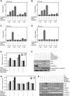
PIAS3 and SENP2 antagonistically regulate cell-cycle arrest. (A, B) SENP2 knockdown promotes p53 transactivity. HeLa cells transfected with shNS or shSENP2 alone or together with shhnRNP-K were incubated for 48 h. They were then transfected with PG13-Luc (A) or P21-Luc (B) and further incubated for the next 24 h. After exposure to UV, cells were incubated for 6 h. Cell lysates were assayed for luciferase. The enzyme activity seen in cells transfected with shNS only but without UV treatment was expressed as 1.0 and the others were as its relative values. (C, D) PIAS3 knockdown ablates p53 transactivity. Experiments were performed as above, except that cells were transfected with shPIAS3 in place of shSENP2. (E, F) SUMOylation of hnRNP-K is required for p21-mediated cell-cycle arrest. Cells transfected with shNS or shhnRNP-K were complemented with shhnRNP-K-insensitive Flag-tagged hnRNP-K or K422R. After exposure to UV, they were incubated for 6 h followed by flow cytometry (E) or immunoblot analysis (F). (G, H) PIAS3 and SENP2 inversely regulate cell-cycle arrest. Cells were transfected with shNS, shSENP2, or shPIAS3 alone or together with shhnRNP-K. After exposure to UV, cells were incubated for 6 h followed by flow cytometry (G) or immunoblot analysis (H). The data in (AE) and (G) represent the mean±s.d. of four experiments. Figure source data can be found with the Supplementary data.
Figure 9
Figure 9
SUMOylation of hnRNP-K is ATR dependent. (A) Caffeine inhibits hnRNP-K SUMOylation. After exposure of HeLa cells to UV, they were incubated with and without 5 mM caffeine for the indicated periods. Cell lysates were subjected to immunoprecipitation with anti-hnRNP-K antibody followed by immunoblot with anti-SUMO1, anti-SENP2, anti-PIAS3, or anti-hnRNP-K antibody. (B) ATR knockdown prevents hnRNP-K SUMOylation. Cells transfected with shNS or shATR were incubated for 48 h. After exposure to UV, cells were incubated for the indicated periods. Cell lysates were subjected to immunoprecipitation as in (A). Note that MG132 was treated 4 h before cell lysis in (A, B). (C) A model for the role of hnRNP-K SUMOylation in UV-induced cell-cycle arrest. Figure source data can be found with the Supplementary data.

References

    1. Abraham RT (2001) Cell cycle checkpoint signaling through the ATM and ATR kinases. Genes Dev 15: 2177–2196 - PubMed
    1. Abraham RT (2004) PI 3-kinase related kinases: 'big' players in stress-induced signaling pathways. DNA Repair (Amst) 3: 883–887 - PubMed
    1. Altmannova V, Eckert-Boulet N, Arneric M, Kolesar P, Chaloupkova R, Damborsky J, Sung P, Zhao X, Lisby M, Krejci L (2010) Rad52 SUMOylation affects the efficiency of the DNA repair. Nucleic Acids Res 38: 4708–4721 - PMC - PubMed
    1. Baptiste-Okoh N, Barsotti AM, Prives C (2008) Caspase 2 is both required for p53-mediated apoptosis and downregulated by p53 in a p21-dependent manner. Cell Cycle 7: 1133–1138 - PubMed
    1. Bartek J, Lukas J (2001) Mammalian G1- and S-phase checkpoints in response to DNA damage. Curr Opin Cell Biol 13: 738–747 - PubMed

Publication types

MeSH terms

Substances