Review
. 2013 Jan;33(1):22-32.
doi: 10.1038/jcbfm.2012.158. Epub 2012 Oct 24.

The dynamics of the mitochondrial organelle as a potential therapeutic target

Affiliations
Review

The dynamics of the mitochondrial organelle as a potential therapeutic target

R Anne Stetler et al. J Cereb Blood Flow Metab. 2013 Jan.

Abstract

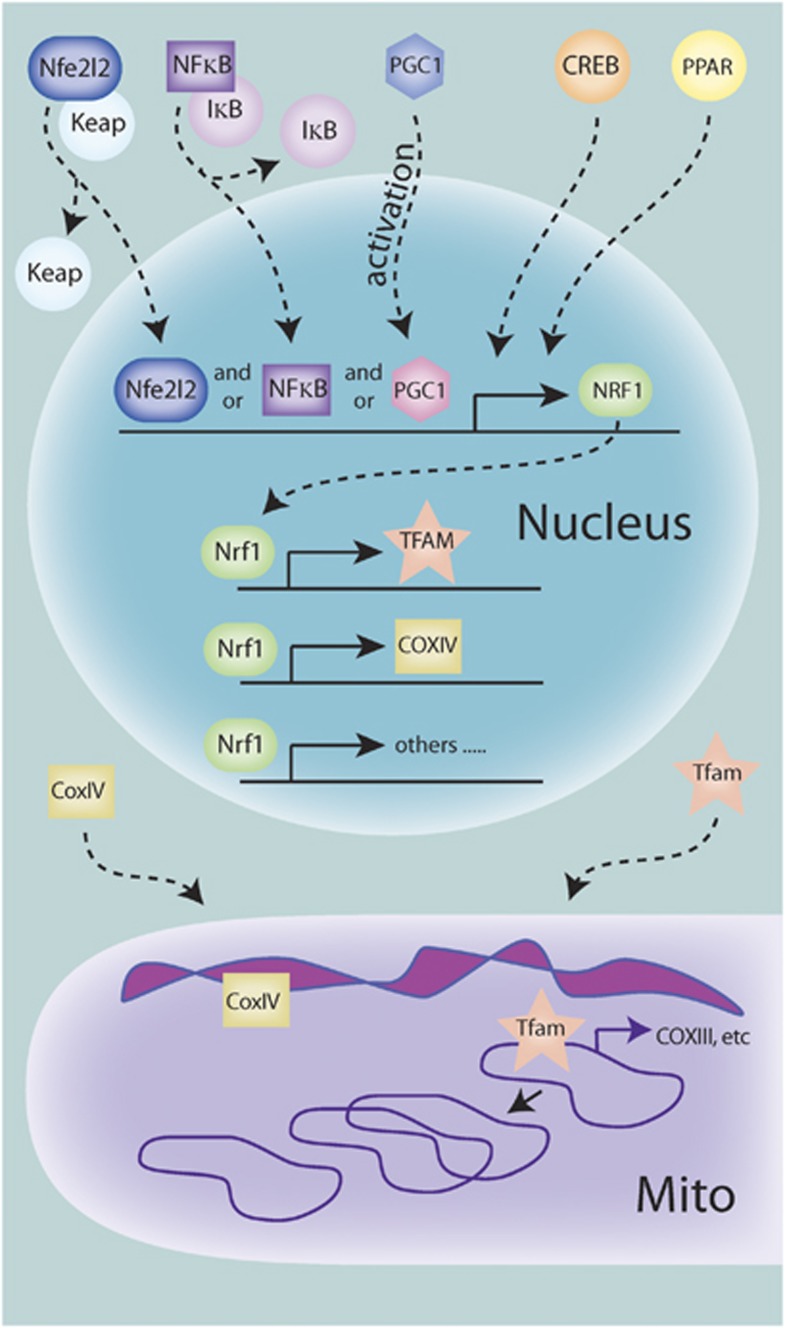
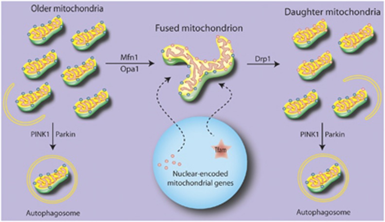
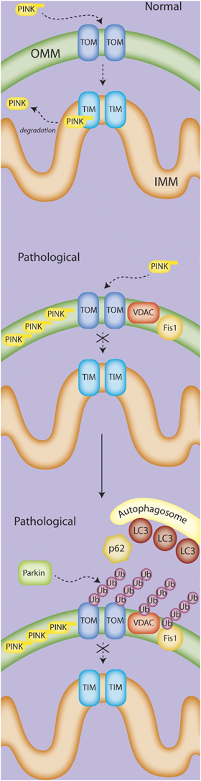
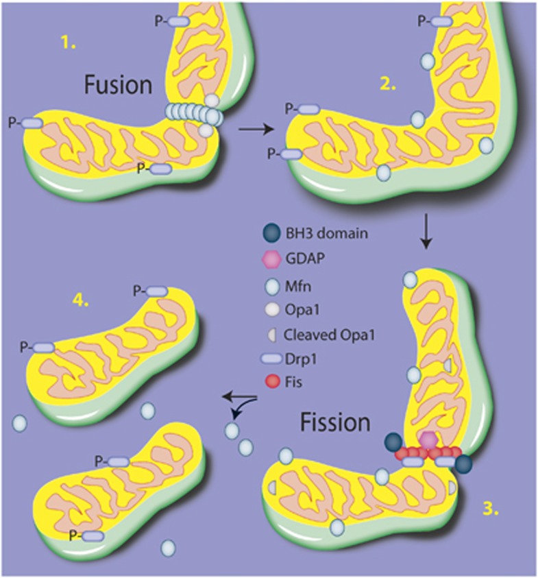
Mitochondria play a central role in cell fate after stressors such as ischemic brain injury. The convergence of intracellular signaling pathways on mitochondria and their release of critical factors are now recognized as a default conduit to cell death or survival. Besides the individual processes that converge on or emanate from mitochondria, a mitochondrial organellar response to changes in the cellular environment has recently been described. Whereas mitochondria have previously been perceived as a major center for cellular signaling, one can postulate that the organelle's dynamics themselves affect cell survival. This brief perspective review puts forward the concept that disruptions in mitochondrial dynamics--biogenesis, clearance, and fission/fusion events--may underlie neural diseases and thus could be targeted as neuroprotective strategies in the context of ischemic injury. To do so, we present a general overview of the current understanding of mitochondrial dynamics and regulation. We then review emerging studies that correlate mitochondrial biogenesis, mitophagy, and fission/fusion events with neurologic disease and recovery. An overview of the system as it is currently understood is presented, and current assessment strategies and their limitations are discussed.


Figures

Figure 1
Figure 1
Signaling leading to transcriptional regulation of mitochondrial biogenesis. Various upstream signaling pathways can contribute to the activation of the transcriptional program necessary for biogenesis. So far, PGC1-α (peroxisome proliferator-activated receptor (PPAR)-γ coactivator 1-α), nuclear factor-κB (NF-κB), and Nfe2l2 can translocate to the nucleus either by dissociation of tethering proteins or posttranslational modification, and are then capable of binding to the promoter region and promoting transcription of nuclear respiratory factor 1 (NRF-1). Nrf-1 can then bind to and promote the transcription of mitochondrial transcription factor A (TFAM) and many other nuclear-encoded mitochondrial proteins, such as subunits of the electron transport machinery. These proteins are then translated and imported into the mitochondria. Tfam then can bind to and initiate transcription of the mitochondrial genomes, which includes mitochondrial-encoded subunits of the electron transport machinery. Tfam, in addition to other proteins, also acts in the control of mitochondrial DNA (mtDNA) copy number, including replication when needed.
Figure 2
Figure 2
Proposed flux for mitochondrial replacement. Under normal circumstances, older elements of existing mitochondria may be sequestered and either targeted for degradation or fused to the mitochondrial network. Newly translated proteins are upregulated by activation of gene expression in the nucleus, and transported to the mitochondrial network to allow for the splitting of daughter mitochondria. These daughter mitochondria are heterogeneous, and those failing to maintain membrane potential are targeted for degradation.
Figure 3
Figure 3
The PINK/Parkin mitophagy pathway. In the current model, normal mitochondria fully import PINK into the mitochondria via the TOM/TIM transport system, where it is cleaved and eventually degraded. Under pathologic conditions, PINK is only partially transported to the mitochondria, and accumulates on the outer membrane. The accumulation of PINK recruits Parkin to the mitochondria, where Parkin then acts via its E3 ubiquitinase activity to add ubiquitin chains to various mitochondrial proteins such as VCAC, Fis1, and Mfn. In an as-yet-undefined mechanism, this activity appears to lead to the recruitment of the autophagosome membrane and subsequent engulfment of the targeted mitochondria.
Figure 4
Figure 4
Fusion and fission. The overall execution of mitochondrial structural changes lies primarily with the dynamin-like GTPases Opa1 for fusion and Drp1 for fission. Briefly, Mfn dimerizes on the adjacent outer mitochondrial membranes and creates a tether between the fusing membranes. Cardiolipin is hydrolyzed, resulting in a curvature of the inner membrane, and the inner membrane protein Opa1 then acts via a GTPase motor activity to coordinate the fusion of the membranes. To progress to fission, Drp1 translocates from the cytosol to the outer membrane and associates with Fis1, forming an oligomeric ring-like structure. Using a GTPase motor activity, Drp1 constricts the membrane, leading to separation of the mitochondria.

References

    1. Goldenthal MJ, Marin-Garcia J. Mitochondrial signaling pathways: a receiver/integrator organelle. Mol Cell Biochem. 2004;262:1–16. - PubMed
    1. Kanamaru Y, Sekine S, Ichijo H, Takeda K. The phosphorylation-dependent regulation of mitochondrial proteins in stress responses. J Signal Transduct. 2012;2012:931215. - PMC - PubMed
    1. Akepati VR, Muller EC, Otto A, Strauss HM, Portwich M, Alexander C. Characterization of OPA1 isoforms isolated from mouse tissues. J Neurochem. 2008;106:372–383. - PubMed
    1. Dennis EA, Kennedy EP. Intracellular sites of lipid synthesis and the biogenesis of mitochondria. J Lipid Res. 1972;13:263–267. - PubMed
    1. Michel S, Wanet A, De Pauw A, Rommelaere G, Arnould T, Renard P. Crosstalk between mitochondrial (dys)function and mitochondrial abundance. J Cell Physiol. 2012;227:2297–2310. - PubMed

Publication types

MeSH terms

Substances