

Assessment of tumor heterogeneity: an emerging imaging tool for clinical practice?
- PMID: 23093486
- PMCID: PMC3505569
- DOI: 10.1007/s13244-012-0196-6
Assessment of tumor heterogeneity: an emerging imaging tool for clinical practice?
Abstract
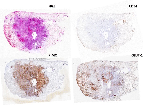
Background: Tumor spatial heterogeneity is an important prognostic factor, which may be reflected in medical images
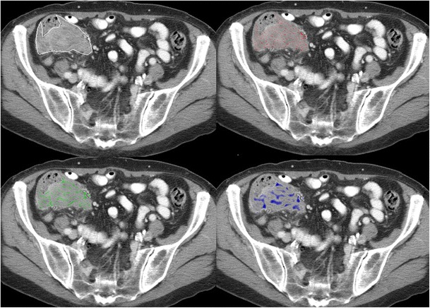
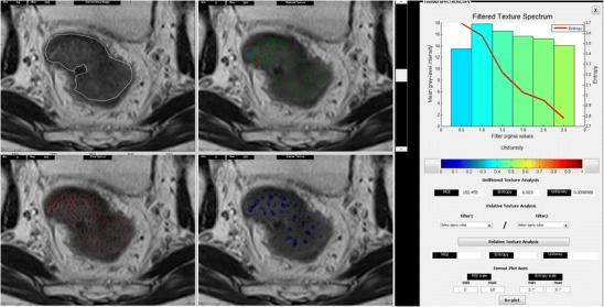
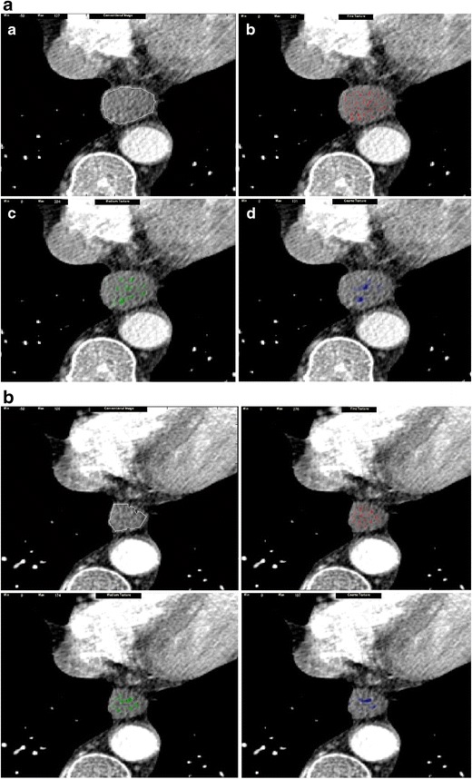
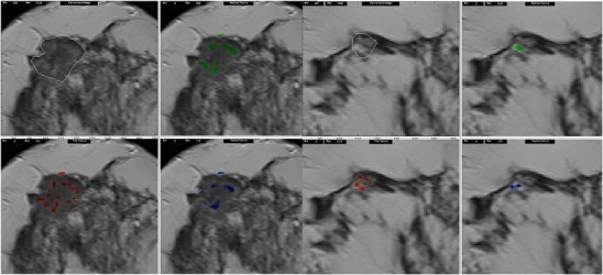
Methods: Image texture analysis is an approach of quantifying heterogeneity that may not be appreciated by the naked eye. Different methods can be applied including statistical-, model-, and transform-based methods.
Results: Early evidence suggests that texture analysis has the potential to augment diagnosis and characterization as well as improve tumor staging and therapy response assessment in oncological practice.
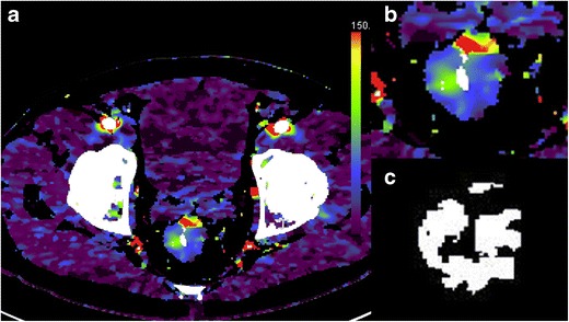
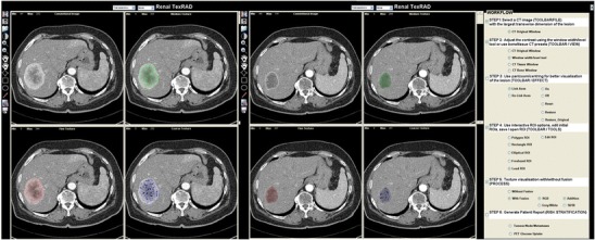
Conclusion: This review provides an overview of the application of texture analysis with different imaging modalities, CT, MRI, and PET, to date and describes the technical challenges that have limited its widespread clinical implementation so far. With further efforts to refine its application, image texture analysis has the potential to develop into a valuable clinical tool for oncologic imaging. TEACHING POINTS : • Tumor spatial heterogeneity is an important prognostic factor. • Image texture analysis is an approach of quantifying heterogeneity. • Different methods can be applied, including statistical-, model-, and transform-based methods. • Texture analysis could improve the diagnosis, tumor staging, and therapy response assessment.
Figures
References
-
- Yang Z, Tang LH, Klimstra DS. Effect of tumor heterogeneity on the assessment of Ki67 labeling index in well-differentiated neuroendocrine tumors metastatic to the liver: implications for prognostic stratification. Am J Surg Pathol. 2011;35(6):853–860. doi: 10.1097/PAS.0b013e31821a0696. - DOI - PubMed
-
- Hockel M, Schlenger K, Aral B, Mitze M, Schaffer U, Vaupel P. Association between tumor hypoxia and malignant progression in advanced cancer of the uterine cervix. Cancer Res. 1996;56(19):4509–4515. - PubMed
LinkOut - more resources
Full Text Sources
Other Literature Sources
Medical
