. 2012 Nov;122(11):4213-7.
doi: 10.1172/JCI65157. Epub 2012 Oct 24.

Targeted deletion of Vegfa in adult mice induces vision loss

Affiliations

Targeted deletion of Vegfa in adult mice induces vision loss

Toshihide Kurihara et al. J Clin Invest. 2012 Nov.

Abstract

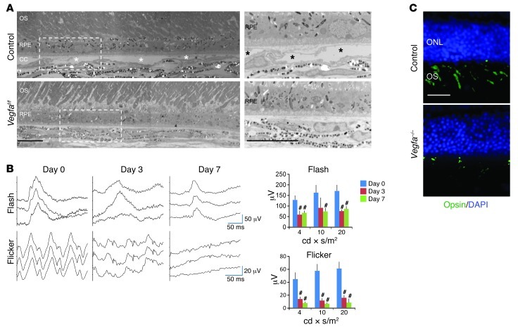
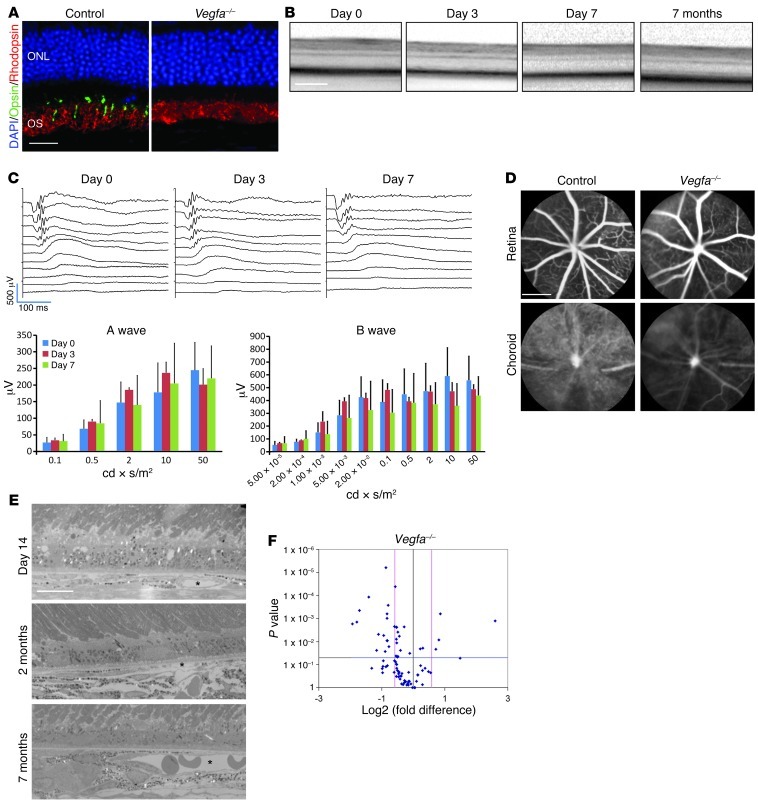
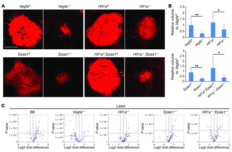
Current therapies directed at controlling vascular abnormalities in cancers and neovascular eye diseases target VEGF and can slow the progression of these diseases. While the critical role of VEGF in development has been well described, the function of locally synthesized VEGF in the adult eye is incompletely understood. Here, we show that conditionally knocking out Vegfa in adult mouse retinal pigmented epithelial (RPE) cells, which regulate retinal homeostasis, rapidly leads to vision loss and ablation of the choriocapillaris, the major blood supply for the outer retina and photoreceptor cells. This deletion also caused rapid dysfunction of cone photoreceptors, the cells responsible for fine visual acuity and color vision. Furthermore, Vegfa deletion showed significant downregulation of multiple angiogenic genes in both physiological and pathological states, whereas the deletion of the upstream regulatory transcriptional factors HIFs did not affect the physiological expressions of angiogenic genes. These results suggest that endogenous VEGF provides critical trophic support necessary for retinal function. Targeting factors upstream of VEGF, such as HIFs, may be therapeutically advantageous compared with more potent and selective VEGF antagonists, which may have more off-target inhibitory trophic effects.


Figures

Figure 1
Figure 1. Inducible Vegfa deletion in adult RPE cells promotes rapid choriocapillaris degeneration and vision loss.
(A) Electron micrographs of control (upper panel, Vegfaf/f without Cre) and mutant (lower panels, Vegfaf/f with VMD2-Cre) murine retinas 3 days after Vegfa deletion in adult RPE. Right panels are enlargements of the boxed regions of the left panels. Note that Vegfa mutants lack choriocapillaris normally observed in controls (asterisks). (B) ERG of Vegfa mutant eyes shows loss of photopic and flicker signals. b-wave amplitudes from flash, or first peaks from flicker, ERG in photopic light-adapted conditions captured in Vegfa mutants are significantly attenuated compared with the same retina prior to Vegfa gene deletion (n = 4 for each time point). This dramatic attenuation is consistent with vision loss. (C) Immunohistochemical analyses for cone-opsin in Vegfa mutants and controls 7 days after induction. The absence of cone outer segments is apparent in the Vegfa mutants. #P < 0.01; 2-tailed Student’s t tests. Error bars indicate mean ± SD. Scale bars: 10 μm (A ); 20 μm (C). ONL, outer nuclear layer; OS, outer segments; CC,choriocapillaris.
Figure 2
Figure 2. Cone but not rod photoreceptor dysfunction in RPE-specific Vegfa mutants.
(A) Immunohistochemistry for rhodopsin (rods) and cone opsin (cones) 45 days after induction. Note that rhodopsin expression in Vegfa mutants is comparable to that observed in controls. (B) Optical coherence tomographic analysis indicates that retinal thicknesses remain relatively unchanged at all stages through 7 months. (C) Scotopic dark-adapted ERG captured in Vegfa mutants 3 or 7 days after induction. Note that no significant reduction in the a- and b-wave amplitudes is observed (n = 4). (D) Indocyanine green angiography for Vegfa mutants 45 days after induction. Note that choroidal circulation is significantly diminished in the mutants, while the retinal circulation is normal. (E) Electron micrograph of a cross-sectioned Vegfa mutant retina 14 days, 2 months, and 7 months after induction. Note that a few deep choroidal vessels are observed (asterisk). (F) mRNA array for angiogenic genes in Vegfa mutant RPE/choroids compared with controls 3 days after injection (n = 3). Fold-change (x axis) and P value (y axis) of gene expression compared with controls are shown. Note that 32 of 84 genes are significantly downregulated. Error bars indicate mean ± SD. Scale bars: 2,000 μm (D); 200 μm (B); 20 μm (A); 10 μm (E).
Figure 3
Figure 3. Attenuated angiogenic gene regulation in pathological states of Vegfa conditional mutants.
(A) Representative Z projections of laser-CNV lesions (stained with GS lectin). (B) Quantified lesion volumes of laser-CNV (n = 8–21). Note that laser-CNV is only partially reduced in the Hif1a mutants, while it is completely inhibited in the Epas1 and Hif1a;Epas1 mutants (comparable to that observed in the Vegfa mutants). (C) mRNA array for angiogenic genes in Hif1a, Epas1, and Hif1a;Epas1 mutant RPE/choroids compared with controls in laser-irradiated C57BL/6 wild-type, Vegfa, Hif1a, Epas1, and Hif1a;Epas1 mutants compared with naive state controls (n = 3). Fold-change (x axis) and P value (y axis) of gene expression compared with controls. Note that few or no significantly dysregulated genes are observed in naive state Hif1a, Epas1, or Hif1a;Epas1 mutants. Twenty-two upregulated and 3 downregulated angiogenic genes are observed in laser-irradiated B6 wild-type mice, whereas we observed 7 (up) and 23 (down) in Vegfa mutants, 6 (up) and 1 (down) in Hif1a mutants, 6 (up) and 1 (down) in Epas1 mutants, and 10 (up) and 4 (down) in Hif1a;Epas1 mutants. *P < 0.05; **P < 0.01; 2-tailed Student’s t tests. Error bars indicate mean ± SD. Scale bar: 100 μm.

Comment in

References

    1. Dorrell MI, Aguilar E, Friedlander M. Retinal vascular development is mediated by endothelial filopodia, a preexisting astrocytic template and specific R-cadherin adhesion. Invest Ophthalmol Vis Sci. 2002;43(11):3500–3510. - PubMed
    1. Gerhardt H, et al. VEGF guides angiogenic sprouting utilizing endothelial tip cell filopodia. J Cell Biol. 2003;161(6):1163–1177. doi: 10.1083/jcb.200302047. - DOI - PMC - PubMed
    1. Le YZ, Bai Y, Zhu M, Zheng L. Temporal requirement of RPE-derived VEGF in the development of choroidal vasculature. J Neurochem. 2010;112(6):1584–1592. doi: 10.1111/j.1471-4159.2010.06573.x. - DOI - PMC - PubMed
    1. Marneros AG, et al. Vascular endothelial growth factor expression in the retinal pigment epithelium is essential for choriocapillaris development and visual function. Am J Pathol. 2005;167(5):1451–1459. doi: 10.1016/S0002-9440(10)61231-X. - DOI - PMC - PubMed
    1. Krzystolik MG, et al. Prevention of experimental choroidal neovascularization with intravitreal anti-vascular endothelial growth factor antibody fragment. Arch Ophthalmol. 2002;120(3):338–346. doi: 10.1001/archopht.120.3.338. - DOI - PubMed

Publication types

MeSH terms

Substances