. 2012 Nov;122(11):4218-30.
doi: 10.1172/JCI59176. Epub 2012 Oct 24.

Hypoxic pulmonary vasoconstriction requires connexin 40-mediated endothelial signal conduction

Affiliations

Hypoxic pulmonary vasoconstriction requires connexin 40-mediated endothelial signal conduction

Liming Wang et al. J Clin Invest. 2012 Nov.

Abstract

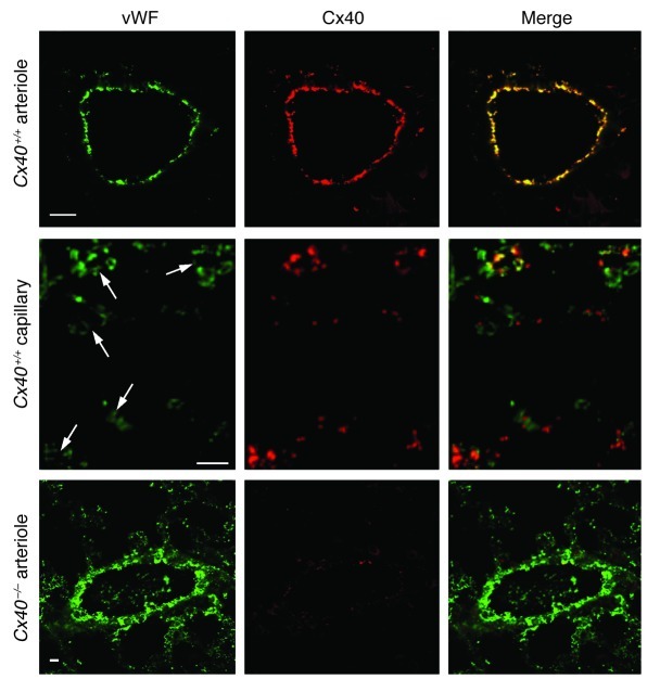
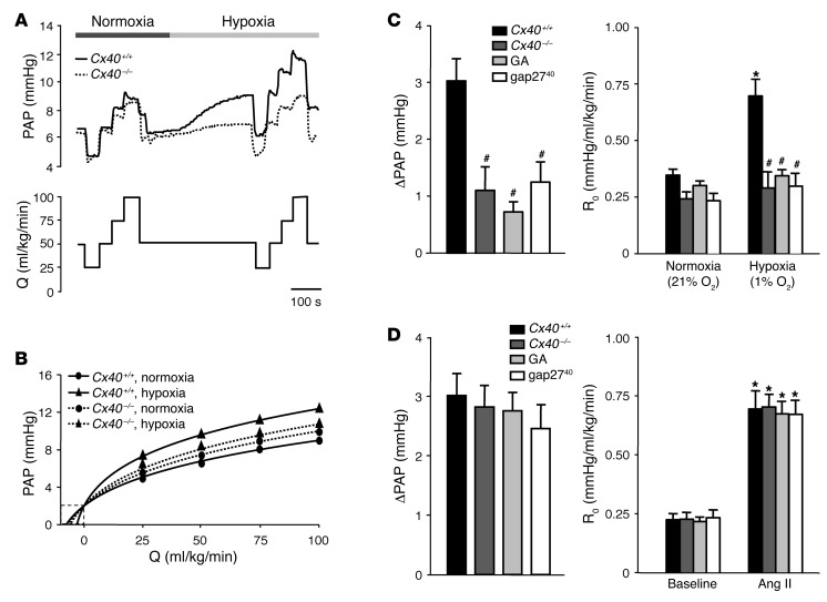
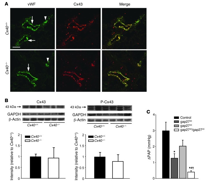
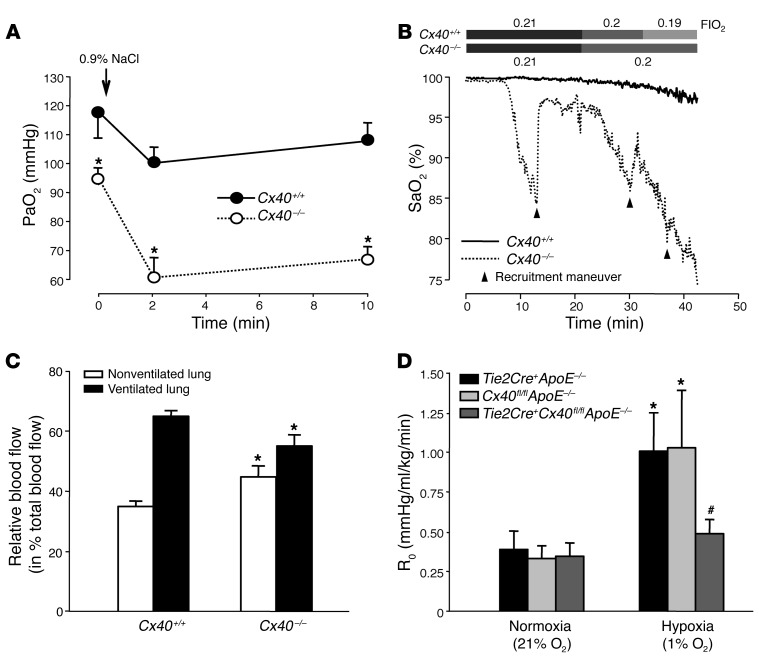
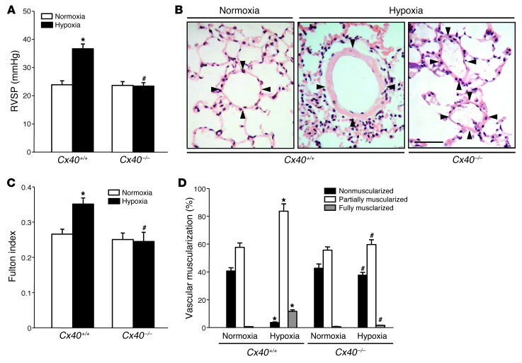
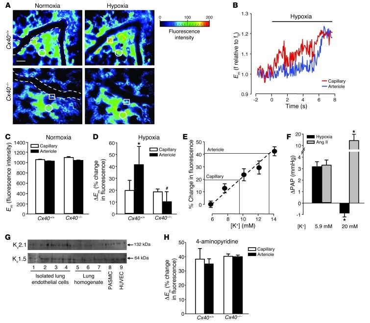
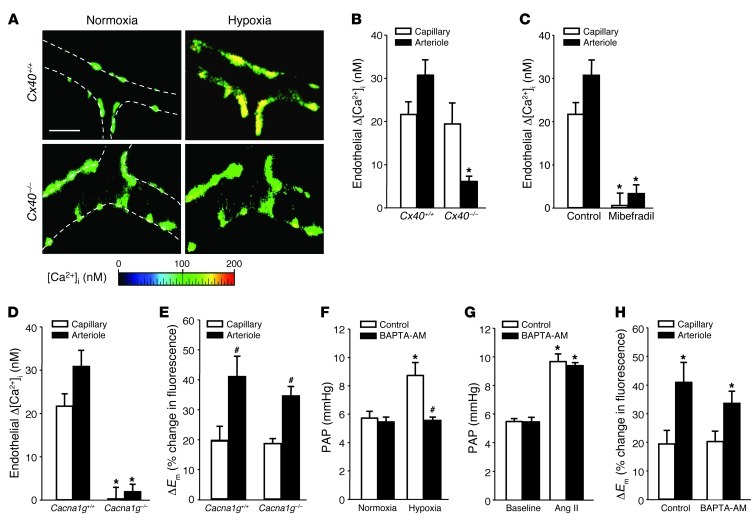
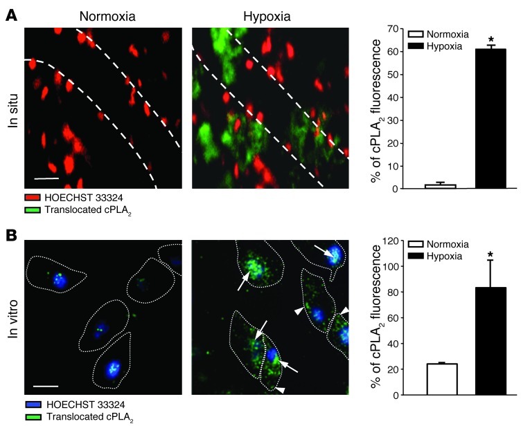
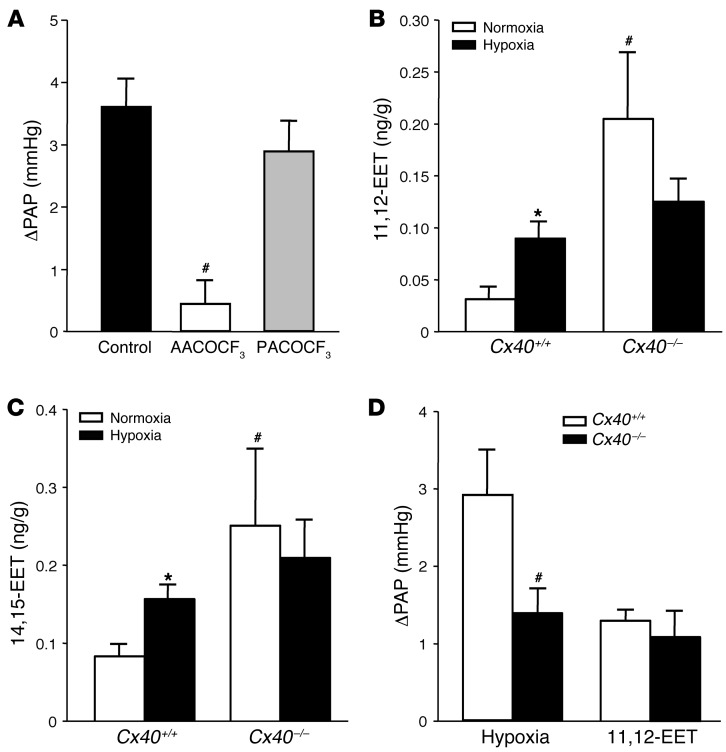
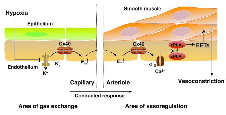
Hypoxic pulmonary vasoconstriction (HPV) is a physiological mechanism by which pulmonary arteries constrict in hypoxic lung areas in order to redirect blood flow to areas with greater oxygen supply. Both oxygen sensing and the contractile response are thought to be intrinsic to pulmonary arterial smooth muscle cells. Here we speculated that the ideal site for oxygen sensing might instead be at the alveolocapillary level, with subsequent retrograde propagation to upstream arterioles via connexin 40 (Cx40) endothelial gap junctions. HPV was largely attenuated by Cx40-specific and nonspecific gap junction uncouplers in the lungs of wild-type mice and in lungs from mice lacking Cx40 (Cx40-/-). In vivo, hypoxemia was more severe in Cx40-/- mice than in wild-type mice. Real-time fluorescence imaging revealed that hypoxia caused endothelial membrane depolarization in alveolar capillaries that propagated to upstream arterioles in wild-type, but not Cx40-/-, mice. Transformation of endothelial depolarization into vasoconstriction involved endothelial voltage-dependent α1G subtype Ca2+ channels, cytosolic phospholipase A2, and epoxyeicosatrienoic acids. Based on these data, we propose that HPV originates at the alveolocapillary level, from which the hypoxic signal is propagated as endothelial membrane depolarization to upstream arterioles in a Cx40-dependent manner.


Figures

Figure 1
Figure 1. Expression of Cx40 in pulmonary microvessels.
Expression of vWF (green) and Cx40 (red) and merged images for pulmonary arterioles and capillaries (arrows) of Cx40+/+ mice and for arterioles of Cx40–/– mice. Scale bars: 10 μm. Note colocalization of vWF and Cx40 in pulmonary arterioles and capillaries of Cx40+/+ mice, indicative of endothelial Cx40 expression, was absent in Cx40–/– mice.
Figure 2
Figure 2. Cx40 is required for an intact HPV response.
(A) Representative tracings of PAP in isolated perfused lungs of Cx40+/+ and Cx40–/– mice obtained during stepwise changes in lung perfusion (Q) at normoxia (21% O2) or hypoxia (1% O2). Note the attenuated HPV response in Cx40–/– mice. (B) Nonlinear regression analysis according to the distensible vessel model yielded representative pressure-flow curves for lungs of Cx40+/+ and Cx40–/– mice at normoxia and hypoxia. The pressure at 0 ml/kg/min flow (Q) reflects left arterial pressure of 2 mmHg, while the slope of the pressure-flow curve at 0 ml/kg/min flow reflects R0. (C) Group data showing acute HPV response, determined as ΔPAP and as R0 10 minutes after hypoxia onset versus normoxia, in untreated isolated perfused lungs of Cx40+/+ and Cx40–/– mice and in lungs of Cx40+/+ mice treated with 18α-glycyrrhetinic acid (GA; 50 μM) or gap2740 (200 μM). (D) Group data (n = 5 lungs each) showing acute HPV response to Ang II (1 μg bolus infusion), determined as ΔPAP and as R0 versus baseline, in untreated isolated perfused lungs of Cx40+/+ and Cx40–/– mice and in lungs of Cx40+/+ mice treated with 18α-glycyrrhetinic acid or gap2740. *P < 0.05 vs. normoxia or baseline control; #P < 0.05 vs. untreated Cx40+/+.
Figure 3
Figure 3. The role of Cx40 in HPV is independent of Cx43.
(A) Expression of vWF (green) and Cx43 (red) and merged images for pulmonary arterioles (arrows) and capillaries (arrowhead) of Cx40+/+ and Cx40–/– mice. Scale bar: 50 μm. (B) Representative Western blots and quantification showing expression and Ser368 phosphorylation of Cx43 in fresh lung homogenate from Cx40+/+ and Cx40–/– mice. GAPDH and β-actin are shown as loading controls. (C) Group data (n = 5 lungs each) showing acute HPV response, determined as ΔPAP 10 minutes after onset of hypoxia (1% O2) versus normoxia (21% O2) in isolated perfused lungs of untreated Cx40+/+ mice and of Cx40+/+ mice treated with gap2740 (200 μM), gap2743 (200 μM), or both in combination. *P < 0.05 vs. control; #P < 0.05 vs. gap2740; P < 0.05 vs. gap2743.
Figure 4
Figure 4. Cx40 optimizes oxygenation and V/Q matching in hypoxic lungs in vivo.
(A) Group data (n = 5 lungs each) showing arterial PaO2 in anesthetized Cx40+/+ and Cx40–/– mice prior to (0 minutes) and after partial occlusion of larger airways by tracheal instillation of 25 μl saline. PaO2 was already significantly lower in Cx40–/– versus Cx40+/+ mice at baseline; this difference was further amplified after induction of V/Q mismatches by saline instillation. *P < 0.05 vs. Cx40+/+. (B) Representative tracings (of 5 replicates) showing SaO2 in anesthetized Cx40+/+ and Cx40–/– mice during stepwise decrements in FIO2 starting at 0.21. In the case of sudden SaO2 decrements, recruitment maneuvers were performed (arrowheads) to counteract atelectases. (C) Group data (n = 5 lungs each) showing relative blood flow (percent total pulmonary blood flow) to the nonventilated right and ventilated left lung during 1-lung ventilation in Cx40+/+ and Cx40–/– mice, assessed by fluorescent microsphere technique. *P < 0.05 vs. Cx40+/+. (D) Group data (n = 5 lungs each) showing acute HPV response, determined as R0 10 minutes after hypoxia (1% O2) onset versus normoxia (21% O2), in isolated perfused lungs of Tie2Cre+Cx40fl/flApoE–/– mice and Tie2Cre+ApoE–/– and Cx40fl/flApoE–/– controls. *P < 0.05 vs. normoxia; #P < 0.05 vs. Tie2Cre+ApoE–/– and Cx40fl/flApoE–/–.
Figure 5
Figure 5. Cx40 deficiency attenuates chronic hypoxic pulmonary hypertension.
Group data showing (A) right ventricular systolic pressure (RVSP) and (B) right ventricular weight relative to septal and left ventricular weight (Fulton index) in Cx40+/+ and Cx40–/– mice housed in either normoxia (21% O2) or hypoxia (10% O2) for 5 weeks. (C) Representative images of H&E-stained lung sections showing pulmonary arterioles (denoted by 4 arrowheads each). Scale bar: 50 μm. (D) Group data (n = 8 mice each) showing degree of vascular muscularization in pulmonary arterioles 20–50 μm in diameter, expressed as proportion of non-, partially, or fully muscularized vessels, in lungs from normoxic and hypoxic Cx40+/+ and Cx40–/– mice. *P < 0.05 vs. normoxia; #P < 0.05 vs. Cx40+/+.
Figure 6
Figure 6. Endothelial membrane depolarization.
(A) Representative images (of 5 replicates) showing endothelial di-8-ANEPPS fluorescence in capillaries and arterioles of Cx40+/+ and Cx40–/– lungs at normoxia (21% O2) and after 10 minutes of hypoxia (1% O2). Arteriolar vessel margins are shown by dotted lines, and representative capillary and arteriolar endothelium are denoted by circles and squares, respectively. Scale bar: 50 μm. (B) Representative tracings (of 5 replicates) of di-8-ANEPPS fluorescence. Hypoxia-induced ΔEm in capillaries preceded arteriole response. Group data (n = 5 lungs each) showing (C) comparable baseline endothelial di-8-ANEPPS fluorescence (reflecting Em) and (D) hypoxia-induced increases in endothelial di-8-ANEPPS fluorescence (reflecting ΔEm) in capillaries and arterioles of Cx40+/+ and Cx40–/– lungs. *P < 0.05 vs. capillaries; #P < 0.05 vs. Cx40+/+. (E) Calibration of endothelial di-8-ANEPPS fluorescence by lung perfusion with different [K+]. n = 3 lungs each. (F) Group data (n = 5 lungs each) showing ΔPAP in response to hypoxia (1% O2) or Ang II (1 μg bolus) in isolated Cx40+/+ lungs perfused with 5.9 or 20 mM [K+]. *P < 0.05 vs. 5.9 mM. (G) Representative Western blots showing Kv1.5 and Kv2.1 expression in freshly isolated pulmonary endothelial cells; whole lung homogenate, rat PASMCs, and HUVECs served as controls. (H) Group data (n = 5 lungs each) showing endothelial ΔEm in capillaries and arterioles of Cx40+/+ and Cx40–/– lungs in response to 10 mM 4-aminopyridine. No significant differences.
Figure 7
Figure 7. Role of endothelial [Ca2+]i in acute HPV.
(A) Representative images (of 5 replicates) of fura-2–loaded lung arterioles showing endothelial [Ca2+]i at normoxia (21% O2) and after 10 minutes of hypoxia (1% O2) in Cx40+/+ and Cx40–/– lungs. Vessel margins are denoted by dotted lines. Scale bar: 50 μm. Group data (n = 5 lungs each) show endothelial Δ[Ca2+]i in response to acute hypoxia in pulmonary capillaries and arterioles of (B) Cx40+/+ and Cx40–/– mice or (C) Cx40+/+ lungs in the absence (control) or presence of the VDCC blocker mibefradil (10 μM). *P < 0.05 vs. Cx40+/+ or control. Group data (n = 5 lungs each) showing (D) endothelial Δ[Ca2+]i or (E) ΔEm in response to acute hypoxia in capillaries and arterioles of Cacna1g+/+ and Cacna1g–/– lungs. *P < 0.05 vs. Cacna1g+/+; #P < 0.05 vs. capillary. Group data (n = 5 lungs each) showing PAP in isolated perfused Cx40+/+ lungs (F) at normoxia and after 10 minutes of hypoxia or (G) at baseline and after Ang II (1 μg bolus) in control lungs or after endothelial Ca2+ chelation by BAPTA-AM (40 μM). *P < 0.05 vs. normoxia or baseline; #P < 0.05 vs. control. (H) Group data (n = 5 lungs each) showing endothelial ΔEm in response to acute hypoxia in pulmonary capillaries and arterioles in the absence (control) or presence of BAPTA-AM. *P < 0.05 vs. capillary.
Figure 8
Figure 8. Translocation of endothelial cPLA2 in acute hypoxia.
(A) Representative merged epifluorescence images showing endothelial cell nuclei in pulmonary arterioles, as stained by HOECHST 33324 (red), and translocated cPLA2, as detected by indirect immunofluorescence (green), in intact lungs of Cx40+/+ mice. Images were obtained at normoxia (21% O2) or after 10 minutes of hypoxia (1 % O2); vessel margins are denoted by dotted lines. Scale bar: 50 μm. Group data (n = 5 lungs each) show cPLA2 translocation as a ratio of cPLA2 immunostaining relative to HOECHST 33324 fluorescence. *P < 0.05 vs. normoxia. (B) Representative merged confocal fluorescence images showing endothelial cell nuclei in cultured PAECs, as stained by HOECHST 33324 (blue), and cPLA2 that had been translocated to the nuclear envelope (arrows) or the cell membrane (arrowheads), as detected by indirect immunofluorescence (green). Images were obtained at normoxia or after 10 minutes of hypoxia; cell margins identified by brightfield microscopy are denoted by dotted lines. Scale bar: 20 μm. Group data show cPLA2 translocation from n = 5 independent experiments. *P < 0.05 vs. normoxia.
Figure 9
Figure 9. Role of cPLA2 and EETs in acute HPV.
(A) Group data (n = 5 lungs each) showing acute HPV response, determined as ΔPAP 10 minutes after hypoxia onset (1% O2) versus normoxia (21% O2) in untreated isolated perfused lungs of Cx40+/+ mice (control) and in lungs of Cx40+/+ mice treated with either the cPLA2-specific inhibitor AACOCF3 or the iPLA2-specific inhibitor PACOCF3 (both 1 μM). #P < 0.05 vs. control. Group data showing concentrations of (B) 11,12-EET and (C) 14,15-EET in isolated lungs of Cx40+/+ and Cx40–/– mice lungs at normoxia or after 10 minutes of hypoxia. *P < 0.05 vs. normoxia; #P < 0.05 vs. Cx40+/+. (D) Group data (n = 5 lungs each) showing acute pulmonary vasoconstrictor response to hypoxia and exogenous infusion of 11,12-EET (3 μM), determined as ΔPAP, in isolated lungs from Cx40+/+ and Cx40–/– mice. #P < 0.05 vs. Cx40+/+.
Figure 10
Figure 10. Proposed concept for a conducted response propagated via endothelial Cx40 in HPV.
The scheme encompasses (i) proposed signaling events at the level of the alveolar site of gas exchange (left), in which hypoxia induces endothelial membrane depolarization (Em↑) in lung capillaries, potentially by inhibition of oxygen-sensitive Kv channels; (ii) retrograde propagation of endothelial membrane depolarization from alveolar capillaries to upstream arterioles via Cx40 (middle), and (iii) elicitation of a vasoconstrictive response at the level of the upstream arteriole (right) through activation of the α1G subtype T type VDCC, subsequent activation of cPLA2, and formation of EETs, which may serve as either direct (intercellular) or indirect (intracellular) mediators of smooth muscle cell contraction.

Comment in

Similar articles

Cited by

References

    1. Ward JP, McMurtry IF. Mechanisms of hypoxic pulmonary vasoconstriction and their roles in pulmonary hypertension: new findings for an old problem. Curr Opin Pharmacol. 2009;9(3):287–296. doi: 10.1016/j.coph.2009.02.006. - DOI - PMC - PubMed
    1. Maggiorini M, et al. High-altitude pulmonary edema is initially caused by an increase in capillary pressure. Circulation. 2001;103(16):2078–2083. doi: 10.1161/01.CIR.103.16.2078. - DOI - PubMed
    1. Dawson CA. Role of pulmonary vasomotion in physiology of the lung. Physiol Rev. 1984;64(2):544–616. - PubMed
    1. Paddenberg R, Konig P, Faulhammer P, Goldenberg A, Pfeil U, Kummer W. Hypoxic vasoconstriction of partial muscular intra-acinar pulmonary arteries in murine precision cut lung slices. Respir Res. 2006;7:93. doi: 10.1186/1465-9921-7-93. - DOI - PMC - PubMed
    1. Hislop A, Reid L. Normal structure and dimensions of the pulmonary arteries in the rat. J Anat. 1978;125(pt 1):71–83. - PMC - PubMed

Publication types

MeSH terms