

In vivo imaging of brain ischemia using an oxygen-dependent degradative fusion protein probe
- PMID: 23094105
- PMCID: PMC3477118
- DOI: 10.1371/journal.pone.0048051
In vivo imaging of brain ischemia using an oxygen-dependent degradative fusion protein probe
Abstract
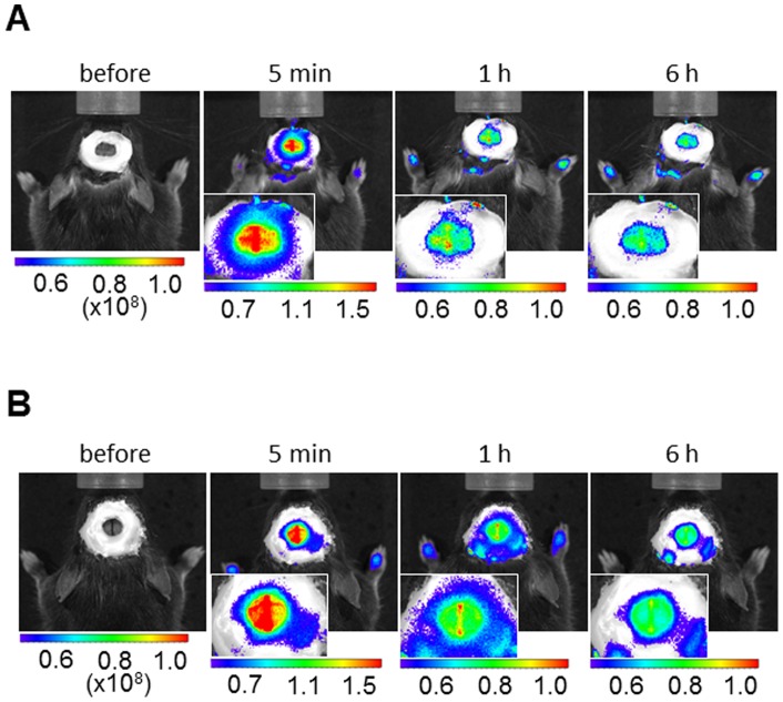
Within the ischemic penumbra, blood flow is sufficiently reduced that it results in hypoxia severe enough to arrest physiological function. Nevertheless, it has been shown that cells present within this region can be rescued and resuscitated by restoring perfusion and through other protective therapies. Thus, the early detection of the ischemic penumbra can be exploited to improve outcomes after focal ischemia. Hypoxia-inducible factor (HIF)-1 is a transcription factor induced by a reduction in molecular oxygen levels. Although the role of HIF-1 in the ischemic penumbra remains unknown, there is a strong correlation between areas with HIF-1 activity and the ischemic penumbra. We recently developed a near-infrared fluorescently labeled-fusion protein, POH-N, with an oxygen-dependent degradation property identical to the alpha subunit of HIF-1. Here, we conduct in vivo imaging of HIF-active regions using POH-N in ischemic brains after transient focal cerebral ischemia induced using the intraluminal middle cerebral artery occlusion technique in mice. The results demonstrate that POH-N enables the in vivo monitoring and ex vivo detection of HIF-1-active regions after ischemic brain injury and suggest its potential in imaging and drug delivery to HIF-1-active areas in ischemic brains.
Conflict of interest statement
Figures


Similar articles
-
In vivo imaging of HIF-active tumors by an oxygen-dependent degradation protein probe with an interchangeable labeling system.PLoS One. 2010 Dec 23;5(12):e15736. doi: 10.1371/journal.pone.0015736. PLoS One. 2010. PMID: 21203417 Free PMC article.
-
Morg1(+/-) heterozygous mice are protected from experimentally induced focal cerebral ischemia.Brain Res. 2012 Oct 30;1482:22-31. doi: 10.1016/j.brainres.2012.09.017. Epub 2012 Sep 13. Brain Res. 2012. PMID: 22982595
-
Neuron-specific prolyl-4-hydroxylase domain 2 knockout reduces brain injury after transient cerebral ischemia.Stroke. 2012 Oct;43(10):2748-56. doi: 10.1161/STROKEAHA.112.669598. Epub 2012 Aug 28. Stroke. 2012. PMID: 22933585
-
The role and regulation of hypoxia-inducible factor-1alpha expression in brain development and neonatal hypoxic-ischemic brain injury.Brain Res Rev. 2009 Dec 11;62(1):99-108. doi: 10.1016/j.brainresrev.2009.09.006. Epub 2009 Sep 25. Brain Res Rev. 2009. PMID: 19786048 Review.
-
The immunosuppressant drug FK506 ameliorates secondary mitochondrial dysfunction following transient focal cerebral ischemia in the rat.Neurobiol Dis. 1997;4(3-4):288-300. doi: 10.1006/nbdi.1997.0146. Neurobiol Dis. 1997. PMID: 9361306 Review.
Cited by
-
A Bio-inspired Hypoxia Sensor using HIF1a-Oxygen-Dependent Degradation Domain.Sci Rep. 2019 May 8;9(1):7117. doi: 10.1038/s41598-019-43618-4. Sci Rep. 2019. PMID: 31068630 Free PMC article.
References
-
- Bergeron M, Yu AY, Solway KE, Semenza GL, Sharp FR (1999) Induction of hypoxia-inducible factor-1 (HIF-1) and its target genes following focal ischemia in rat brain. Eur J Neurosci 11: 4159–4170. - PubMed
-
- Semenza GL (2000) HIF-1: mediator of physiological and pathophysiological responses to hypoxia. J Appl Physiol 88: 1474–1480. - PubMed
-
- Kaelin WG (2005) Proline hydroxylation and gene expression. Annu Rev Biochem 74: 115–128. - PubMed
-
- Schofield CJ, Ratcliffe PJ (2004) Oxygen sensing by HIF hydroxylases. Nat Rev Mol Cell Biol 5: 343–354. - PubMed
Publication types
MeSH terms
Substances
LinkOut - more resources
Full Text Sources
Medical
