

Mechanism of experimental autoimmune encephalomyelitis in Lewis rats: recent insights from macrophages
- PMID: 23094201
- PMCID: PMC3472139
- DOI: 10.5115/acb.2012.45.3.141
Mechanism of experimental autoimmune encephalomyelitis in Lewis rats: recent insights from macrophages
Abstract
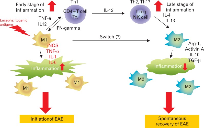
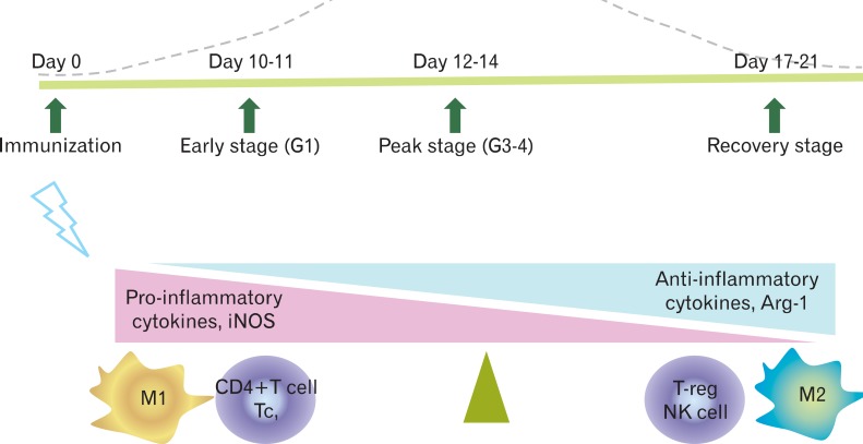
Experimental autoimmune encephalomyelitis (EAE) in Lewis rats is an acute monophasic paralytic central nervous system disease, in which most rats spontaneously recover from paralysis. EAE in Lewis rats is induced by encephalitogenic antigens, including myelin basic protein. EAE is mediated by CD4(+) Th1 cells, which secrete pro-inflammatory mediators, and spontaneous recovery is mediated by regulatory T cells. Recently, it was established that classically activated macrophages (M1 phenotype) play an important role in the initiation of EAE, while alternatively activated macrophages (M2 phenotype) contribute to spontaneous recovery from rat EAE. This review will summarize the neuroimmunological aspects of active monophasic EAE, which manifests as neuroinflammation followed by neuroimmunomodulation and/or neuroprotection, with a focus on the role of alternatively activated macrophages.
Keywords: Experimental autoimmune encephalomyelitis; Lewis rats; Macrophages; Neuroimmunomodulation; Regulatory T lymphocytes.
Figures
References
-
- Stadelmann C, Wegner C, Brück W. Inflammation, demyelination, and degeneration: recent insights from MS pathology. Biochim Biophys Acta. 2011;1812:275–282. - PubMed
-
- Herz J, Zipp F, Siffrin V. Neurodegeneration in autoimmune CNS inflammation. Exp Neurol. 2010;225:9–17. - PubMed
-
- Kapadia M, Sakic B. Autoimmune and inflammatory mechanisms of CNS damage. Prog Neurobiol. 2011;95:301–333. - PubMed
-
- Wekerle H. Lessons from multiple sclerosis: models, concepts, observations. Ann Rheum Dis. 2008;67(Suppl 3):iii56–iii60. - PubMed
LinkOut - more resources
Full Text Sources
Other Literature Sources
Research Materials