. 2012 Jul 1;2(3):185-196.
doi: 10.4161/spmg.21711.

Cytokinesis in Drosophila male meiosis

Affiliations

Cytokinesis in Drosophila male meiosis

Maria Grazia Giansanti et al. Spermatogenesis. .

Abstract

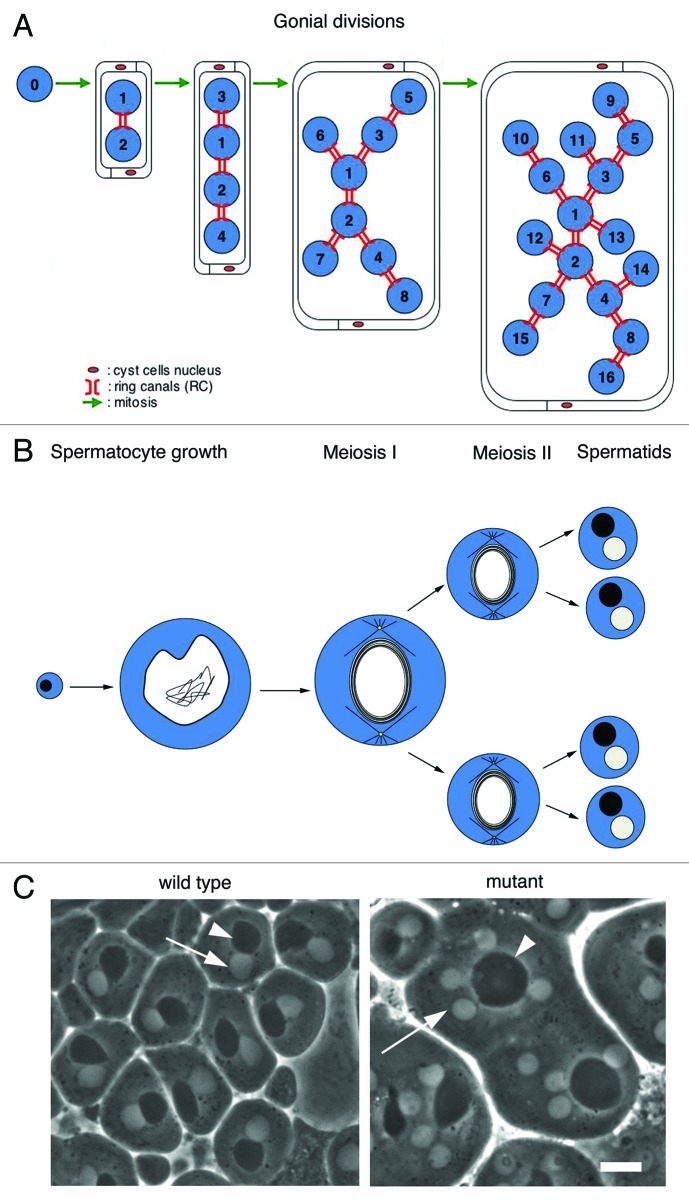
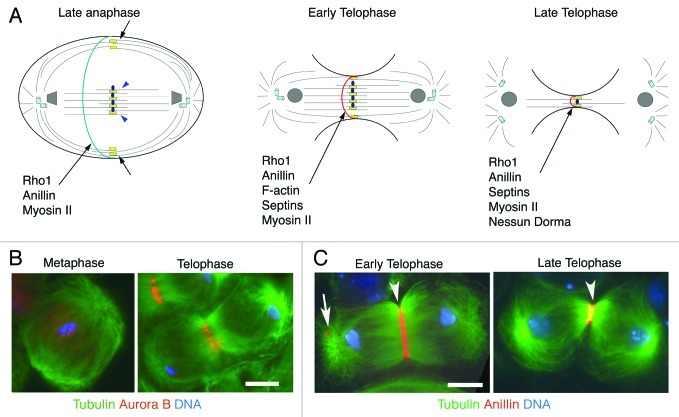
Cytokinesis separates the cytoplasm and the duplicated genome into two daughter cells at the end of cell division. This process must be finely regulated to maintain ploidy and prevent tumor formation. Drosophila male meiosis provides an excellent cell system for investigating cytokinesis. Mutants affecting this process can be easily identified and spermatocytes are large cells particularly suitable for cytological analysis of cytokinetic structures. Over the past decade, the powerful tools of Drosophila genetics and the unique characteristics of this cell system have led researchers to identify molecular players of the cell cleavage machinery and to address important open questions. Although spermatocyte cytokinesis is incomplete, resulting in formation of stable intercellular bridges, the molecular mechanisms are largely conserved in somatic cells. Thus, studies of Drosophila male meiosis will shed new light on the complex cell circuits regulating furrow ingression and substantially further our knowledge of cancer and other human diseases.


Figures

None
Figure 1. Spermatogenesis in Drosophila melanogaster. (A, B) Schematic representation of spermatogenesis in D. melanogaster. (A) A single primary spermatogonium undergoes four mitotic divisions. Numbers indicate gonial cells, number 0 indicates the primary spermatogonium, the mitotic founder of a cluster of dividing secondary spermatogonia connected by ring canals. Two cyst cells engulf the progeny of the primary spermatogonium throughout spermatogenesis. Blue, spermatogonia. (B) Each primary spermatocyte undergoes a growth phase, which lasts 90 h before undergoing two meiotic divisions. A wild type spermatid cyst at the so called onion stage contains 64 spermatids connected by 63 ring canals (not shown). Each spermatid contains a single nucleus (white) associated with a nebenkern (black) of similar size. Only four spermatids are represented. Blue, cytoplasm. (C) Spermatids at the onion-stage viewed by phase-contrast microscopy. Each wild type spermatid contains a single light nucleus (arrow) associated with a dark nebenkern (arrowhead). Spermatids from mutants defective in male meiotic cytokinesis, contain large nebenkerne (arrowhead) associated with 2 or 4 nuclei of similar size (arrow). Bar, 10μm.
None
Figure 2. Cytokinesis in Drosophila spermatocytes. (A) Schematic representation of different stages of male meiotic cytokinesis. During late anaphase the central spindle is made up of two distinct populations of microtubule bundles, the “peripheral” astral microtubules (short black arrows) and the “interior” central spindle microtubules (blue arrowheads). The kinesin Pavarotti (Pav) is enriched at the central spindle midzone and associates with both populations of microtubule bundles (yellow rectangles). The microtubule plus-end stabilizing protein Orbit specifically localizes to the interior central spindle microtubules but does not associate with the peripheral astral microtubules (blue ovals). Other microtubule associated proteins, required for central spindle formation, such as Fascetto, KLP3A, KLP67A and the Chromosomal Passenger Complex (CPC) are not depicted. Rho1, Anillin and Myosin II concentrate in a narrow cortical ring during late anaphase, before the recruitment of F-actin. During early- and mid-telophase, peripheral astral microtubule bundles merge with interior microtubule bundles. At this stage Rho1, Anillin, F-actin, Septins and Myosin II colocalize in the contractile rings. Nessun Dorma is recruited to the cleavage furrow during late telophase; this protein, like Septins, Anillin and Myosin II, is a ring canal component after the completion of cytokinesis. (B) Localization of the CPC protein Aurora B in dividing spermatocytes. Primary spermatocytes were stained for tubulin (green), Aurora B (red) and DNA (blue). Note that Aurora B is enriched at metaphase centromeres and concentrates at the central spindle midzone during telophase. (C) Localization of Anillin in early telophase spermatocytes. Spermatocytes were stained for tubulin (green), Anillin (red) and DNA (blue). Arrowhead points to the contractile ring, arrow points to a ring canal. Bars, 10 μm.

References

    1. Lacroix B, Maddox AS. Cytokinesis, ploidy and aneuploidy. J Pathol. 2012;226:338–51. doi: 10.1002/path.3013. - DOI - PubMed
    1. Glotzer M. The molecular requirements for cytokinesis. Science. 2005;307:1735–9. doi: 10.1126/science.1096896. - DOI - PubMed
    1. Schweitzer JK, D’Souza-Schorey C. Finishing the job: cytoskeletal and membrane events bring cytokinesis to an end. Exp Cell Res. 2004;295:1–8. doi: 10.1016/j.yexcr.2003.12.023. - DOI - PubMed
    1. Fuller MT. Spermatogenesis. In Bate M and Martinez-Arias A., eds. The Development of Drosophila melanogaster Cold Spring Harbor, NY: Cold Spring Harbor Press,1993:71-147.
    1. Hime GR, Brill JA, Fuller MT. Assembly of ring canals in the male germ line from structural components of the contractile ring. J Cell Sci. 1996;109:2779–88. - PubMed

LinkOut - more resources