. 2012 Oct 24;6(1):20.
doi: 10.1186/1754-1611-6-20.

Directed evolution of bright mutants of an oxygen-independent flavin-binding fluorescent protein from Pseudomonas putida

Affiliations

Directed evolution of bright mutants of an oxygen-independent flavin-binding fluorescent protein from Pseudomonas putida

Arnab Mukherjee et al. J Biol Eng. .

Abstract

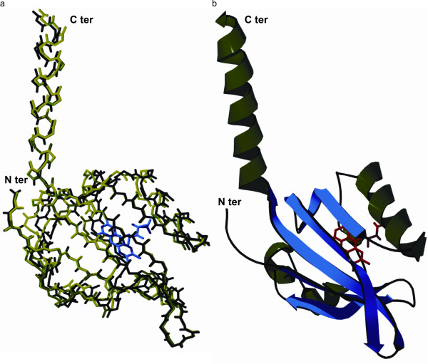
Background: Fluorescent reporter proteins have revolutionized our understanding of cellular bioprocesses by enabling live cell imaging with exquisite spatio-temporal resolution. Existing fluorescent proteins are predominantly based on the green fluorescent protein (GFP) and related analogs. However, GFP-family proteins strictly require molecular oxygen for maturation of fluorescence, which precludes their application for investigating biological processes in low-oxygen environments. A new class of oxygen-independent fluorescent reporter proteins was recently reported based on flavin-binding photosensors from Bacillus subtilis and Pseudomonas putida. However, flavin-binding fluorescent proteins show very limited brightness, which restricts their utility as biological imaging probes.

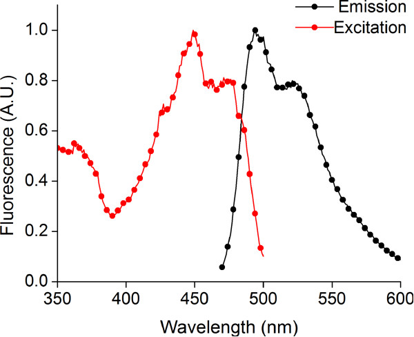
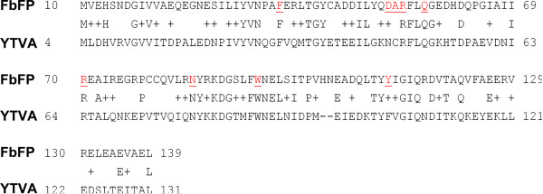
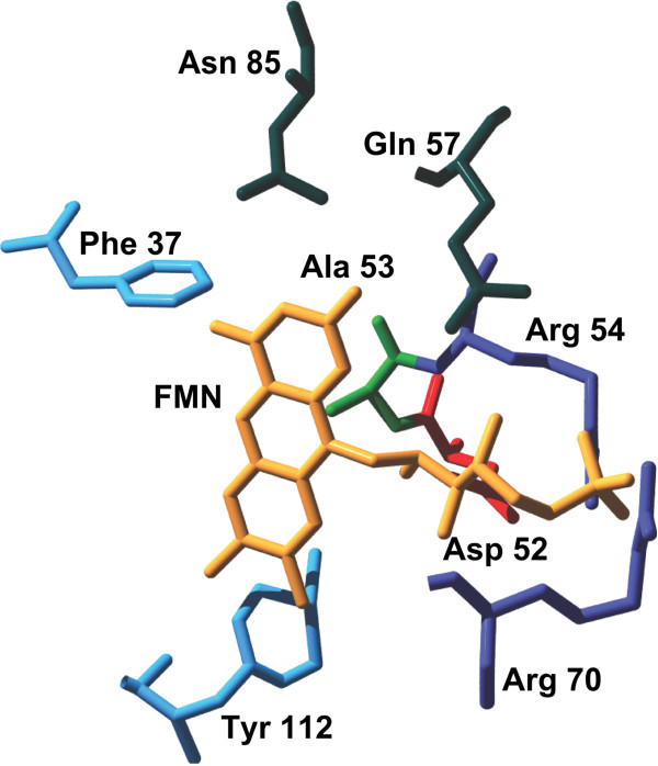
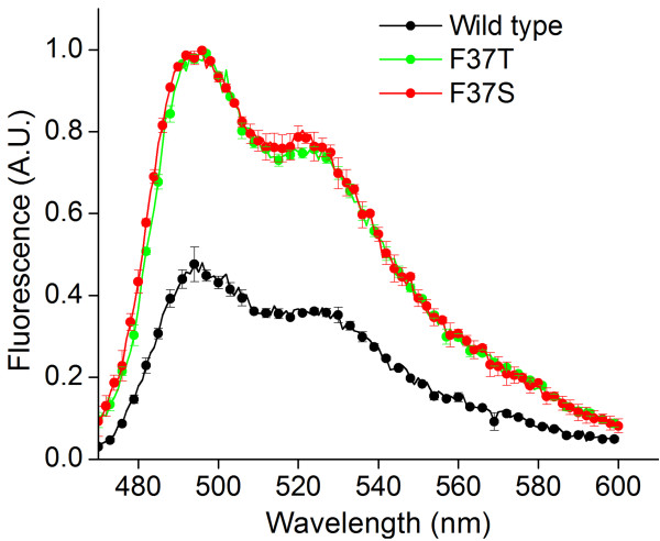
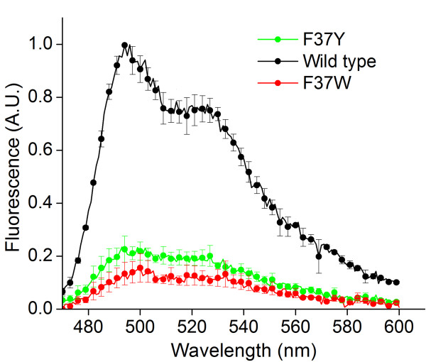
Results: In this work, we report the discovery of bright mutants of a flavin-binding fluorescent protein from P. putida using directed evolution by site saturation mutagenesis. We discovered two mutations at a chromophore-proximal amino acid (F37S and F37T) that confer a twofold enhancement in brightness relative to the wild type fluorescent protein through improvements in quantum yield and holoprotein fraction. In addition, we observed that substitution with other aromatic amino acids at this residue (F37Y and F37W) severely diminishes fluorescence emission. Therefore, we identify F37 as a key amino acid residue in determining fluorescence.

Conclusions: To increase the scope and utility of flavin-binding fluorescent proteins as practical fluorescent reporters, there is a strong need for improved variants of the wild type protein. Our work reports on the application of site saturation mutagenesis to isolate brighter variants of a flavin-binding fluorescent protein, which is a first-of-its-kind approach. Overall, we anticipate that the improved variants will find pervasive use as fluorescent reporters for biological studies in low-oxygen environments.


Figures

Figure 1
Figure 1
Fluorescence excitation and emission spectra for purified wild type FbFP protein. (a) Excitation spectrum of purified FbFP in Buffer E (25 mM sodium phosphate, 1 M sodium chloride, pH 7.4). Excitation spectrum was determined by monitoring fluorescence emission at 500 nm while scanning the excitation light between 350 to 480 nm. (b) Emission spectrum of purified FbFP in Buffer E. Emission spectrum was determined by monitoring fluorescence emission between 470 and 600 nm while exciting the sample at 450 nm.
Figure 2
Figure 2
Sequence alignment between FbFP and YTVA (B. subtilis). Sequence alignment reveals close agreement between FbFP from P. putida and YTVA from B. subtilis, with 37% identical and 62% similar amino acids shared between the two proteins. In this way, the YTVA structure serves as an ideal template for molecular modeling of the FbFP. Amino acids selected for site saturation mutagenesis are indicated in red and underlined.
Figure 3
Figure 3
Homology derived structure of FbFP. (a) Alignment between the backbone Cα atoms of the YTVA template (green) and the FbFP model (black). The structures are well aligned, with a root mean squared deviation of 0.5 nm over 126 Cα atoms. The FMN cofactor is depicted in blue. (b) Ribbon model of homology derived three dimensional structure of FbFP. The buried FMN cofactor is indicated in red. Alpha helices and beta strands in the protein structure are represented in green and blue respectively. Images were generated by ray tracing with POV-Ray version 3.6.
Figure 4
Figure 4
Flavin-binding cavity in FbFP. Amino acids upto a distance of 0.3 nm from FMN are shown. Also shown are A52 and F37 which are at a distance of 0.4 nm from FMN. Amino acids are colored according to their property (blue – aromatic; red - negatively charged; violet - positively charged; green – hydrophobic; deep green - polar). The W94 target lies outside the vicinity of the chromophore and does not appear in the structure.
Figure 5
Figure 5
Emission spectra of purified F37S and F37T mutant proteins. FbFP F37S and F37T mutant proteins have approximately twofold enhanced emission yields at 500 nm, similar to fluorescence measurements in whole cell based assays. Excitation was performed at 450 nm, and values are normalized to the protein concentration as determined by the Bradford assay.
Figure 6
Figure 6
SDS PAGE analysis for wild type and mutant FbFPs. Lane 1: F37W, Lane 2: F37E, Lane 3: F37Y, Lane 4: F37T, Lane 5: F37S, Lane 6: Wild type FbFP. Selected mutants (F37W, F37E, F37Y, F37S, and F37T) of the FbFP F37X saturation mutagenesis library were purified and run on a 10% denaturing polyacrylamide gel (160 V, 45 minutes). Fluorescence enhanced mutants FbFP F37S and F37T comigrate with the wild type band (WT) at an approximate molecular mass of 17 kDa. Additional mutants correspond to neutral (F37E, lane 2) or deleterious mutants (F37W, lane 1 and F37Y, lane 3).
Figure 7
Figure 7
Oligomeric state of improved FbFP F37S mutant. Oligomeric states of native and mutant FbFPs were determined by size exclusion chromatography. Chromatograms of (a) wild type FbFP and (b) FbFP F37S from size exclusion chromatography. The F37S mutant and wild type FbFP were run through the Superdex 200 column under conditions identical to those used for the protein standards. FbFP F37S and wild type FbFP elute at similar volumes, thereby confirming that the native and mutant proteins have identical oligomeric conformations (dimeric). (c) Calibration plot for estimation of molecular mass using size exclusion chromatography. Globular proteins of known molecular weights were loaded onto a Superdex 200 gel filtration column and protein elution volumes corresponding to peak absorbance at 280 nm were recorded. A semi-logarithmic plot of molecular weight against elution volume yielded a straight line whose slope was used to calculate the molecular mass of the FbFP wild type and mutants.
Figure 8
Figure 8
Emission spectra of purified F37Y and F37W mutant proteins. F37Y and F37W mutants show severely quenched fluorescence emission yields at 500 nm relative to the wild type protein. Samples were excited at 450 nm, and all values were normalized to protein concentrations as determined by the Bradford assay.

References

    1. Zhao HM. Directed evolution of novel protein functions. Biotechnol Bioeng. 2007;98(2):313–317. doi: 10.1002/bit.21455. - DOI - PubMed
    1. Bloom JD, Arnold FH. In the light of directed evolution: Pathways of adaptive protein evolution. Proc Natl Acad Sci USA. 2009;106:9995–10000. doi: 10.1073/pnas.0901522106. - DOI - PMC - PubMed
    1. Brustad EM, Arnold FH. Optimizing non-natural protein function with directed evolution. Curr Opin Chem Biol. 2011;15(2):201–210. doi: 10.1016/j.cbpa.2010.11.020. - DOI - PMC - PubMed
    1. Cobb RE, Sun N, Zhao H. Directed evolution as a powerful synthetic biology tool. Methods. 2012. - DOI - PMC - PubMed
    1. Cormack BP, Valdivia RH, Falkow S. FACS-optimized mutants of the green fluorescent protein (GFP) Gene. 1996;173(1):33–38. doi: 10.1016/0378-1119(95)00685-0. - DOI - PubMed