. 2012 Oct 24;2(1):36.
doi: 10.1186/2045-3701-2-36.

Strategy for eliciting antigen-specific CD8+ T cell-mediated immune response against a cryptic CTL epitope of merkel cell polyomavirus large T antigen

Affiliations

Strategy for eliciting antigen-specific CD8+ T cell-mediated immune response against a cryptic CTL epitope of merkel cell polyomavirus large T antigen

Bianca P Gomez et al. Cell Biosci. .

Abstract

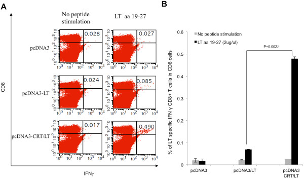
Background: Merkel cell carcinoma (MCC) is a relatively new addition to the expanding category of oncovirus-induced cancers. Although still comparably rare, the number of cases has risen dramatically in recent years. Further complicating this trend is that MCC is an extremely aggressive neoplasm with poor patient prognosis and limited treatment options for advanced disease. The causative agent of MCC has been identified as the merkel cell polyomavirus (MCPyV). The MCPyV-encoded large T (LT) antigen is an oncoprotein that is theorized to be essential for virus-mediated tumorigenesis and is therefore, an excellent MCC antigen for the generation of antitumor immune responses. As a foreign antigen, the LT oncoprotein avoids the obstacle of immune tolerance, which normally impedes the development of antitumor immunity. Ergo, it is an excellent target for anti-MCC immunotherapy. Since tumor-specific CD8+ T cells lead to better prognosis for MCC and numerous other cancers, we have generated a DNA vaccine that is capable of eliciting LT-specific CD8+ T cells. The DNA vaccine (pcDNA3-CRT/LT) encodes the LT antigen linked to a damage-associated molecular pattern, calreticulin (CRT), as it has been demonstrated that the linkage of CRT to antigens promotes the induction of antigen-specific CD8+ T cells.

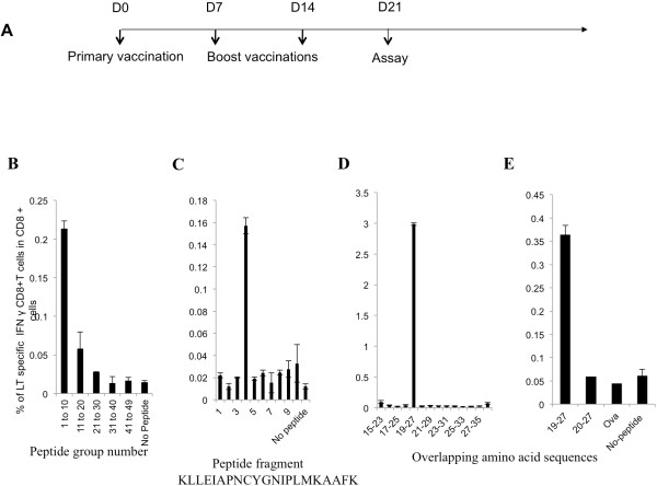
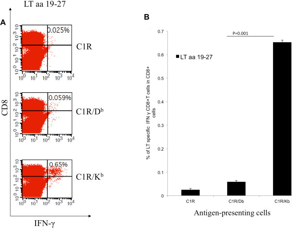
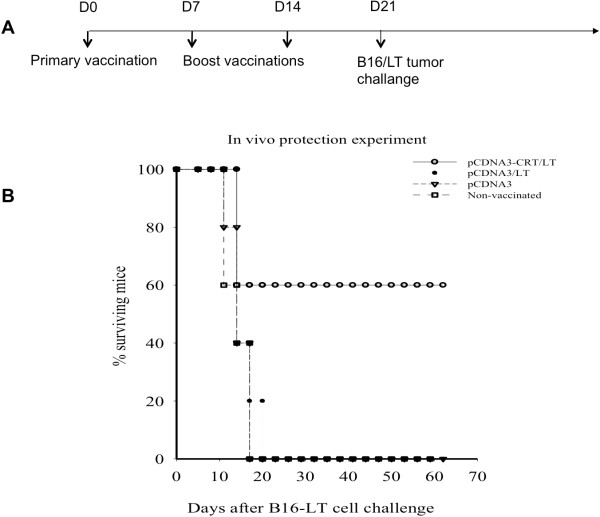
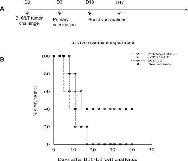
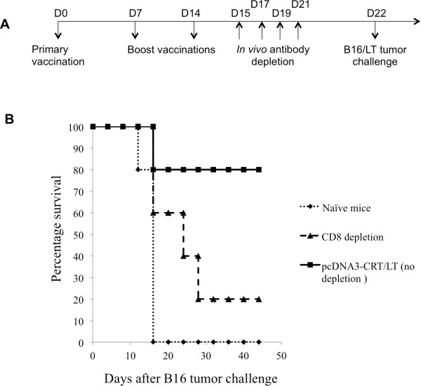
Results: The present study shows that DNA vaccine-induced generation of LT-specific CD8+ T cells is augmented by linking CRT to the LT antigen. This is relevant since the therapeutic effects of the pcDNA3-CRT/LT DNA vaccine is mediated by LT-specific CD8+ T cells. Mice vaccinated with the DNA vaccine produced demonstrably more LT-specific CD8+ T cells. The DNA vaccine was also able to confer LT-specific CD8+ T cell-mediated protective and therapeutic effects to prolong the survival of mice with LT-expressing tumors. In the interest of determining the LT epitope which most MCC-specific CD8+ T cells recognize, we identified the amino acid sequence of the immunodominant LT epitope as aa19-27 (IAPNCYGNI) and found that it is H-2kb-restricted.

Conclusion: The results of this study can facilitate the development of other modes of MCC treatment such as peptide-based vaccines and adoptive transfer of LT-specific CD8+ T cells. Likewise, the MCC DNA vaccine has great potential for clinical translation as the immunologic specificity is high and the treatment strategy can be exported to address other virus-induced tumors.


Figures

Figure 1
Figure 1
Identification of MHC class I-restricted immunodominant LT epitope using overlapping peptides and splenocytes from mice vaccinated with pcDNA3-CRT/LT. (A) Schematic diagram of vaccination schedule in vivo. C57BL/6 mice (5 mice/group) intradermally received pcDNA, pcDNA3-LT, or pcDNA3-CRT/LT 3 times by gene gun at a 7-day interval. Vaccination with pcDNA3-CRT/LT produced the most LT-specific CD8+ T cells therefore pooled splenocytes from pcDNA3-CRT/LT vaccinated mice were cultured in vitro with various overlapping LT peptides to find the immunodominant LT epitope. (B) Representative bar graph of flow cytometric data indicating the pool containing peptides #1-10 activated the most LT-specific CD8+ T cells. (C) Representative bar graph of flow cytometric data indicating that out of peptides #1 through 10, LT peptide #4 was able to activate the most LT-specific CD8+ T cells. (D) Representative bar graph of flow cytometric data suggesting amino acid 19–27 (IAPNCYGNI) of the LT antigen may be the immunodominant epitope determining the specificity of the vast majority of LT-specific CD8+ T cells. (E) Representative bar graph of flow cytometric data confirming that the suggested amino acid 19–27 (IAPNCYGNI) of the LT antigen may be the immunodominant epitope determining the specificity of the vast majority of LT-specific CD8+ T cells, and not the peptide occupying positions 20–27 (APNCYGNI). Ova (SIINFEKL) was used a negative control.
Figure 2
Figure 2
MHC class I H-2b binding restriction of LT aa 19–27 epitope identified by intracellular cytokine staining and flow cytometry. MHC class I binding restriction was determined by C1R transfectants, C1R/Db and C1R/Kb , that were pulsed with immunodominant LT peptide (aa 19–27). (A) Representative flow cytometric data showing the amounts of LT-specific IFN-γ+ CD8+ T-cells after splenocytes from pcDNA3-CRT/LT vaccinated mice are stimulated by LT peptide-pulsed C1R, C1R/Db, or C1R/Kb cells in vitro. (B) Representative bar graph of flow cytometric data showing the proportions of LT-specific IFN-γ + CD8+ T cells per 3 × 105 splenocytes following in vitro stimulation as described above. Note that LT epitope (aa 19–27) is H-2kb-restricted.
Figure 3
Figure 3
pcDNA3-CRT/LT generates the most LT-specific (aa 19–27) CD8+ T cells. C57BL/6 mice (5 mice/group) were immunized with DNA vaccines by gene gun in the same schedule as Figure 1A. Pooled splenocytes from mice vaccinated with pcDNA3 vector (control), pcDNA3-LT, and pcDNA3-CRT/LT were collected and cultured in vitro with either no peptide or amino acid 19–27 then stained for intracellular IFN-gamma and CD8+ T cell surface marker. (A) Representative flow cytometry dot plot of LT-specific CD8+ T cell activation after stimulation with amino acid sequence 19–27. (B) Representative bar graph of flow cytometric data.
Figure 4
Figure 4
In vivo tumor protection experiment examining the antitumor effects generated by pcDNA3-CRT/LT against LT-expressing tumor. (A) Schematic diagram of experiment schedule. (B) Survival curve of vaccinated mice subjected to tumor challenge. Note that vaccination with pcDNA3-CRT/LT greatly prolonged mouse survival after tumor challenge.
Figure 5
Figure 5
In vivo tumor treatment experiment with DNA vaccines. (A) Schematic diagram of experiment schedule. (B) Survival curve of tumor-bearing mice treated with various DNA vaccines. Note that pcDNA3-CRT/LT had the greatest therapeutic effect and extended the survival of B16/LT tumor-bearing mice.
Figure 6
Figure 6
The effect of CD8 T cells on tumor protection of the pCDNA3-CRT/LT vaccine. (A) Schematic diagram of the vaccination regimen for in vivo antibody depletion experiments. C57BL/6 mice (5 per group) were vaccinated by gene gun with pcDNA3-CRT/LT DNA vaccine on D0. Vaccinated mice were boosted two times at the same dose and regimen at one week intervals. Beginning 1 day after last vaccination, vaccinated mice were intraperitoneally injected with anti-CD8 monoclonal antibody other day. Antibody-depleted mice were then challenged with B16/LT tumor (1 × 105 cells/mouse) subcutaneously in the right flank on D22. Mice were monitored for evidence of tumor growth by inspection, palpation and tumor size was measured twice a week. (B) Survival analysis of B16/LT tumor-bearing mice treated with pcDNA3-CRT/LT DNA vaccine.

References

    1. Albores-Saavedra J, Batich K, Chable-Montero F, Sagy N, Schwartz AM, Henson DE. Merkel cell carcinoma demographics, morphology, and survival based on 3870 cases: a population based study. J Cutan Pathol. 2010;37(1):20–27. doi: 10.1111/j.1600-0560.2009.01370.x. Epub 2009/07/30. - DOI - PubMed
    1. Hodgson NC. Merkel cell carcinoma: changing incidence trends. J Surg Oncol. 2005;89(1):1–4. doi: 10.1002/jso.20167. Epub 2004/12/22. - DOI - PubMed
    1. Herbst A, Haynes HA, Nghiem P. The standard of care for Merkel cell carcinoma should include adjuvant radiation and lymph node surgery. J Am Acad Dermatol. 2002;46(4):640–642. Epub 2002/03/22. - PubMed
    1. Agelli M, Clegg LX, Becker JC, Rollison DE. The etiology and epidemiology of merkel cell carcinoma. Curr Probl Canc. 2010;34(1):14–37. doi: 10.1016/j.currproblcancer.2010.01.001. Epub 2010/04/08. - DOI - PubMed
    1. Heath M, Jaimes N, Lemos B, Mostaghimi A, Wang LC, Penas PF. et al.Clinical characteristics of Merkel cell carcinoma at diagnosis in 195 patients: the AEIOU features. J Am Acad Dermatol. 2008;58(3):375–381. doi: 10.1016/j.jaad.2007.11.020. Epub 2008/02/19. - DOI - PMC - PubMed