Review
. 2012 Dec 1;11(23):4328-38.
doi: 10.4161/cc.22485. Epub 2012 Oct 24.

Barrett esophagus: what a mouse model can teach us about human disease

Affiliations
Review

Barrett esophagus: what a mouse model can teach us about human disease

Michael Quante et al. Cell Cycle. .

Abstract

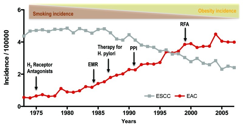
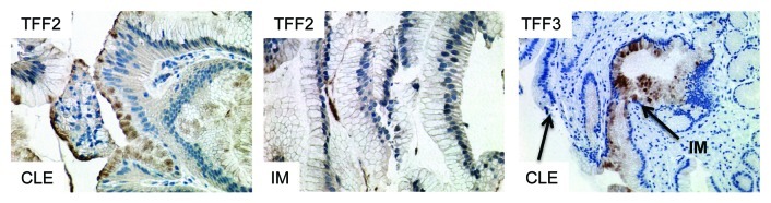
The incidence of esophageal adenocarcinoma (EAC) is rapidly rising in the western world and accounts for 2% of all cancer-related deaths. The precursor lesion for EAC is Barrett esophagus (BE), which is strongly associated with gastresophageal reflux disease. A major limitation to the study of EAC has been the absence of tractable and genetically modifiable preclinical models of BE. A mouse model of BE and EAC that resembles human disease could provide novel insights into the origins and molecular pathogenesis of BE. In addition, validated animal models could help stratify BE patients given the limited predictive power of current standard endoscopic measures and clinical assessment. Here, we review the findings from recently developed mouse models of BE and EAC and their impact on clinical decision making, surveillance programs and therapeutic options. The data, taken together, suggest potential origins of BE from the gastric cardia, a role of bile acid and hypergatrinemia for carcinogenesis, a growing importance for columnar-like epithelium and a critical role for Notch signaling.


Figures

None
Figure 1. The increasing incidence of esophageal adenocarcinoma (EAC) between 1975 and 2005 and associated factors. During this period of time, esophageal squamous cell carcinoma (ESCC) has declined in incidence, as has its major risk factor, tobacco use. EAC incidence has risen concomitantly with obesity, which is one risk factor for the disease. Despite the advent of medical therapies such as acid inhibition through H2 receptor antagonists and proton pump inhibitors (PPIs), and the treatment of H pylori, EAC has continued to rise in incidence. Techniques such as endoscopic mucosal resection (EMR) and RFA (radiofrequency ablation) have also failed to stem the rise in EAC incidence. Data from the Surveillance Epidemiology and End Results (SEER) database of the National Cancer Institute.
None
Figure 2. The L2-IL-1β mouse model of Barrett esophagus (BE) and esophageal adenocarcinoma (EAC). (A) Histological appearance of BE lesions, consisting of columnar mucosa and mucus-producing cells at the squamocolumnar junction (SCJ) in the L2-IL-1β mouse. (B) Anatomical differences between human and mouse stomach. The SCJ is distal to the esophago-gastric junction and comprises the border between the squamous forestomach and the columnar-lined stomach in the mouse. In the figure on the far right, the mouse stomach has been opened along the greater curvature to demonstrate the location of the SCJ. The vertical line demonstrates the location of the sagittal section seen in Figure 2A. (with kind permission from Cancer Cell / Elsevier, Quante, Bhagat et al. 2012) (C) Human Barrett esophagus, containing columnar cells and goblet cells. (D) Electron microscopic appearance of mouse BE lesions demonstrating columnar appearance, mucin granules, and surface microvilli. (E) Tumor at the SCJ seen during upper endoscopy of an L2-IL-1β mouse. (F) Macroscopic appearance of tumor at the SCJ in the L2-IL-1β mouse.
None
Figure 3. Schematic diagram of hypothesized cells of origin for BE. (A) Lgr5+ stem cells migrate proximally from the cardia into the esophagus in response to proinflammatory stimuli (Quante, Bhagat et al. 2012). (B) Car4+/Krt7+ residual embryonic cells expand proximally from the SCJ into the esophagus (Wang, Ouyang et al. 2011). (C) Esophageal squamous epithelial cells de-differentiate and expand to form squamous metaplasia. (D) Stem cells of the submucosal esophageal glands expand and give rise to BE (Leedham SJ et al., 2008).
None
Figure 4. (A) Pooled analysis of studies that characterized the mucosa adjacent to esophageal adenocarcinoma in patients as intestinal metaplasia (IM, in %) or columnar-lined esophagus (CLE, %). In these nine studies comprising 1328 patients, there was no significant difference between the presence of IM or CLE as the tissue adjacent to EAC (p = 0.88). (with kind permission from Cancer Cell / Elsevier, Quante, Bhagat et al. 2012) (B) Histopathologic evaluation of the mucosa adjacent to EAC in 31 specimens in a German cohort. CLE was most commonly seen, compared with IM, cardia-type tissue and squamous mucosa (SQ) (*p < 0.05).
None
Figure 5. Expression of TFF2 and TFF3 in human columnar-lined epithelium (CLE) and intestinal metaplasia (IM). TFF2 marks an early precursor cell in the columnar lineage; staining is seen in CLE, but (B) with the development of IM, TFF2 staining decreases. (C) Conversely, TFF3 staining is not seen in CLE but increases with the development of IM.

References

    1. Kahrilas PJ. The problems with surveillance of Barrett’s esophagus. N Engl J Med. 2011;365:1437–8. doi: 10.1056/NEJMe1108435. - DOI - PubMed
    1. Xian W, Ho KY, Crum CP, McKeon F. Cellular origin of Barrett’s esophagus: controversy and therapeutic implications. Gastroenterology. 2012;142:1424–30. doi: 10.1053/j.gastro.2012.04.028. - DOI - PMC - PubMed
    1. Quante M, Bhagat G, Abrams JA, Marache F, Good P, Lee MD, et al. Bile acid and inflammation activate gastric cardia stem cells in a mouse model of Barrett-like metaplasia. Cancer Cell. 2012;21:36–51. doi: 10.1016/j.ccr.2011.12.004. - DOI - PMC - PubMed
    1. Spechler SJ, Fitzgerald RC, Prasad GA, Wang KK. History, molecular mechanisms, and endoscopic treatment of Barrett’s esophagus. Gastroenterology. 2010;138:854–69. doi: 10.1053/j.gastro.2010.01.002. - DOI - PMC - PubMed
    1. Everhart JE, Ruhl CE. Burden of digestive diseases in the United States part I: overall and upper gastrointestinal diseases. Gastroenterology. 2009;136:376–86. doi: 10.1053/j.gastro.2008.12.015. - DOI - PubMed

MeSH terms

LinkOut - more resources