Case Reports
. 2012 Oct 24:10:220.
doi: 10.1186/1477-7819-10-220.

Long-term survival of patients suffering from glioblastoma multiforme treated with tumor-treating fields

Affiliations
Case Reports

Long-term survival of patients suffering from glioblastoma multiforme treated with tumor-treating fields

Aaron Michael Rulseh et al. World J Surg Oncol. .

Abstract

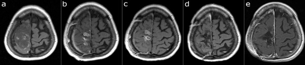
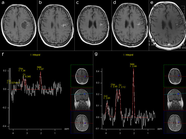
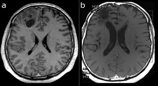
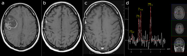
Glioblastoma multiforme (GBM) is the most common and malignant primary intracranial tumor, and has a median survival of only 10 to 14 months with only 3 to 5% of patients surviving more than three years. Recurrence (RGBM) is nearly universal, and further decreases the median survival to only five to seven months with optimal therapy. Tumor-treating fields (TTField) therapy is a novel treatment technique that has recently received CE and FDA approval for the treatment of RGBM, and is based on the principle that low intensity, intermediate frequency electric fields (100 to 300 kHz) may induce apoptosis in specific cell types. Our center was the first to apply TTField treatment to histologically proven GBM in a small pilot study of 20 individuals in 2004 and 2005, and four of those original 20 patients are still alive today. We report two cases of GBM and two cases of RGBM treated by TTField therapy, all in good health and no longer receiving any treatment more than seven years after initiating TTField therapy, with no clinical or radiological evidence of recurrence.


Figures

Figure 1
Figure 1
Serial MR imaging in Case 1. T1-weighted image after application of contrast agent. a) April 2004, before surgery. GBM located in the right central region. b) July 2004, post-operative radiotherapy and chemotherapy. Two enhancing lesions present. c) September 2004, one month after the start of TTField therapy. The dorsal enhancing lesion increased in size, highly suspicious of tumor recurrence. d) June 2005, TTField treatment. No enhancing lesion present. e) August 2011. No enhancing lesion present. GBM, glioblastoma multiforme; MR, magnetic resonance; TTField, tumor-treating fields.
Figure 2
Figure 2
Serial MR imaging in Case 2. T1-weighted image after application of contrast agent. a) January 2004, before surgery. GBM located in the left frontal region. b) August 2004, post-operative radiotherapy and chemotherapy. Start of TTField treatment. An enhancing lesion suspected to be recurrent or residual tumor. c) March 2005, TTF treatment. The enhancing lesion became partly cystic. d) June 2005, regression of the cystic part. A subtle enhancing lesion still present. e) November 2011. A subtle enhancing lesion without progression. f) Proton MR spectroscopy of the enhancing lesion with dominant noise signal, suggesting gliosis rather than tumor. g) Neighboring spectrum is practically normal, demonstrating that MR spectroscopy provided reliable data from the selected slice. GBM, Glioblastoma multiforme; MR, magnetic resonance; TTField, tumor-treating fields.
Figure 3
Figure 3
Serial MR imaging in case 3. T1-weighted image after application of contrast agent. a) January 2005 before surgery. GBM located in the right frontal region. b) December 2011. No tumor recurrence detected. GBM, glioblastoma multiforme; MR, magnetic resonance.
Figure 4
Figure 4
Serial MR imaging in case 4. T1-weighted image after application of contrast agent. a) November 2005, before surgery. GBM located in the right frontal region. b) May 2006, a small, extra-axial enhancing lesion. c) September 2011, no change of the enhancing lesion. d) FLAIR and MR spectroscopy images, September 2011. The small volume of increased signal intensity on FLAIR images did not show a tumor-like pattern on MR spectroscopy. FLAIR, Fluid Attenuated Inversion Recovery; GBM, glioblastoma multiforme; MR, magnetic resonance.
Figure 5
Figure 5
Kaplan-Meier survival curves. Kaplan-Meier survival curves for all 20 participants in the original pilot study, both as a single group and divided into recurrent and newly diagnosed glioblastoma multiforme. Two patients were lost to follow-up in the newly diagnosed group and are represented by censor marks.

References

    1. Dobes M, Khurana VG, Shadbolt B, Jain S, Smith SF, Smee R, Dexter M, Cook R. Increasing incidence of glioblastoma multiforme and meningioma, and decreasing incidence of Schwannoma (2000–2008): findings of a multicenter Australian study. Surg Neurol Int. 2011;2:176. doi: 10.4103/2152-7806.90696. - DOI - PMC - PubMed
    1. Dubrow R, Darefsky AS. Demographic variation in incidence of adult glioma by subtype, United States, 1992–2007. BMC Cancer. 2011;11:325. doi: 10.1186/1471-2407-11-325. - DOI - PMC - PubMed
    1. Johnson DR, O’Neill BP. Glioblastoma survival in the United States before and during the temozolomide era. J Neurooncol. 2012;107:359–364. doi: 10.1007/s11060-011-0749-4. - DOI - PubMed
    1. Krex D, Klink B, Hartmann C, von Deimling A, Pietsch T, Simon M, Sabel M, Steinbach JP, Heese O, Reifenberger G, Weller M, Schackert G. Long-term survival with glioblastoma multiforme. Brain. 2007;130:2596–2606. doi: 10.1093/brain/awm204. - DOI - PubMed
    1. Deb P, Sharma MC, Mahapatra AK, Agarwal D, Sarkar C. Glioblastoma multiforme with long term survival. Neurol India. 2005;53:329–332. doi: 10.4103/0028-3886.16934. - DOI - PubMed

Publication types

MeSH terms