. 2012 Dec 1;139(23):4449-60.
doi: 10.1242/dev.084871. Epub 2012 Oct 24.

Endothelial deletion of murine Jag1 leads to valve calcification and congenital heart defects associated with Alagille syndrome

Affiliations

Endothelial deletion of murine Jag1 leads to valve calcification and congenital heart defects associated with Alagille syndrome

Jennifer J Hofmann et al. Development. .

Abstract

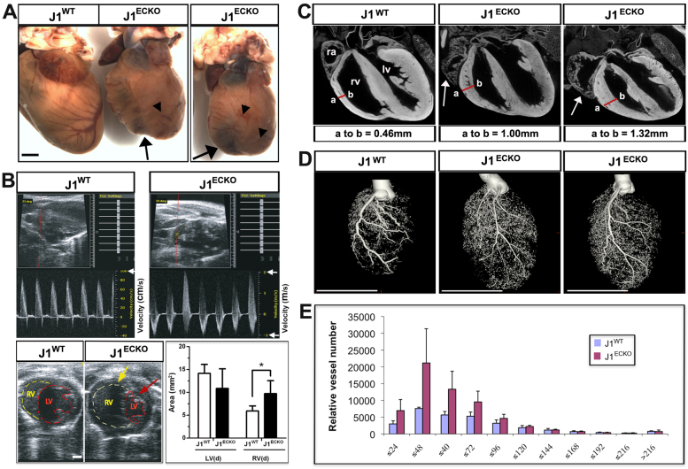
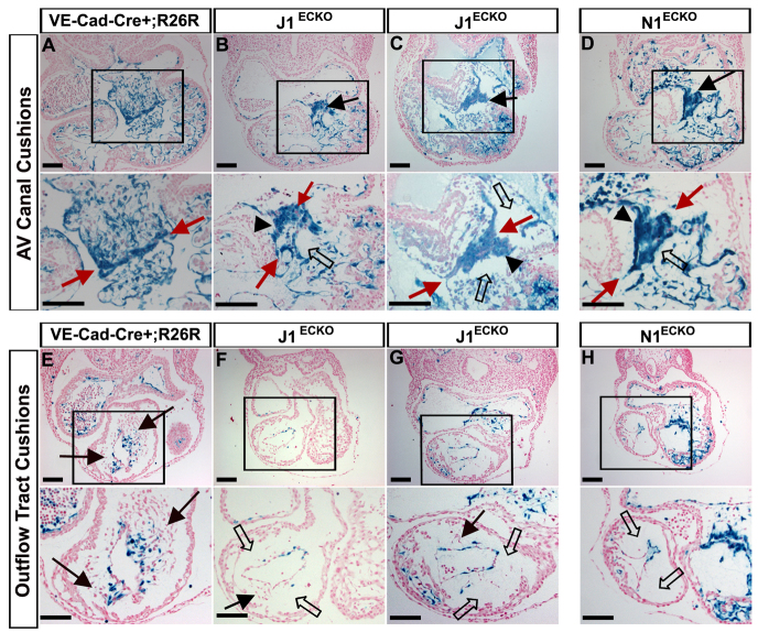
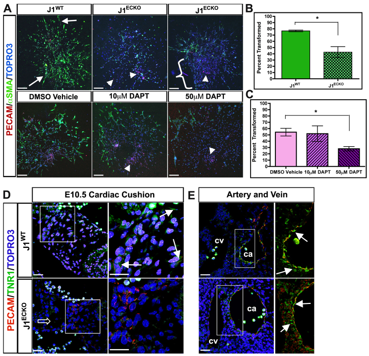
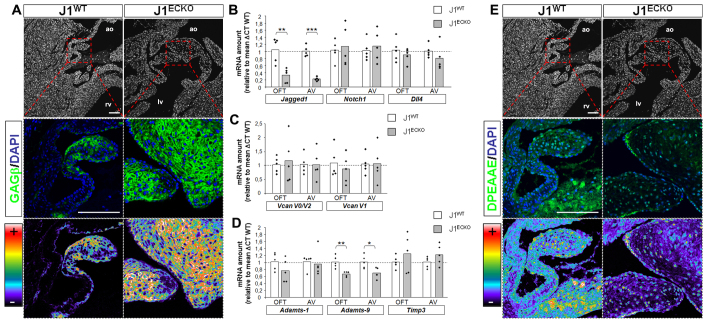
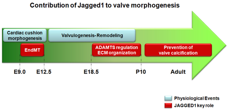
The Notch signaling pathway is an important contributor to the development and homeostasis of the cardiovascular system. Not surprisingly, mutations in Notch receptors and ligands have been linked to a variety of hereditary diseases that impact both the heart and the vasculature. In particular, mutations in the gene encoding the human Notch ligand jagged 1 result in a multisystem autosomal dominant disorder called Alagille syndrome, which includes tetralogy of Fallot among its more severe cardiac pathologies. Jagged 1 is expressed throughout the developing embryo, particularly in endothelial cells. Here, we demonstrate that endothelial-specific deletion of Jag1 leads to cardiovascular defects in both embryonic and adult mice that are reminiscent of those in Alagille syndrome. Mutant mice display right ventricular hypertrophy, overriding aorta, ventricular septal defects, coronary vessel abnormalities and valve defects. Examination of mid-gestational embryos revealed that the loss of Jag1, similar to the loss of Notch1, disrupts endothelial-to-mesenchymal transition during endocardial cushion formation. Furthermore, adult mutant mice exhibit cardiac valve calcifications associated with abnormal matrix remodeling and induction of bone morphogenesis. This work shows that the endothelium is responsible for the wide spectrum of cardiac phenotypes displayed in Alagille Syndrome and it demonstrates a crucial role for Jag1 in valve morphogenesis.


Figures

Fig. 1.
Fig. 1.
Deletion of Jag1 in the endothelium. (A) Jag1 immunostaining (green) in the endothelium (red) of J1WT and J1ECKO mice. Mosaic Jag1 expression in E10.5 J1ECKO vessels compared with J1WT mice, with areas of Jag1 loss (arrowheads) adjacent to areas of Jag1 expression (arrows). By P0, endothelial Jag1 deletion was highly penetrant (arrowheads) in J1ECKO mice, whereas Jag1 was maintained in veins and some smooth muscle cells (arrows). (B) Western blot on fluorescence-activated cell sorting (FACS)-isolated endothelial cells from J1WT and J1ECKO embryos at progressive developmental stages showed decreases in Jag1 levels over time in J1ECKO mice compared with J1WT. Tubulin levels were used as loading controls. Several faint lower bands in embryonic J1ECKO samples were degradation/processing fragments not seen in other equivalent samples. (C) Evaluation of VE-Cadherin-Cre activity at E10.5 revealed animals with high (upper panels) and low (lower panels) recombination, as shown by β-galactosidase staining (blue). Arrows indicate recombined cells and arrowheads non-recombined endothelium. Cardiac cushions at high magnification show both recombined (red arrows) and non-recombined (red arrowheads) mesenchymal cells resulting from EndMT cells. a, atrium; v, ventricle. Scale bars: 50 μm in A; 100 μm in C.
Fig. 2.
Fig. 2.
Cardiac defects in adult Jag1 endothelial mutants. (A) J1ECKO mice demonstrated morphological defects in the adult heart. Hearts from mutant mice frequently exhibited defective coronary vessel branching (arrowheads) and abnormal heart structure near the apex (arrows), suggesting dilation or right ventricular hypertrophy (arrows) (n=20). (B) Representative 2D echo and Doppler images of adult male mice, measured near the right ventricular outflow tract, showed severe regurgitation at the pulmonary semilunar valve and an increase in systolic blood velocity during right ventricular ejection in J1ECKO hearts. Note different scales of velocity (white arrows) in J1WT (cm/second) and J1ECKO (m/second) measurements. The lower images of the same panel display a left parasternal transverse section with 2D measurement of adult hearts, demonstrating an increase in the right ventricular chamber (yellow dotted line and yellow arrow) and a decrease in the left ventricular chamber (red dotted line and red arrow) in J1ECKO mice (n=6) compared with J1WT (n=6). Measurement of ventricular chamber area at diastolic demonstrated a significant increase in the size of J1ECKO right ventricles (*P=0.024). (C) μCT imaging of adult J1WT and J1ECKO hearts showed right ventricular hyperplasia in mutants, as per measurements of the right ventricular wall (red line between a and b). J1ECKO hearts also showed enlarged right atria (arrows). (D) Additional μCT imaging of adult J1ECKO hearts revealed increased coronary vessel density in many mice. (E) Quantification of vessel number according to vessel diameter of J1ECKO and J1WT hearts using μCT (n=11). lv, left ventricle; ra, right atrium; rv, right ventricle. Error bars represent s.d. Scale bars: 1 mm in A,B; 5 mm in D.
Fig. 3.
Fig. 3.
AGS-related cardiac defects in embryonic and neonatal J1ECKO mice. (A-C) Light microscopy of the back (A) and front (B) of neonatal J1WT and J1ECKO hearts (n≥20). (A) Compared with J1WT mice, hearts of P0 J1ECKO mice displayed a variety of cardiac defects, such as abnormal morphology and increased atrial and ventricular size (A, arrows), in addition to dilated, hemorrhaging cardiac vessels (A, arrowheads). (B) P1 hearts of J1WT and J1ECKO mutants, with corresponding H&E-stained histological sections of the respective hearts below (C). An overriding aorta was noted in the J1ECKO heart (B, arrow). Dotted lines delineate great vessels and highlight outflow tract defects. (C) Histological sections of these J1ECKO hearts, with high magnification images below, revealed ventricular septal defects (arrows) and hypertrophic valves (asterisk). (D) Light microscopy of β-galactosidase-stained histological sections of E14.5 VE-Cadherin-Cre;Rosa26R wild-type and J1ECKO hearts revealed VSDs in the mutant hearts (D, arrow) 24 hours after the septum normally closes, as seen in the wild-type heart (D). (E) Large bulges were occasionally found near the valve annulus between the left atria and ventricle in P5 J1ECKO H&E-stained hearts (E, arrow). ao, aorta; la, left atrium; lv, left ventricle; pa, pulmonary artery; ra, right atrium; rv, right ventricle. Scale bars: 200 μm.
Fig. 4.
Fig. 4.
Loss of Notch signaling impairs EndMT in cardiac and outflow cushions. (A-H) β-Galactosidase- and Nuclear Fast Red-stained sections of E10.5 control (A,E), J1ECKO (B,C,F,G), and N1ECKO (D,H) mouse hearts (n=4 or more samples evaluated per cohort). Loss of endothelial Jag1 (B,C) leads to delayed EMT in E10.5 cardiac cushions (magnified view indicated by box) compared with control (A). Defects in N1ECKO hearts (D) phenocopy those of the J1ECKO hearts (B,C). Rounded cells (arrowheads) were attached to the cushion interface (red arrows), and both N1ECKO and J1ECKO hearts displayed areas of decreased cellular density within the cardiac jelly (B-D, open arrows) compared with control (A). (E-H) The OFT cushions of E10.5 J1ECKO and N1ECKO mutants were also hypocellular (F-H, open arrows) in contrast to extensive EndMT observed in control littermates (E, black arrows) (magnified view indicated by box). Mesenchymal cells were transformed in J1ECKO mutant OFT cushions mostly in places with unrecombined endothelium, revealed by absence of β-galactosidase staining (F,G, black arrows). Scale bars: 100 μm.
Fig. 5.
Fig. 5.
Jag1 is a crucial ligand for Notch signaling during EndMT in endocardial cushions. (A) Representative images of E9.5 cardiac cushion explants cultured for 48 hours, fixed, stained with anti-αSMA (in green, arrows) and anti-PECAM1 (in red, arrowheads). Explants from J1ECKO hearts showed a reduction in the number of transformed, αSMA+ cells compared with controls (A, top panel), and exhibited sheets of Pecam1+ endothelial cells (A, arrowheads) on gel surface (A, bracket). (B) Quantification of transformed cells (calculated by αSMA+ cells/total nuclei) obtained from J1WT and J1ECKO explants. Bars represent the mean ± s.e.m. The percentage of transformed cells was significantly reduced in J1ECKO explants (*P=0.012) compared with J1WT explants (P<0.05). Blocking Notch signaling in wild-type E9.5 explants with the γ-secretase inhibitor DAPT mimicked the J1ECKO phenotype with a dose-dependent reduction of transformed cells (arrowheads in A, lower panels). (C) Quantification of transformed cells from explants exposed to either vehicle (DMSO) or DAPT for 48 hours after isolation from E9.5 wild-type embryos. Bars represent the mean ± s.e.m. Although the P-value of 0.09 did not reach significance by one-way ANOVA owing to increased variance in the 10 μM DAPT-treated group, application of the Bonferroni correction with an adjusted P-value (P=0.0167) to the pairwise t-test showed a significant difference between the 50 μM DAPT-treated group (*P=0.009) and DMSO vehicle-only control. For AV explants for each group, n=4-5 from different litters of 20- to 28-somite embryos. (D) Evaluation of Notch signaling in J1WT and J1ECKO hearts using GFP reporter from TNR1 transgenic crosses. Cardiac cushions at E10.5 showed prominent Notch activity (green) in J1WT mice (arrows) and low levels of Pecam1 (red). By contrast, cushions from J1ECKO exhibited high levels of Pecam1 (red) and a conspicuous absence of Notch reporter (green). (E) When the carotid artery (ca) and cardinal vein (cv) from the same animals were evaluated, no significant differences in Notch reporter levels/location (green) were noted between J1WT and J1ECKO mice. Scale bars: 150 μm in A; 25 μm in D,E.
Fig. 6.
Fig. 6.
Cardiac valve abnormalities in J1ECKO animals. (A,B) H&E-stained cardiac valves from J1WT and J1ECKO neonates. (A) The AV valves (tricuspid and mitral) of neonatal J1ECKO mice display a lack of remodeling and thickened valves compared with littermate controls (A, arrows). (B) Aortic valves of P1 J1WT hearts are functionally compliant, keeping blood from returning to the lower compartment (B, arrowhead), in contrast to thickened valves from J1ECKO mice (B, arrow). Inadequate valve remodeling is evident in J1ECKO hearts in the P10 aortic valves (B, arrows). (C) Nuclear density of valves from J1WT and J1ECKO P0 and P10 neonates. No difference in the number of nuclei was observed in J1WT (n=10) and J1ECKO (n=8) neonates; however, nuclear density appeared decreased in J1ECKO valves (n=4) compared with J1WT (n=4) by P10. Horizontal bars represent the mean. (D) DAPI staining of cardiac valves at P10. The graphs below correspond to pixels intensity counts (maximum 250) performed by Zen software. Each peak in the graph represents a single nucleus. Seven nuclei are detected in J1WT valve width (100 μm, one red arrow beneath graph). The larger valves of J1ECKO mice showed similar cellular density, in this case ten nuclei in a 200 μm thick valve (two red arrows beneath graph). **P<0.01. ao, aortic valve; lv, left ventricle; m, mitral; pa, pulmonary artery; rv, right ventricle; t, tricuspid. Scale bars: 100 μm.
Fig. 7.
Fig. 7.
Valve thickening in J1ECKO animals is associated with increased versican deposition and decreased versican cleavage. (A) Versican GAGβ immunodetection (green) of valves from P10 mice is present throughout the J1ECKO valve leaflet whereas it is mainly near the periphery of the J1WT leaflet. A pseudocolor scale used for GAGβ detection revealed increased staining intensity in J1ECKO valves, as illustrated by the increase of warm range pixels (orange to white), compared with J1WT. (B-D) qPCR analysis of OFT and AV valves mRNA at P10. mRNA levels were normalized using Hprt as a housekeeping gene. The data are expressed relative to mean ΔCT of control animals (J1WT n=5; J1ECKO n=5). (B) Notch signaling pathway-related genes. Jagged 1 mRNA is significantly decreased in J1ECKO OFT and AV valves compared with J1WT, whereas Notch1 and Dll4 do not vary. (C) Versican isoforms (V0/V2 and V1) mRNA expression showed no change when comparing valves from J1WT and J1ECKO mice. (D) Expression of versican-processing proteases and inhibitors. Only minor changes were detected in Adamts1 expression, whereas Adamts9 expression was significantly decreased in both J1ECKO OFT and AV valves. Although not statistically significant, the inhibitor Timp3 was increased in the J1ECKO valves. (E) Versican neoepitope DPEAAE immunodetection of P10 valves. Immunostaining intensity was lower in J1ECKO valves compared with J1WT. *P<0.02, **P<0.01, ***P<0.001. Scale bars: 100 μm.
Fig. 8.
Fig. 8.
Jagged 1 deletion in endothelial cells is associated with calcification of adult heart valves. (A,B) J1WT and J1ECKO mice between 5 and 13 months old were randomly selected (J1WT n=10; J1ECKO n=10). (A) H&E staining of valves showed cartilage-like nodules (A, red arrowhead) and bone tissue (A, black arrowhead) in OFT and AV valves of J1ECKO adult mice. These features were absent from valves of J1WT mice. Also, note that OFT valve leaflets appeared thicker in J1ECKO compared with J1WT animals (A, brackets). The annulus is shown as a dashed circle. (B) Von Kossa staining confirmed the presence of calcium accumulation in the bone-like tissue observed in J1ECKO valves, particularly in the annulus region (black arrowheads). (C) β-Galactosidase staining of valves from adult VE-Cadherin-Cre;R26R animals (high magnification images indicated by black and red boxes) revealed Cre activity in both valve annulus and leaflets (red arrows), highlighting the endothelial origin of cells that populate these regions. β-Galactosidase-negative cells are noted by black arrows. OFT and J1WT AV valve leaflets are shown from a three-leaflet view, whereas the J1ECKO AV valve is a four-chamber view of the section. A, annulus; L, leaflet. Scale bars: 100 μm.
Fig. 9.
Fig. 9.
Contribution of Jag1 to cardiac development. At mid-gestation (E9.5-14.5), Jag1 is crucial for EndMT with essential contributions to morphogenesis of the interventricular septum and AV, aortic and pulmonary valves. In addition, Jag1 regulates valve remodeling postnatally by affecting ECM dynamics and by preventing valve calcification.

Similar articles

Cited by

References

    1. Acharya A., Hans C. P., Koenig S. N., Nichols H. A., Galindo C. L., Garner H. R., Merrill W. H., Hinton R. B., Garg V. (2011). Inhibitory role of Notch1 in calcific aortic valve disease. PLoS ONE 6, e27743 - PMC - PubMed
    1. Alva J. A., Zovein A. C., Monvoisin A., Murphy T., Salazar A., Harvey N. L., Carmeliet P., Iruela-Arispe M. L. (2006). VE-Cadherin-Cre-recombinase transgenic mouse: a tool for lineage analysis and gene deletion in endothelial cells. Dev. Dyn. 235, 759-767 - PubMed
    1. Armstrong E. J., Bischoff J. (2004). Heart valve development: endothelial cell signaling and differentiation. Circ. Res. 95, 459-470 - PMC - PubMed
    1. Bauer R. C., Laney A. O., Smith R., Gerfen J., Morrissette J. J., Woyciechowski S., Garbarini J., Loomes K. M., Krantz I. D., Urban Z., et al. (2010). Jagged1 (JAG1) mutations in patients with tetralogy of Fallot or pulmonic stenosis. Hum. Mutat. 31, 594-601 - PMC - PubMed
    1. Benedito R., Roca C., Sörensen I., Adams S., Gossler A., Fruttiger M., Adams R. H. (2009). The notch ligands Dll4 and Jagged1 have opposing effects on angiogenesis. Cell 137, 1124-1135 - PubMed

Publication types

MeSH terms