. 2013 Feb;21(1):11-20.
doi: 10.1007/s10787-012-0153-5. Epub 2012 Oct 25.

The musculoskeletal abnormalities of the Similaun Iceman ("ÖTZI"): clues to chronic pain and possible treatments

Affiliations

The musculoskeletal abnormalities of the Similaun Iceman ("ÖTZI"): clues to chronic pain and possible treatments

Walter F Kean et al. Inflammopharmacology. 2013 Feb.

Abstract

Background and introduction: In 1991, a deceased human male was found frozen in a glacier pool in the Italian Alps in north west Italy, and is now carefully preserved in the South Tyrol Museum of Archaeology, in Bolzano, Italy. The bodily tissues of the 5,300 year old male (colloquially referred to as the Iceman or Ötzi) were well preserved despite damage related to freezing, and glacial movement. Associated articles of well-preserved clothing, tools, weapons and other devices were also present and have been studied in detail. Clinical examination and imaging investigations have also shown that the Icemen had experienced possible illnesses in his lifetime and had identifiable areas of arthritis and musculoskeletal injury. This report includes some key observations on the musculoskeletal state of Ötzi and reference to the involvement of tattoo markings. Some aspects about the aetiology of his abnormalities and inflammatory arthritis are considered along with possible treatments that he might have employed.

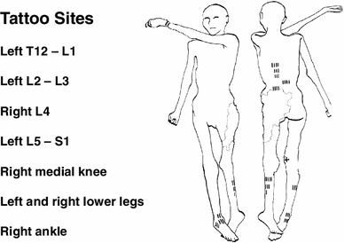
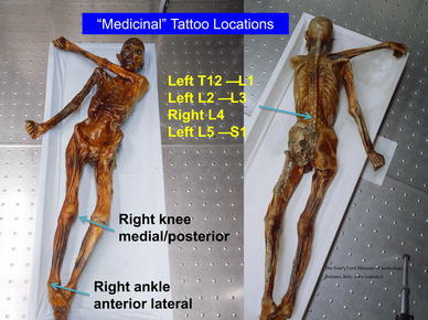
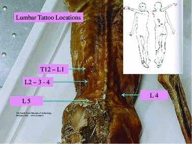
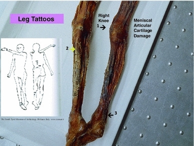
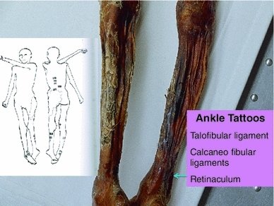
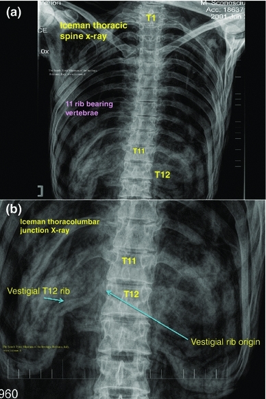
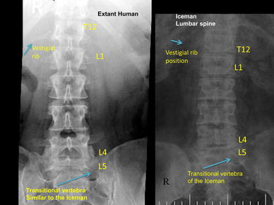
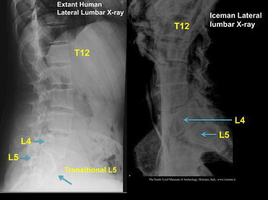
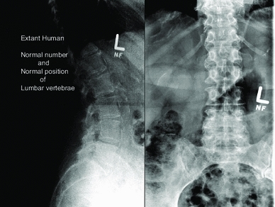
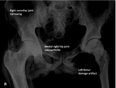
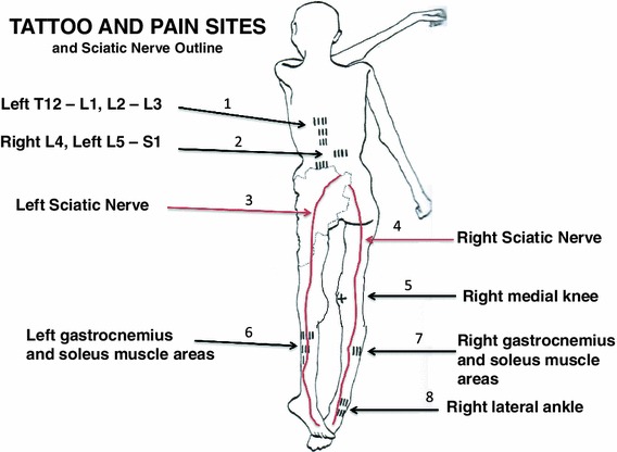
Methods and results: We (WFK and MK) undertook a clinical musculoskeletal examination of the Iceman, details of which with available photographs and radiographic imaging pertaining to the musculoskeletal findings of the Iceman are reported here. The skin of the Iceman has numerous linear carbon tattoos, which are not of a decorative type. These have been presumed to possibly be "medicinal" tattoos administered for therapeutic reasons and may have been used in acupuncture-like treatment of pain. Spinal imaging identified areas of spinal damage and our observations have provided clues as to possible sites of spinal initiated pain and hence sites for administration of the "medicinal" tattoos. We observed body areas of the Iceman, in which imaging demonstrated arthritis and other forms of long-term musculoskeletal damage, but which do not have adjacent or corresponding "medicinal" tattoos. We contend that the back and leg "medicinal" tattoos correspond directly to sites of chronic right knee and right ankle pain, and left thoracolumbar pain. They also correspond to lower lumbar and sciatic referred radicular pain which may have a contributory cause related to the presence of a transitional lumbar 5 vertebra. Using recent published data (Keller et al. in Nature Commun 3:698, 2012. doi: 10.1038/ncomms1701 ) of the genome structure of the Iceman, we suggest some potential causes of the osteoarthritis or inflammatory joint injury may relate to presence of coronary heart disease (CHD) and Lyme disease (Borrelia burgdorferi) infection. We speculate on possible medical applications of natural products for self-medication.

Conclusions: These observations highlight several diagnostic features of musculoskeletal conditions in the Iceman with the possibility that tattoos may have been used for diagnosis or location of his painful states. The origins of his musculoskeletal conditions are unclear but there are indications that Lyme disease and CHD may have been factors. The associations or use of natural products may give insights into their applications at the time of the life of the Iceman.


Figures

Fig. 1
Fig. 1
A graphic outline of the prone and supine positions of the Iceman. The small linear strokes identify the approximate locations of skin impregnated carbon tattoo sites. Adapted from Fleckinger (2005)
Fig. 2
Fig. 2
The prone and supine position of the Iceman with the arrows showing location of “medicinal” tattoo sites at: vertebrae thoracic 1 (T1) and lumbar (L) levels 1, 2, 3, 4, 5, and Sacral (S) 1; the right medial posterior knee; and right anterior lateral ankle
Fig. 3
Fig. 3
Prone position of the Iceman, which shows the tattoos in the region of the lumbar spine and adjacent thoracic and sacral areas
Fig. 4
Fig. 4
Position of leg tattoos at: 1 the right knee, which may represent medial meniscal cartilage and or articular cartilage damage, and/or medial collateral ligament damage, 2 the level of gastrocnemius and soleus muscles, which are common sites of expression of sciatic radicular pain in humans, and 3 the right ankle tattoo sites which may represent local ligamentous structure damage
Fig. 5
Fig. 5
Position of ankle tattoos, which may represent local ligamentous structure damage at any or all of the talofibular ligament, calcaneo fibular ligaments, and retinaculum. The ankle region is also a common site of expression of sciatic radicular pain in humans
Fig. 6
Fig. 6
a Iceman thoracic spine X-ray which shows the thoracic vertebrae with 11 normal sized right and left ribs. The location of thoracic (T) vertebrae 1, 11 and 12 are shown as T1, T11, T12, b the X-ray of the lower thoracic spine and upper lumbar spine known as the thoracolumbar junction is shown to identify origin and position of the right thoracic 12 vestigial rib
Fig. 7
Fig. 7
Iceman anteroposterior lumbar spine X-rays in comparison to an extant human with the same configuration of a right side thoracic 12 vestigial rib and lumbar 5 transitional vertebra. The Iceman’s vestigial rib is not as clearly seen in this image as in Fig. 6b
Fig. 8
Fig. 8
Iceman lateral lumbar X-rays in comparison to an extant human with the same configuration of a lumbar 5 transitional vertebra. The transitional L5 of the extant human and the Iceman are incorporated in the upper sacrum
Fig. 9
Fig. 9
Extant human lumbar X-rays which show the normal number (5) and position of the lumber vertebrae
Fig. 10
Fig. 10
Iceman pelvis and hip joints: this shows right hip medial joint space loss and juxta articular sclerosis of osteoarthritis; there is narrowing of the sacroiliac joints; the left hip abnormalities are due to artifact when the body was extracted from the glacier ice pool
Fig. 11
Fig. 11
A graphic outline of the prone and supine positions of the Iceman is shown. The small linear strokes identify the approximate locations of skin impregnated carbon tattoo sites adapted from Fleckinger (2005). This image has been overlayed with a linear caricature of the left and right sciatic nerves from their origin in the lumbar 3, 4, 5 and sacral 3, 4, 5 nerve root positions, to the distal distribution in the posterior upper and lower legs and the ankles and feet. The gastrocnemius and soleus muscle areas and sometimes the ankles are sites of sciatic nerve radicular pain expression in humans (Kimura 1989), and may provide some explanation of the location of these presumed “medicinal” tattoos

References

    1. Adatia A, Rainsford KD, Kean WF. Osteoarthritis of the knee and hip. Part I: aetiology and pathogenesis as a basis for pharmacotherapy. J Pharm Pharmacol. 2012;2012(64):617–625. doi: 10.1111/j.2042-7158.2012.01458.x. - DOI - PubMed
    1. Bertolotti M. Contribute alla conoscenza dei vizi di differenzazione del rachide con speciale reguardo all assimilazione sacrale della v lombare. Radiol Med (Torino) 1917;4:113–144.
    1. Buchanan WW, Kean WF (2002) Osteoarthritis III: radiological and clinical definition. Inflammopharmacolgy 10:53–78
    1. Castellvi AE, Goldstein LA, Chan DP. Lumbosacral transitional vertebrae and their relationship with lumbar extradural defects. Spine. 1984;9(5):493–495. doi: 10.1097/00007632-198407000-00014. - DOI - PubMed
    1. De Leo JA, Tawfik JL, LaCroix-Fralish ML. The tetra-partite synapse: path to CNS sensitization and chronic pain. Pain. 2006;122(1–2):17–21. doi: 10.1016/j.pain.2006.02.034. - DOI - PubMed