

Influence of tibial slope asymmetry on femoral rotation in patients with lateral patellar instability
- PMID: 23096490
- PMCID: PMC3751338
- DOI: 10.1007/s00167-012-2247-4
Influence of tibial slope asymmetry on femoral rotation in patients with lateral patellar instability
Abstract
Purpose: The geometry of the tibial plateau and its influence on the biomechanics of the tibiofemoral joint has gained increased significance. However, no quantitative data are available regarding the inclination of the medial and lateral tibial slope in patients with patellar instability. It was therefore the purpose of this study to evaluate tibial slope characteristics in patients with patellar dislocations and to assess the biomechanical effect of medial-to-lateral tibial slope asymmetry on lateral patellar instability.
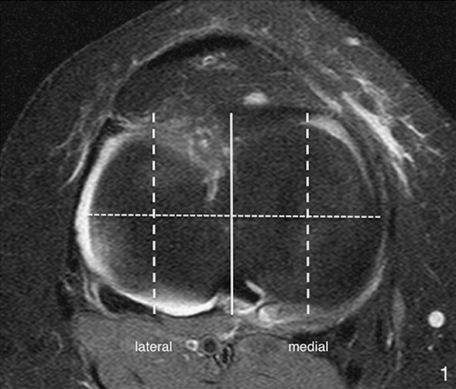
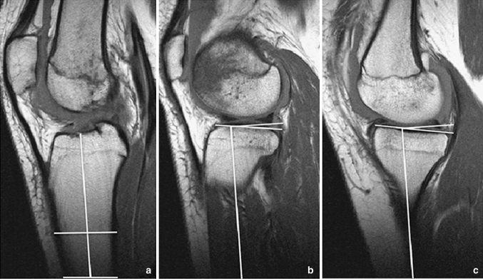
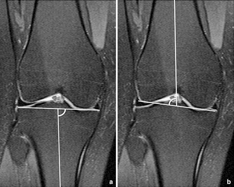
Methods: Medial and lateral tibial slope was measured on knee magnetic resonance images in 107 patients and in 83 controls. The medial-to-lateral tibial slope asymmetry was assessed as the intra-individual difference between the medial and lateral tibial plateau inclination considering severity of trochlear dysplasia. The effect of tibial slope asymmetry on femoral rotation was calculated by means of radian measure.
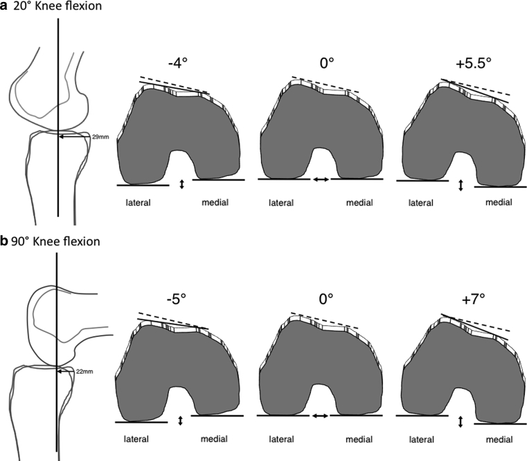
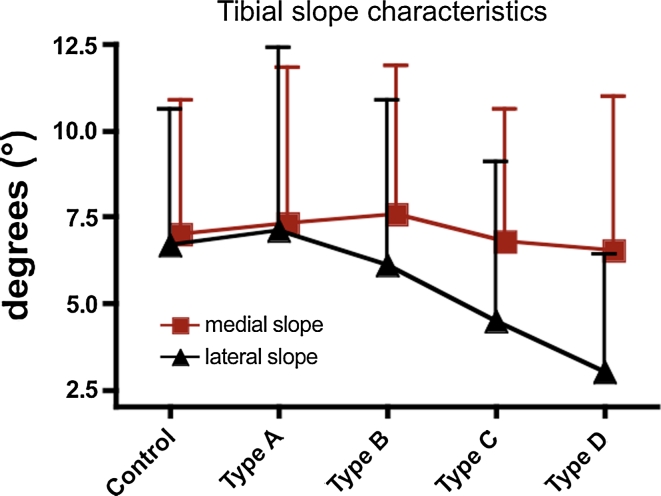
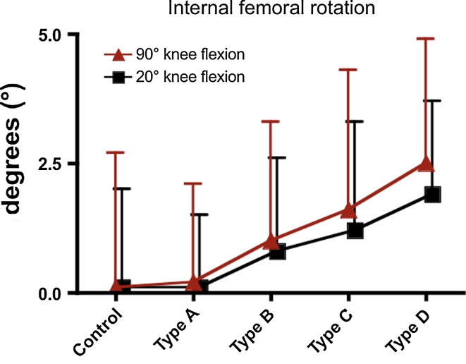
Results: Severity of trochlear dysplasia was significantly associated with an asymmetric inclination of the tibial plateau. Whereas the medial tibial slope showed identical values between controls and study patients (n.s.), lateral tibial plateau inclination becomes flatter with increasing severity of trochlear dysplasia (p < 0.01). Consequently, the intra-individual tibial slope asymmetry increased steadily (p < 0.01) and increased internal femoral rotation in 20° and 90° of knee flexion angles in patients with severe trochlear dysplasia (p < 0.01). In addition, the extreme values of internal femoral rotation were more pronounced in patients with patellar instability, whereas the extreme values of external femoral rotation were more pronounced in control subjects (p = 0.024).
Conclusion: Data of this study indicate an association between tibial plateau configuration and internal femoral rotation in patients with lateral patellar instability and underlying trochlear dysplasia. Thereby, medial-to-lateral tibial slope asymmetry increased internal femoral rotation during knee flexion and therefore might aggravate the effect of femoral antetorsion in patients with patellar instability.
Level of evidence: III.
Figures
Comment in
-
Influence of tibial slope asymmetry on femoral rotation in patients with lateral patellar instability.Knee Surg Sports Traumatol Arthrosc. 2014 Dec;22(12):3198. doi: 10.1007/s00167-013-2765-8. Epub 2013 Nov 12. Knee Surg Sports Traumatol Arthrosc. 2014. PMID: 24217717 No abstract available.
Similar articles
-
Characteristics of femorotibial joint geometry in the trochlear dysplastic femur.J Anat. 2014 Sep;225(3):367-73. doi: 10.1111/joa.12214. Epub 2014 Jul 10. J Anat. 2014. PMID: 25040233 Free PMC article.
-
Combined deepening trochleoplasty and supracondylar external rotation osteotomy for recurrent patellar instability in patients with trochlear dysplasia and increased femoral antetorsion.Knee. 2020 Aug;27(4):1158-1166. doi: 10.1016/j.knee.2020.05.002. Epub 2020 Jun 27. Knee. 2020. PMID: 32711877
-
The effect of native knee rotation on the tibial-tubercle-trochlear-groove distance in patients with patellar instability: an analysis of MRI and CT measurements.Arch Orthop Trauma Surg. 2022 Nov;142(11):3149-3155. doi: 10.1007/s00402-021-03947-4. Epub 2021 May 12. Arch Orthop Trauma Surg. 2022. PMID: 33978809
-
[Radiologic assessment of femoro-patellar instability. Personal experience and review of the literature].Radiol Med. 2001 Jan-Feb;101(1-2):66-74. Radiol Med. 2001. PMID: 11360756 Review. Italian.
-
How to Deal With Chronic Patellar Instability: What Does the Literature Tell Us?Sports Health. 2016 Jan-Feb;8(1):86-90. doi: 10.1177/1941738115604156. Epub 2015 Aug 28. Sports Health. 2016. PMID: 26733595 Free PMC article. Review.
Cited by
-
Relationship between Anatomical Risk Factors, Articular Cartilage Lesions, and Patient Outcomes Following Medial Patellofemoral Ligament Reconstruction.Cartilage. 2021 Dec;13(1_suppl):993S-1001S. doi: 10.1177/1947603519894728. Epub 2019 Dec 26. Cartilage. 2021. PMID: 31876167 Free PMC article.
-
Steep lateral tibial slope and lateral-to-medial slope asymmetry are risk factors for concomitant posterolateral meniscus root tears in anterior cruciate ligament injuries.Knee Surg Sports Traumatol Arthrosc. 2019 Aug;27(8):2585-2591. doi: 10.1007/s00167-018-5279-6. Epub 2018 Nov 2. Knee Surg Sports Traumatol Arthrosc. 2019. PMID: 30390134
-
When does the patella dislocate? A systematic review of biomechanical & kinematic studies.J Orthop. 2019 Nov 16;20:70-77. doi: 10.1016/j.jor.2019.11.018. eCollection 2020 Jul-Aug. J Orthop. 2019. PMID: 32042233 Free PMC article. Review.
-
Assessment of demographic and pathoanatomic risk factors in recurrent patellofemoral instability.Knee Surg Sports Traumatol Arthrosc. 2017 Dec;25(12):3849-3855. doi: 10.1007/s00167-016-4346-0. Epub 2016 Oct 7. Knee Surg Sports Traumatol Arthrosc. 2017. PMID: 27717972
-
Assessment of the reliability and validity of imaging measurements for patellofemoral instability: an updated systematic review.Skeletal Radiol. 2022 Dec;51(12):2245-2256. doi: 10.1007/s00256-022-04110-9. Epub 2022 Jul 7. Skeletal Radiol. 2022. PMID: 35794393
References
-
- Amiri S, Cooke D, Kim IY, Wyss U. Mechanics of the passive knee joint. Part 2: interaction between the ligaments and the articular surfaces in guiding the joint motion. Proc Inst Mech Eng H. 2007;221:821–832. - PubMed
-
- Balcarek P, Jung K, Ammon J, Walde TA, Frosch S, Schüttrumpf JP, Stürmer KM, Frosch KH. Anatomy of lateral patellar instability: trochlear dysplasia and tibial tubercle-trochlear groove distance is more pronounced in women who dislocate the patella. Am J Sports Med. 2010;38:2320–2327. doi: 10.1177/0363546510373887. - DOI - PubMed
MeSH terms
LinkOut - more resources
Full Text Sources
Research Materials
