

doi: 10.1038/jid.2012.379.
Epub 2012 Oct 25.
Whole-exome sequencing reveals somatic mutations in HRAS and KRAS, which cause nevus sebaceus
Affiliations
- PMID: 23096712
- PMCID: PMC3556376
- DOI: 10.1038/jid.2012.379
Item in Clipboard
Whole-exome sequencing reveals somatic mutations in HRAS and KRAS, which cause nevus sebaceus
J Invest Dermatol.
2013 Mar.
No abstract available
Conflict of interest statement
The authors report no conflict of interest.
Figures
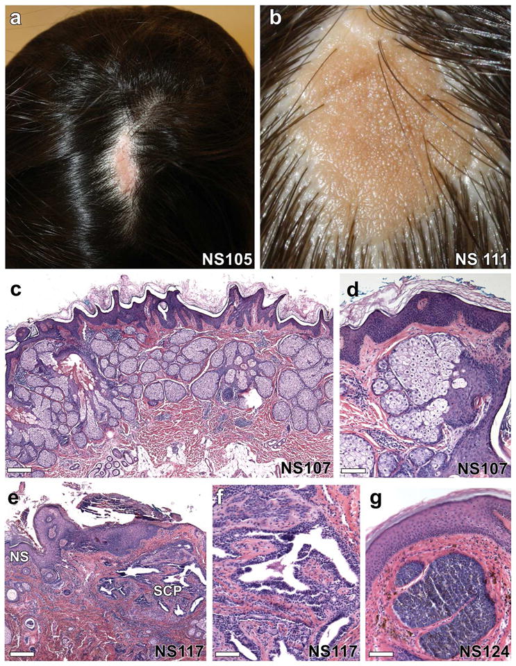
(a, b) Solitary, well-demarcated lesions on the scalp of two individuals show alopecia and a yellow-orange waxy appearance. (c) On histological examination, there is epidermal acanthosis, papillomatosis, and hyperkeratosis with dramatic increase in the number of sebaceous lobules and abortive hair follicles, scale = 288 μm, which is more evident at higher magnification (d), scale = 85 μm. (e–g) Up to 20% of nevus sebaceus lesions develop tumors including syringocystadenomas, trichoblastomas, trichilemmomas and tubular apocrine adenomas. (e) Nevus sebaceus (NS) with syringocystadenoma papilliferum (SCP) composed of villous structures lined by a columnar epithelium with stromal plasma cells, scale = 570 μm, most evident at higher magnification, (f), scale = 92 μm. (g) A trichoblastoma arising within a nevus sebaceus shows a well-circumscribed nodule of basaloid cells with a dense fibrocytic stroma, scale = 92 μm.
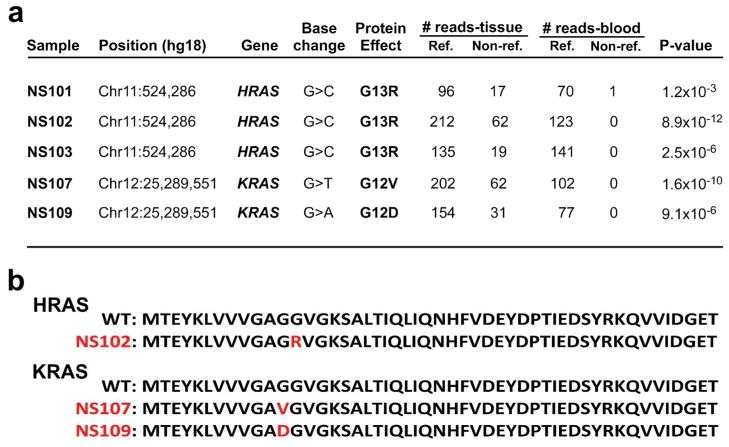
(a) HRAS and KRAS mutation annotation, including genomic position, nucleotide change, protein consequence, and number of reference and non-reference reads obtained from paired sequencing of tissue and blood in 5 independent, unrelated nevus sebaceus cases. Significance of the mutant allele frequency difference between tissue and blood DNA was calculated with a one-tailed Fisher’s exact test. When corrected for multiple testing, 2.4×10⁻⁶ is the threshold for genome wide significance. In each case, HRAS and KRAS mutations showed the lowest P-value. (b) Alignment of the N-termini of HRAS and KRAS reveals identical residues through position 94, with an overall 95% identity and 99% similarity. The first 50 amino acids are shown for the wild-type and each mutant protein, with mutant residues indicated in red.
Comment in
-
Nevus sebaceus is a mosaic RASopathy.J Invest Dermatol. 2013 Mar;133(3):597-600. doi: 10.1038/jid.2012.447. J Invest Dermatol. 2013. PMID: 23399824
References
-
- Blaschko A. Die Nervenverteilung in der Haut in ihrer Beziehung zu den Erkrankungen der Haut. Braumüller, Wien-Leipzig 1901
-
- Cribier B, Scrivener Y, Grosshans E. Tumors arising in nevus sebaceus: A study of 596 cases. J Am Acad Dermatol. 2000;42:263–8. - PubMed
-
- Groesser L, Herschberger E, Ruetten A, et al. Postzygotic HRAS and KRAS mutations cause nevus sebaceous and Schimmelpenning syndrome. Nature genetics 2012 - PubMed
-
- Hafner C, Toll A, Gantner S, et al. Keratinocytic epidermal nevi are associated with mosaic RAS mutations. J Med Genet. 2012;49:249–53. - PubMed
Publication types
MeSH terms
Substances
Grants and funding
LinkOut - more resources
Full Text Sources
Other Literature Sources
Medical
Molecular Biology Databases
Research Materials
Miscellaneous
