. 2012 Dec 15;125(Pt 24):5974-83.
doi: 10.1242/jcs.108886. Epub 2012 Oct 24.

Matrix stiffness reverses the effect of actomyosin tension on cell proliferation

Affiliations

Matrix stiffness reverses the effect of actomyosin tension on cell proliferation

Justin D Mih et al. J Cell Sci. .

Abstract

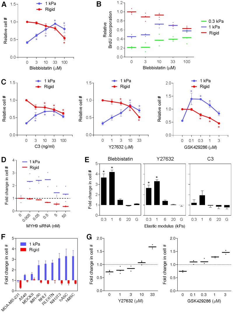
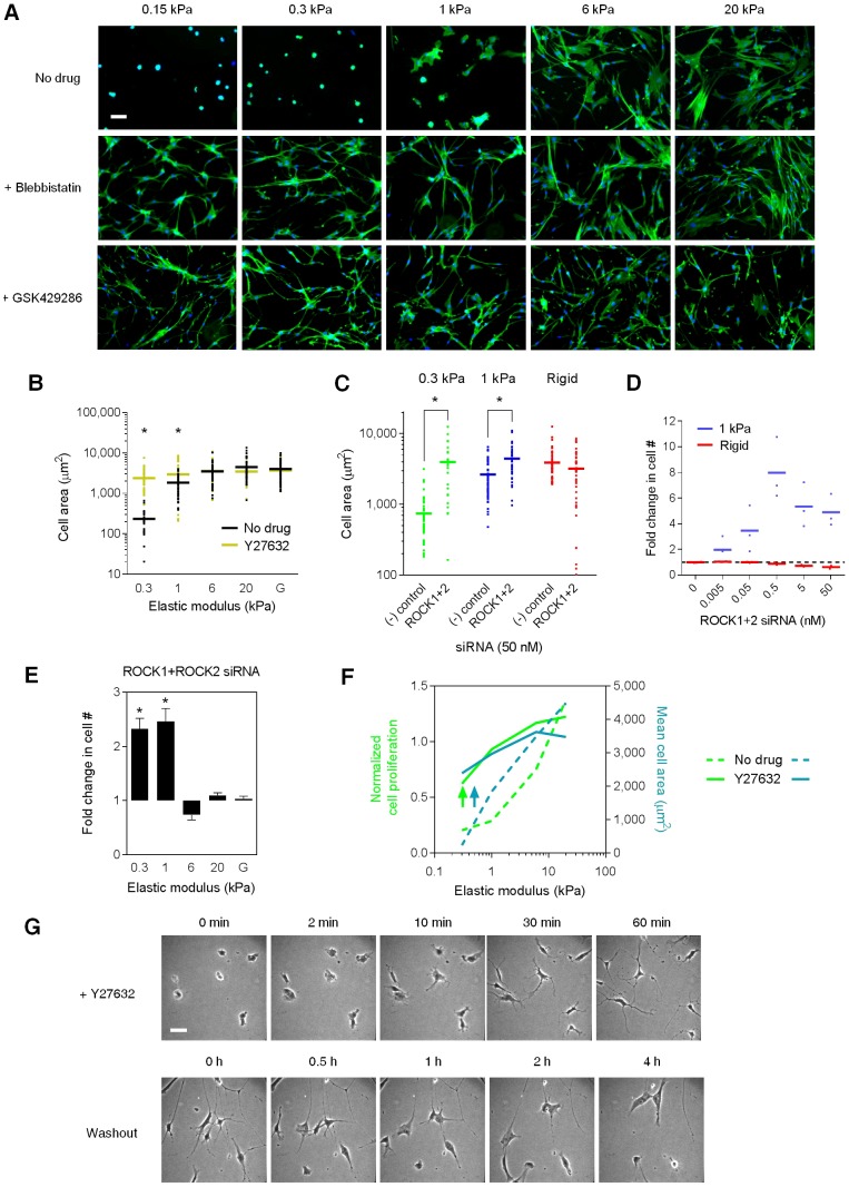
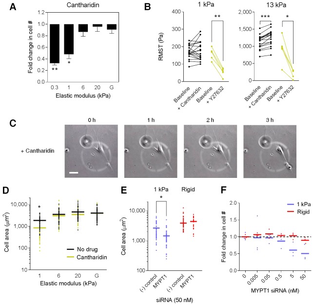
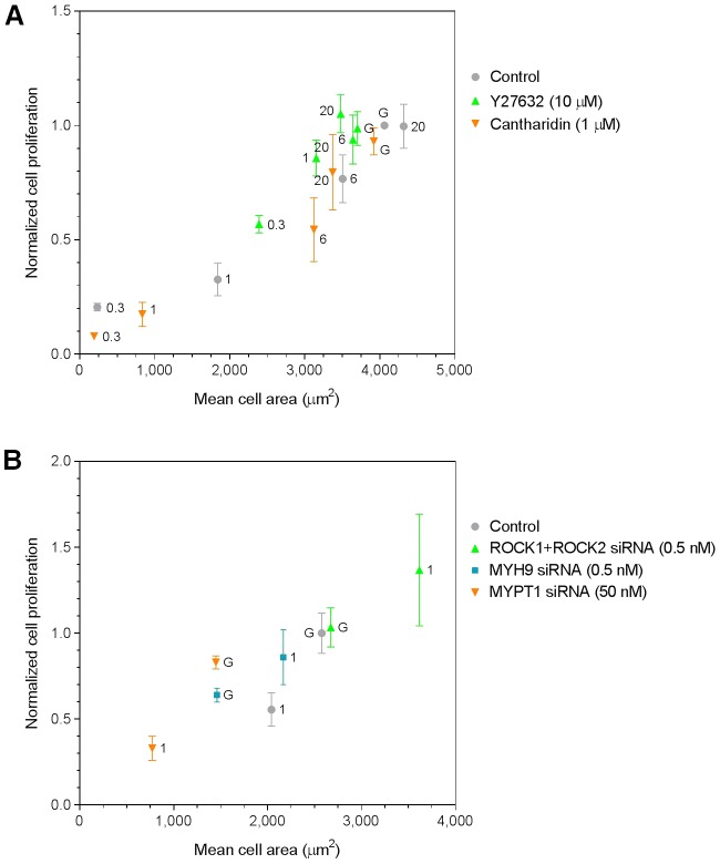
The stiffness of the extracellular matrix exerts powerful effects on cell proliferation and differentiation, but the mechanisms transducing matrix stiffness into cellular fate decisions remain poorly understood. Two widely reported responses to matrix stiffening are increases in actomyosin contractility and cell proliferation. To delineate their relationship, we modulated cytoskeletal tension in cells grown across a physiological range of matrix stiffnesses. On both synthetic and naturally derived soft matrices, and across a panel of cell types, we observed a striking reversal of the effect of inhibiting actomyosin contractility, switching from the attenuation of proliferation on rigid substrates to the robust promotion of proliferation on soft matrices. Inhibiting contractility on soft matrices decoupled proliferation from cytoskeletal tension and focal adhesion organization, but not from cell spread area. Our results demonstrate that matrix stiffness and actomyosin contractility converge on cell spreading in an unexpected fashion to control a key aspect of cell fate.


Figures

Fig. 1.
Fig. 1.
Inhibition of Rho–ROCK–myosin switches from suppression to promotion of cell proliferation as matrix stiffness decreases. (A) Lung fibroblasts were seeded on collagen I-coated 1 kPa and rigid (glass) substrates at low density and cultured in the presence of various concentrations of blebbistatin for 72 hours. Cell numbers are normalized to the rigid, no drug condition. Data are means ± s.e.m. (n = 3). One-way ANOVA followed by Tukey's test: *P<0.05 versus no drug for the same stiffness condition. (B) BrdU incorporation over 48 hours normalized to the no drug, rigid condition. Means and individual data points from one experiment are shown. (C) Fibroblasts were treated with C3 transferase and two ROCK inhibitors for 72 hours. Data are means ± s.e.m. (n≥3). One-way ANOVA followed by Tukey's test: *P<0.05 versus no drug for the same stiffness condition. (D) Fibroblasts on 1 kPa and rigid substrates were transfected with different concentrations of siRNA targeting MYH9 and cultured for 6 days. Fold change in cell number is normalized against a concentration-matched negative siRNA control for each stiffness condition. Means and individual data points from one experiment are shown. (E) Effects of blebbistatin (10 µM), Y27632 (10 µM), and C3 transferase (100 ng/ml) on cell proliferation across five stiffness conditions. Fold change indicates ratio of cell number in the drug versus no drug condition after 72 hours. Data are means ± s.e.m. (n = 3). One-way ANOVA followed by Tukey's test: *P<0.0001 versus rigid (G, glass) condition. (F) Effect of GSK429286 (1 µM) on a panel of normal and immortalized cell types cultured on 1 kPa and rigid substrates. Fold change represents cell number in the drug versus no drug condition following 72 hours of exposure. MDA-MB-231, breast cancer cell line; A549, human lung adenocarcinoma cell line; MDCKII, Madin–Darby canine kidney epithelial cell line; IMR-90, normal human fetal lung fibroblast; NHLF, normal human adult lung fibroblast; RLE6TN, rat lung epithelial cell line; NIH3T3, mouse embryonic fibroblast cell line; hASC, human adipose-derived stem cells; hMSC, human bone marrow-derived stem cells. Data are means ± s.d. (n = 3). (G) Lung fibroblasts were seeded within Geltrex 3D matrices and exposed to Y27632 (10 µM) or GSK429286 (1 µM). Fold change represents cell number after 5 days versus an initial 4 hour time point. Means and individual data points from one experiment are shown.
Fig. 2.
Fig. 2.
Proliferation on soft matrices is decoupled from myosin light chain phosphorylation, cell tractions, and focal adhesions. (A) Immunofluorescence staining of phosphorylated myosin light chain in fibroblasts across matrix stiffness. Y27632 (10 µM) was applied for 24 hours. (B) Fibroblast root-mean-square tractions (RMST) measured by Fourier transform traction microscopy. Y27632 (10 µM) was applied for 45 minutes. Means and data points (n = 5 cells) are shown. Student's t-test: *P<0.0001 versus no drug. (C) Relationship between cell proliferation and tractions with or without Y27632 (10 µM). Proliferation is expressed as cell number relative to the no drug, rigid condition following 72 hours of culture of one representative experiment. (D,E) Immunofluorescence staining of vinculin (D) and talin (E) across matrix stiffness. Images are at 400× magnification; corresponding images of boxed regions are at 1000×. Scale bar: 25 µm. (F) Effect of matrix stiffness and Y27632 (10 µM) on focal adhesion (FA) length. Focal adhesions on 0.3 kPa gels were not observed above the detection threshold (0.2 µm). Data are means ± s.d. (n≥47). Student's t-test: *P<0.001 versus no drug.
Fig. 3.
Fig. 3.
ROCK inhibition triggers cell spreading on soft matrices to promote proliferation. (A) Fibroblasts stained for F-actin (green) and nuclei (blue) across five stiffness conditions following 48 hours of exposure to no drug, blebbistatin (10 µM), or GSK429286 (1 µM). (B) Effect of matrix stiffness and Y27632 (10 µM) on 2D projected cell area. Means and individual data points (n≥29) are shown. Student's t-test: *P<0.0001 versus no drug. (C) Effect of combined ROCK1 and ROCK2 (ROCK1+2) siRNA knockdown and non-targeting [(−) control] siRNA on cell area. Means and individual data points (n≥23) are shown. Student's t-test: *P<0.0001 versus (−) control. (D) Fibroblasts on 1 kPa and rigid substrates were transfected with various concentrations of siRNA targeting both ROCK1 and ROCK2 and cultured for 6 days. Fold change in cell number is relative to the no siRNA condition for each stiffness condition. Means and individual data points (n = 3) from one experiment are shown. (E) Fibroblasts across five stiffness conditions were transfected with 0.25 nM each of ROCK1 and ROCK2 siRNA and cultured for 6 days. For each stiffness condition, fold change in cell number is normalized to a 0.5 nM non-targeting siRNA control. Data are means ± s.d. (n = 6). One-way ANOVA followed by Tukey's test: *P<0.0001 versus rigid (glass, G) condition. (F) Relationship between normalized cell proliferation and mean cell area with or without exposure to Y27632 (10 µM). (G) Time-lapse images of fibroblasts on a 1 kPa substrate following addition of Y27632 (10 µM) and subsequent washout. All images are at 200× magnification. Scale bar: 50 µm.
Fig. 4.
Fig. 4.
Enhancing actomyosin tension leads to cell retraction and reduced proliferation on soft matrices. (A) Effect of cantharidin (1 µM) on cell proliferation across five stiffness conditions. Fold change indicates ratio of cell number in drug versus no drug samples after 72 hours. Data are means ± s.e.m. (n = 5). One-way ANOVA followed by Tukey's test: *P<0.01, **P<0.0001 versus rigid (glass, G) condition. (B) RMST at baseline and following 2 hours of cantharidin (1 µM) or 45 minutes of Y27632 (10 µM) treatment. Paired observations of individual cells (cantharidin, n = 17; Y27632, n = 5) are connected. Paired t-test: *P = 0.0059, **P = 0.0002, ***P<0.0001 versus baseline RMST. (C) Time-lapse sequence of a fibroblast on a 1 kPa substrate following addition of cantharidin (0.3 µM). Images are at 200× magnification. Scale bar: 50 µm. (D) Effect of cantharidin (1 µM) on mean cell area (n≥29). One-way ANOVA followed by Tukey's test: *P<0.0001 versus rigid condition. (E) Effect of MYPT1 and non-targeting [(−) control] siRNA on cell area. Means and individual data points (n≥22) are shown. Student's t-test: *P<0.0001 versus (−) control. (F) Fibroblasts on 1 kPa and rigid substrates were transfected with siRNA targeting MYPT1 and cultured for 6 days. Fold change in cell number is relative to the no siRNA condition for each stiffness condition. Means and individual data points (n = 3) from one experiment are shown.
Fig. 5.
Fig. 5.
Actomyosin tension and matrix stiffness converge on cell spreading to control proliferation. (A) Relationship between cell proliferation and area across five stiffness conditions. Proliferation is expressed as cell number relative to a control, rigid (glass, G) condition following 72 hours of culture. For Y27632 and cantharidin treatment conditions, data are proliferation means ± s.e.m. (n≥4) plotted against mean cell areas obtained in Fig. 3B and Fig. 4D. Elastic moduli (kPa) corresponding to each data point are indicated. (B) Effect of ROCK1+ROCK2, MYH9, and MYPT1 knockdown on cell area and proliferation. Cell numbers in soft (1 kPa) and rigid (G) conditions were determined 6 days following siRNA transfection. Proliferation is expressed as fold change in cell number relative to the rigid condition containing a matched concentration of non-targeting siRNA. Cell areas were determined 3 days following siRNA transfection. Data are proliferation means ± s.d. (n = 3) plotted against mean cell areas from one representative experiment.

References

    1. Aratyn–Schaus Y., Gardel M. L. (2010). Transient frictional slip between integrin and the ECM in focal adhesions under myosin II tension. Curr. Biol. 20, 1145–1153 10.1016/j.cub.2010.05.049 - DOI - PMC - PubMed
    1. Assoian R. K., Klein E. A. (2008). Growth control by intracellular tension and extracellular stiffness. Trends Cell Biol. 18, 347–352 10.1016/j.tcb.2008.05.002 - DOI - PMC - PubMed
    1. Balaban N. Q., Schwarz U. S., Riveline D., Goichberg P., Tzur G., Sabanay I., Mahalu D., Safran S., Bershadsky A., Addadi L.et al. (2001). Force and focal adhesion assembly: a close relationship studied using elastic micropatterned substrates. Nat. Cell Biol. 3, 466–472 10.1038/35074532 - DOI - PubMed
    1. Butcher D. T., Alliston T., Weaver V. M. (2009). A tense situation: forcing tumour progression. Nat. Rev. Cancer 9, 108–122 10.1038/nrc2544 - DOI - PMC - PubMed
    1. Butler J. P., Tolić–Nørrelykke I. M., Fabry B., Fredberg J. J. (2002). Traction fields, moments, and strain energy that cells exert on their surroundings. Am. J. Physiol. Cell Physiol. 282, C595–C605 - PubMed

Publication types

MeSH terms