. 2013:945:13-29.
doi: 10.1007/978-1-62703-125-7_2.

Culture of choroid plexus epithelial cells and in vitro model of blood-CSF barrier

Affiliations

Culture of choroid plexus epithelial cells and in vitro model of blood-CSF barrier

Andrew D Monnot et al. Methods Mol Biol. 2013.

Abstract

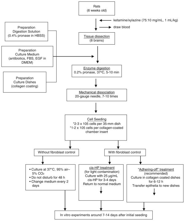
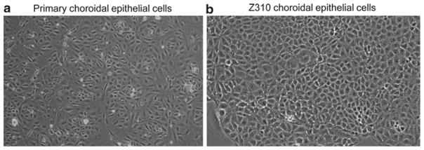
Chemical homeostasis in the extracellular fluid of the central nervous system (CNS) is maintained by two brain barrier systems, i.e., the blood-brain barrier (BBB) that separates the blood circulation from brain interstitial fluid and the blood-cerebrospinal fluid barrier (BCB) that separates the blood from the cerebrospinal fluid (CSF). The choroid plexus, where the BCB is located, is a polarized tissue, with the basolateral side of the choroidal epithelium facing the blood and the apical microvilli in direct contact with the CSF. The tissue plays a wide range of roles in brain development, aging, nutrient transport, endocrine regulation, and pathogenesis of certain neurodegenerative disorders. This chapter describes two in vitro cultures that have been well established to allow for study of the BCB structure and function. The primary choroidal epithelial cell culture can be established from rat choroid plexus tissue, and a similar immortalized murine choroidal epithelial cell culture known as Z310 cells has also been established. Both cultures display a dominant polygonal morphology, and immunochemical studies demonstrate the presence of transthyretin, a thyroxine transport protein known to be exclusively produced by the choroidal epithelia in the CNS. These cultures have been adapted for use on freely permeable Transwell(®) membranes sandwiched between two culture chambers, facilitating transport studies of various compounds across this barrier in vitro. These choroidal epithelia cultures with the Transwell system will perceivably assist blood-CSF barrier research.


Figures

Fig. 1
Fig. 1
Flowchart of procedures in establishing primary culture of choroidal epithelial cells.
Fig. 2
Fig. 2
Morphology of choroidal epithelial cells in culture. (a) Primary culture of choroidal epithelial cells after 5 days in culture (10×). Note the con fluent layer of cells with a predominant polygonal cell type. The choroid plexus tissue was obtained from 6-week-old Sprague-Dawley rats. (b) Immortalized Z310 choroidal epithelial cells in culture (20×). Passage 86.
Fig. 3
Fig. 3
Transepithelial model of the blood–CSF barrier used to study transepithelial transport. Epithelial cells are connected by tight junctions and form a barrier between fluids in the inner and outer chambers. Fluid in the inner chamber is in contact with the apical microvilli on the surface of the cells, while the fluid in the outer chamber has access to the basal surface of the cells.
Fig. 4
Fig. 4
Confocal microscopic depicting TTR staining in the choroid plexus tissue (a) and cultured Z310 cells (b). Tissue was treated with anti-TTR primary antibody followed by secondary antibody conjugated with fluorescein. Note the positive staining primarily along the basolateral side of the choroid plexus tissue. A Nikon C1 series modular confocal microscope was used to view the tissue through a 60× oil immersion objective with a 488 nm laser line for excitation.
Fig. 5
Fig. 5
Expression of TTR mRNA in primary choroidal epithelial cells and in choroidal Z310 cells by RT-PCR analysis. All samples underwent RT-PCR unless otherwise stated. Arrow indicates bands corresponding to TTR mRNA. Lane ID: Lane 1 total RNA for PCR without RT, lane 2 base pair ladder, lane 3 liver mRNA with selected primer, lane 4 cultured primary plexus cells mRNA with selected primer, lane 5 Z310 cells mRNA with selected marker.

Similar articles

Cited by

References

    1. Zheng W, Zhao Q, Graziano JH. Primary culture of rat choroidal epithelial cells: a model for in vitro study of the blood–cerebrospinal fluid barrier. In Vitro Cell Biol Dev. 1998;34:40–45. - PMC - PubMed
    1. Crossgrove JS, Li GJ, Zheng W. The choroid plexus removes beta-amyloid from the cerebrospinal fluid. Exp Biol Med. 2005;230(10):771–776. - PMC - PubMed
    1. Deane R, Zheng W, Zlokovic BV. Brain capillary endothelium and choroid plexus epithelium regulate transport of transferrin-bound and free iron into the rat brain. J Neurochem. 2004;88:813–820. - PMC - PubMed
    1. Zheng W, Deane R, Redzic Z, Preston JE, Segal MB. Transport of L-(125I)thyroxine by in-situ perfused ovine choroid plexus: inhibition by lead exposure. J Toxicol Env Health. 2003;66:435–451. - PMC - PubMed
    1. Zheng W, Perry DF, Nelson DL, Aposhian HV. Protection of cerebrospinal fluid against toxic metals by the choroid plexus. FASEB J. 1991;5:2188–2193. - PubMed