Growth factor transgenes interactively regulate articular chondrocytes
- PMID: 23097312
- PMCID: PMC3994886
- DOI: 10.1002/jcb.24430
Growth factor transgenes interactively regulate articular chondrocytes
Abstract
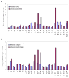
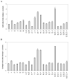
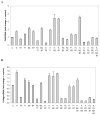
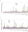
Adult articular chondrocytes lack an effective repair response to correct damage from injury or osteoarthritis. Polypeptide growth factors that stimulate articular chondrocyte proliferation and cartilage matrix synthesis may augment this response. Gene transfer is a promising approach to delivering such factors. Multiple growth factor genes regulate these cell functions, but multiple growth factor gene transfer remains unexplored. We tested the hypothesis that multiple growth factor gene transfer selectively modulates articular chondrocyte proliferation and matrix synthesis. We tested the hypothesis by delivering combinations of the transgenes encoding insulin-like growth factor I (IGF-I), fibroblast growth factor-2 (FGF-2), transforming growth factor beta1 (TGF-β1), bone morphogenetic protein-2 (BMP-2), and bone morphogenetic protien-7 (BMP-7) to articular chondrocytes and measured changes in the production of DNA, glycosaminoglycan, and collagen. The transgenes differentially regulated all these chondrocyte activities. In concert, the transgenes interacted to generate widely divergent responses from the cells. These interactions ranged from inhibitory to synergistic. The transgene pair encoding IGF-I and FGF-2 maximized cell proliferation. The three-transgene group encoding IGF-I, BMP-2, and BMP-7 maximized matrix production and also optimized the balance between cell proliferation and matrix production. These data demonstrate an approach to articular chondrocyte regulation that may be tailored to stimulate specific cell functions, and suggest that certain growth factor gene combinations have potential value for cell-based articular cartilage repair.
Copyright © 2012 Wiley Periodicals, Inc.
Conflict of interest statement
Conflicts of interest: Stephen B. Trippel serves as a paid consultant to Biomeasures and Lilly. The other authors have no conflicts of interest.
Figures


Similar articles
-
Growth factor regulation of growth factor production by multiple gene transfer to chondrocytes.Growth Factors. 2013 Feb;31(1):32-8. doi: 10.3109/08977194.2012.750652. Epub 2013 Jan 9. Growth Factors. 2013. PMID: 23302100 Free PMC article.
-
Role of sox9 in growth factor regulation of articular chondrocytes.J Cell Biochem. 2015 Jul;116(7):1391-400. doi: 10.1002/jcb.25099. J Cell Biochem. 2015. PMID: 25708223 Free PMC article.
-
Regulation of articular chondrocyte aggrecan and collagen gene expression by multiple growth factor gene transfer.J Orthop Res. 2012 Jul;30(7):1026-31. doi: 10.1002/jor.22036. Epub 2011 Dec 16. J Orthop Res. 2012. PMID: 22180348 Free PMC article.
-
Mechanisms of synovial joint and articular cartilage development.Cell Mol Life Sci. 2019 Oct;76(20):3939-3952. doi: 10.1007/s00018-019-03191-5. Epub 2019 Jun 14. Cell Mol Life Sci. 2019. PMID: 31201464 Free PMC article. Review.
-
Regulation of FGF-2, FGF-18 and Transcription Factor Activity by Perlecan in the Maturational Development of Transitional Rudiment and Growth Plate Cartilages and in the Maintenance of Permanent Cartilage Homeostasis.Int J Mol Sci. 2022 Feb 9;23(4):1934. doi: 10.3390/ijms23041934. Int J Mol Sci. 2022. PMID: 35216048 Free PMC article. Review.
Cited by
-
Growth factor regulation of growth factor production by multiple gene transfer to chondrocytes.Growth Factors. 2013 Feb;31(1):32-8. doi: 10.3109/08977194.2012.750652. Epub 2013 Jan 9. Growth Factors. 2013. PMID: 23302100 Free PMC article.
-
Prospective clinical trial of patients who underwent ankle arthroscopy with articular diseases to match clinical and radiological scores with intra-articular cytokines.Int Orthop. 2015 Aug;39(8):1631-7. doi: 10.1007/s00264-015-2797-4. Epub 2015 May 7. Int Orthop. 2015. PMID: 25947905
-
Tissue engineering and future directions in regenerative medicine for knee cartilage repair: a comprehensive review.Croat Med J. 2024 Jun 13;65(3):268-287. doi: 10.3325/cmj.2024.65.268. Croat Med J. 2024. PMID: 38868973 Free PMC article. Review.
-
Harnessing Growth Factor Interactions to Optimize Articular Cartilage Repair.Adv Exp Med Biol. 2023;1402:135-143. doi: 10.1007/978-3-031-25588-5_10. Adv Exp Med Biol. 2023. PMID: 37052852 Review.
-
Biochemical Aspects of Scaffolds for Cartilage Tissue Engineering; from Basic Science to Regenerative Medicine.Arch Bone Jt Surg. 2022 Mar;10(3):229-244. doi: 10.22038/ABJS.2022.55549.2766. Arch Bone Jt Surg. 2022. PMID: 35514762 Free PMC article. Review.
References
-
- Bradham DM, der Wiesche B, Precht P, Balakir R, Horton W. Transrepression of type II collagen by TGF-beta and FGF is protein kinase C dependent and is mediated through regulatory sequences in the promoter and first intron. J of cell phys. 1994;158:61–68. - PubMed
-
- Brower-Toland BD, Saxer RA, Goodrich LR, Mi Z, Robbins PD, Evans CH, Nixon AJ. Direct adenovirus-mediated insulin-like growth factor I gene transfer enhances transplant chondrocyte function. Hum Gene Ther. 2001;12:117–129. - PubMed
-
- Buckwalter JA, Mankin HJ. Articular cartilage repair and transplantation. Arthritis Rheum. 1998;41:1331–1342. - PubMed
Publication types
MeSH terms
Substances
Grants and funding
LinkOut - more resources
Full Text Sources
Other Literature Sources

