. 2013 Jan;87(1):444-53.
doi: 10.1128/JVI.00676-12. Epub 2012 Oct 24.

APOBEC3G restricts HIV-1 to a greater extent than APOBEC3F and APOBEC3DE in human primary CD4+ T cells and macrophages

Affiliations

APOBEC3G restricts HIV-1 to a greater extent than APOBEC3F and APOBEC3DE in human primary CD4+ T cells and macrophages

Chawaree Chaipan et al. J Virol. 2013 Jan.

Abstract

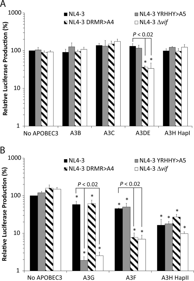
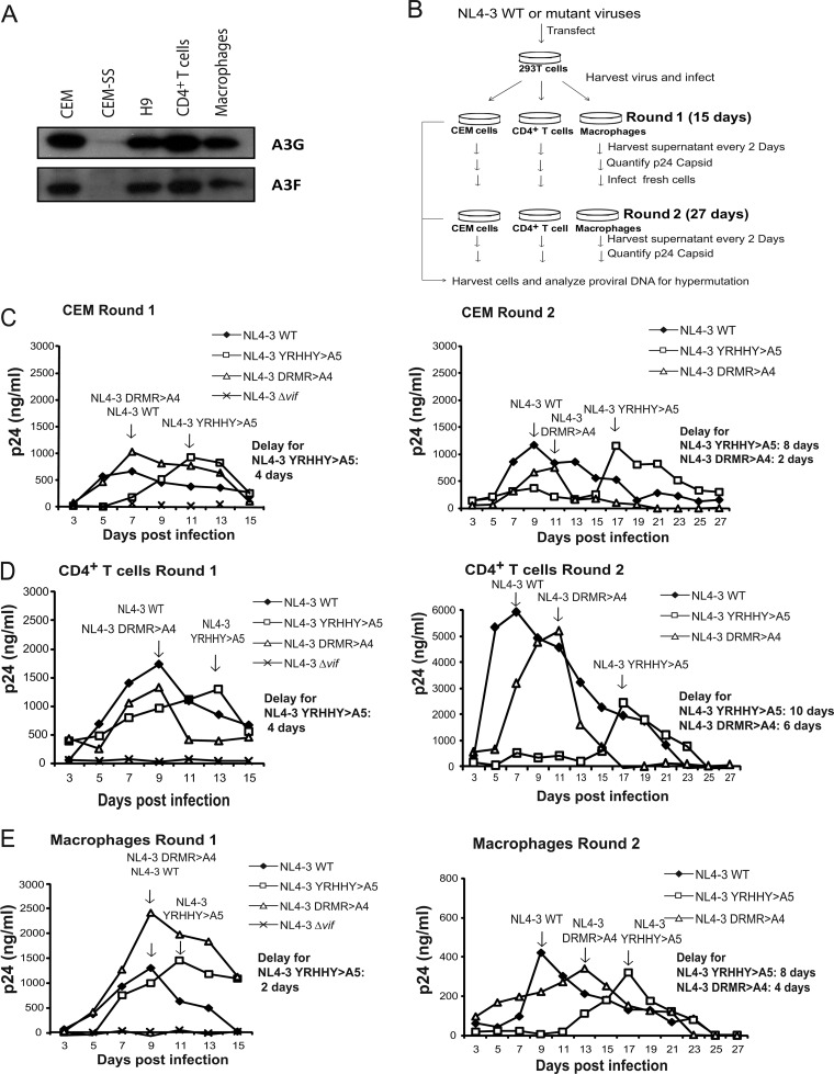
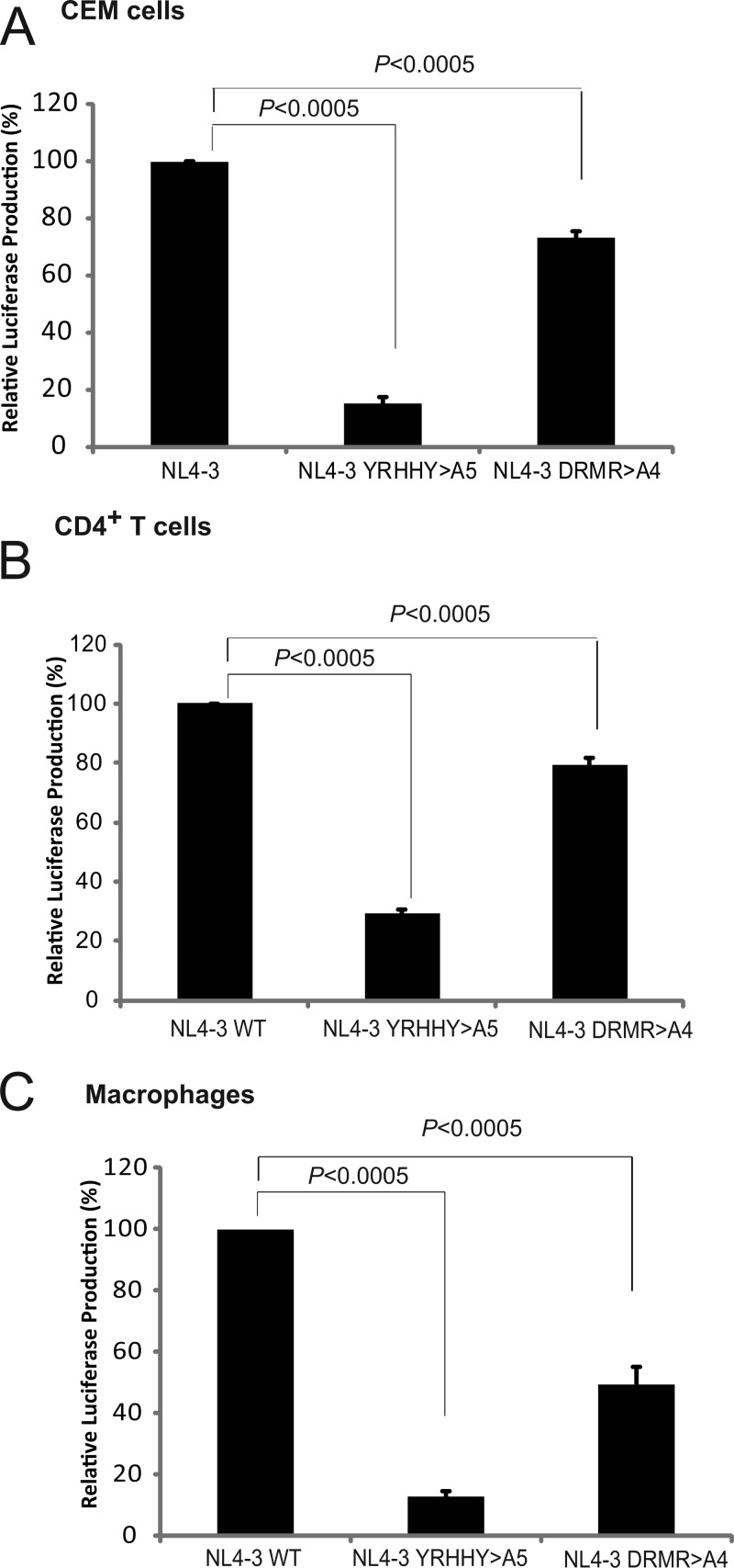
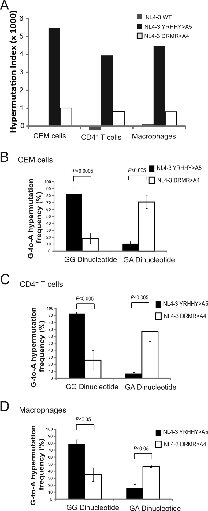
APOBEC3 proteins inhibit HIV-1 replication in experimental systems and induce hypermutation in infected patients; however, the relative contributions of several APOBEC3 proteins to restriction of HIV-1 replication in the absence of the viral Vif protein in human primary CD4(+) T cells and macrophages are unknown. We observed significant inhibition of HIV-1Δvif produced in 293T cells in the presence of APOBEC3DE (A3DE), APOBEC3F (A3F), APOBEC3G (A3G), and APOBEC3H haplotype II (A3H HapII) but not APOBEC3B (A3B), APOBEC3C (A3C), or APOBEC3H haplotype I (A3H HapI). Our previous studies showed that Vif amino acids Y(40)RHHY(44) are important for inducing proteasomal degradation of A3G, whereas amino acids (14)DRMR(17) are important for degradation of A3F and A3DE. Here, we introduced substitution mutations of (40)YRHHY(44) and (14)DRMR(17) in replication-competent HIV-1 to generate vif mutants NL4-3 YRHHY>A5 and NL4-3 DRMR>A4 to compare the antiviral activity of A3G to the combined antiviral activity of A3F and A3DE in activated CD4(+) T cells and macrophages. During the first 15 days (round 1), in which multiple cycles of viral replication occurred, both the NL4-3 YRHHY>A5 and NL4-3 DRMR>A4 mutants replicated in activated CD4(+) T cells and macrophages, and only the NL4-3 YRHHY>A5 mutant showed a 2- to 4-day delay in replication compared to the wild type. During the subsequent 27 days (round 2) of cultures initiated with peak virus obtained from round 1, the NL4-3 YRHHY>A5 mutant exhibited a longer, 8- to 10-day delay and the NL4-3 DRMR>A4 mutant exhibited a 2- to 6-day delay in replication compared to the wild type. The NL4-3 YRHHY>A5 and NL4-3 DRMR>A4 mutant proviruses displayed G-to-A hypermutations primarily in GG and GA dinucleotides as expected of A3G- and A3F- or A3DE-mediated deamination, respectively. We conclude that A3G exerts a greater restriction effect on HIV-1 than A3F and A3DE.


Figures

Fig 1
Fig 1
Single-cycle assays to determine sensitivities of NL4-3 mutants to A3 proteins. Plasmids encoding NL4-3 WT, NL4-3 YRHHY>A5, NL4-3 DRMR>A4, or NL4-3 Δvif were transfected into 293T cells in the presence of the A3 expression plasmids. The ratio of NL4-3 to A3 plasmid DNAs was either 3:1 (A) or 12:1 (B). After 48 h, culture supernatants were harvested, the amounts of p24 CA in the supernatants were determined, and viruses containing equivalent amounts of p24 CA were used to infect TZM-bl indicator cells. Infectivity was determined by measuring the luciferase activity produced in the infected cells at 48 h postinfection. Infectivity of NL4-3 WT in the absence of any A3 protein (no APOBEC3) was set to 100%. The average from three to five independent experiments is shown. Error bars represent the standard error of the mean. Asterisks indicate statistically significant decreases in infectivity compared to the NL4-3 WT/no APOBEC3 control (P < 0.02 by Student's t test).
Fig 2
Fig 2
Replication of NL4-3 WT, NL4-3 YRHHY>A5, NL4-3 DRMR>A4, and NL4-3Δvif in CEM T cells, primary CD4+ T cells, and macrophages. (A) Expression of A3G and A3F in CEM, CEM-SS, H9, and CD4+ T cells and macrophages. Endogenous A3G and A3F were detected by using the anti-A3G Apo-C17 antibody and the anti-A3F C18 antibody, respectively. (B) Protocol for evaluation of viral replication. (C to E) Replication of NL4-3 WT, NL4-3 YRHHY>A5, NL4-3 DRMR>A4, and NL4-3Δvif mutants in CEM cells (C), CD4+ T cells (D), and macrophages (E). Cells were infected with 10 ng of p24 CA-normalized viruses, supernatants were harvested every 2 days, and the p24 CA amounts were determined by ELISA. Multiple cycles of viral replication occurred over a 15-day period during round 1. The viruses from round 1 (left panels) peak time point samples were then p24 CA normalized and used to infected fresh cells (right panels) for round 2 infections, during which multiple cycles of viral replication occurred over an additional 27-day period. Two or three independent experiments were performed to determine the average delay in replication kinetics; results from one representative experiment are shown.
Fig 3
Fig 3
Relative infectivities of NL4-3 WT, NL4-3 YRHHY>A5, and NL4-3 DRMR>A4 mutant viruses in CEM cells (A), CD4+ T cells (B), and macrophages (C). The infectivities of the peak viruses from round 1 were normalized for p24 CA, and equal amounts of p24 CA were used to infect TZM-bl cells. After 72 h, the infectivity, as determined by the luciferase activity, was measured. The average from 3 independent experiments is shown. Error bars represent the standard error of the mean. Statistical significance was determined by Student's t test (P < 0.0005).
Fig 4
Fig 4
Hypermutation in CEM cells, CD4+ T cells, and macrophages. Proviruses from round 1 and round 2 infections were isolated, and a 730-bp sequence containing vif and a portion of vpr were sequenced and analyzed for G-to-A hypermutation. (A) Hypermutation indices for round 2 infections of CEM cells, CD4+ T cells, and macrophages. The hypermutation index was calculated as G-to-A substitutions (bp) − A-to-G substitutions (bp)/sequence length (bp) in CEM cells, CD4+ T cells, and macrophages. (B to D) Dinucleotide contexts of G-to-A hypermutation in CEM cells (B), CD4+ T cells (C), and macrophages (D) for round 2 infections. The error bars represent the standard deviation. Statistical significance was determined by Student's t test (P < 0.0005 to 0.05).

Similar articles

Cited by

References

    1. Liddament MT, Brown WL, Schumacher AJ, Harris RS. 2004. APOBEC3F properties and hypermutation preferences indicate activity against HIV-1 in vivo. Curr. Biol. 14:1385–1391 - PubMed
    1. Rose KM, Marin M, Kozak SL, Kabat D. 2005. Regulated production and anti-HIV type 1 activities of cytidine deaminases APOBEC3B, 3F, and 3G. AIDS Res. Hum. Retroviruses 21:611–619 - PubMed
    1. Sheehy AM, Gaddis NC, Choi JD, Malim MH. 2002. Isolation of a human gene that inhibits HIV-1 infection and is suppressed by the viral Vif protein. Nature 418:646–650 - PubMed
    1. Wiegand HL, Doehle BP, Bogerd HP, Cullen BR. 2004. A second human antiretroviral factor, APOBEC3F, is suppressed by the HIV-1 and HIV-2 Vif proteins. EMBO J. 23:2451–2458 - PMC - PubMed
    1. Zheng YH, Irwin D, Kurosu T, Tokunaga K, Sata T, Peterlin BM. 2004. Human APOBEC3F is another host factor that blocks human immunodeficiency virus type 1 replication. J. Virol. 78:6073–6076 - PMC - PubMed

Publication types

MeSH terms

LinkOut - more resources