

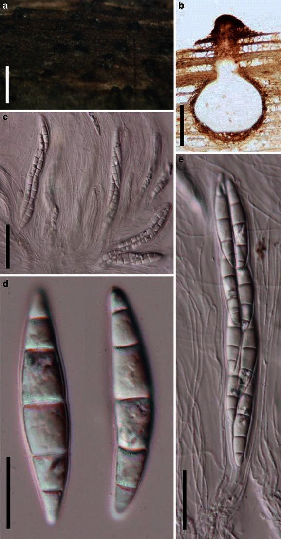
doi: 10.1007/s13225-011-0117-x.
Epub 2011 Oct 9.
Pleosporales
Affiliations
- PMID: 23097638
- PMCID: PMC3477819
- DOI: 10.1007/s13225-011-0117-x
Item in Clipboard
Pleosporales
Fungal Divers.
2012 Mar.
Abstract
One hundred and five generic types of Pleosporales are described and illustrated. A brief introduction and detailed history with short notes on morphology, molecular phylogeny as well as a general conclusion of each genus are provided. For those genera where the type or a representative specimen is unavailable, a brief note is given. Altogether 174 genera of Pleosporales are treated. Phaeotrichaceae as well as Krie-geriella, Zeuctomorpha and Muroia are excluded from Pleosporales. Based on the multigene phylogenetic analysis, the suborder Massarineae is emended to accommodate five families, viz. Lentitheciaceae, Massarinaceae, Montagnulaceae, Morosphaeriaceae and Trematosphaeriaceae.
Figures
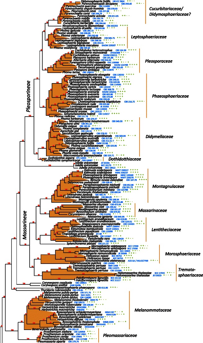
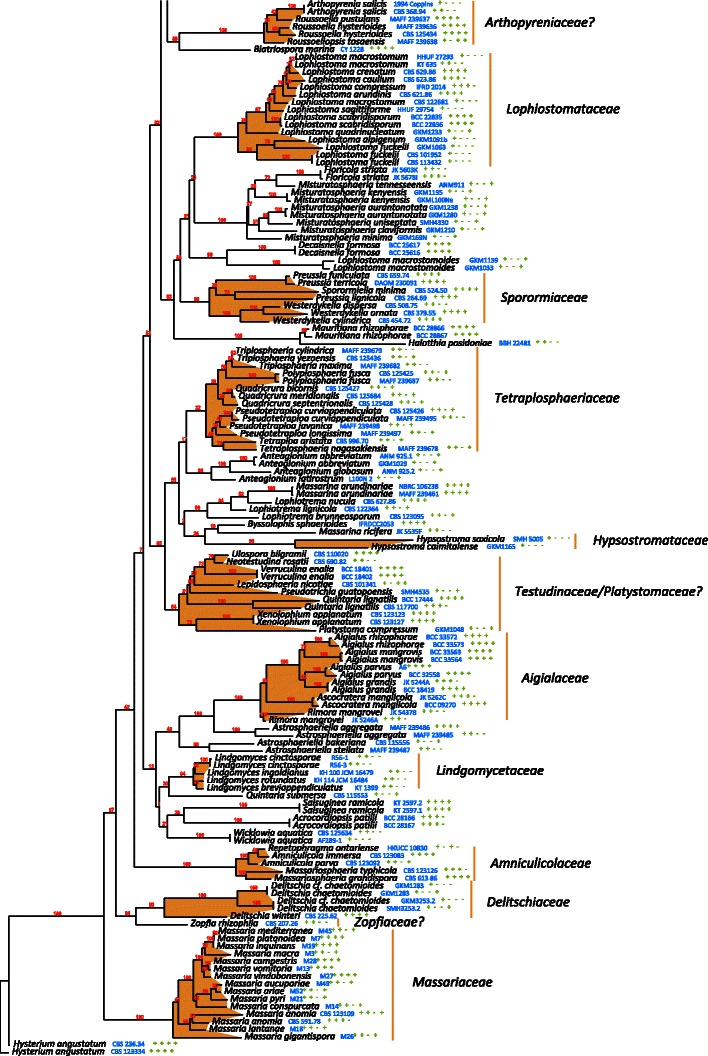
The best scoring likelihood tree of representative Pleosporales obtained with RAxML v. 7.2.7 for a concatenated set of nucleotides from LSU, SSU, RPB2 and TEF1. Family and suborder names are indicated where possible. The percentages of nodes present in 250 bootstrap pseudo replicates are shown above branches. Culture and voucher numbers are indicated after species names and the presence of the genes used in the analysis are indicated by pluses in this order: LSU, SSU, RPB2, TEF1
The best scoring likelihood tree of representative Pleosporales obtained with RAxML v. 7.2.7 for a concatenated set of nucleotides from LSU, SSU, RPB2 and TEF1. Family and suborder names are indicated where possible. The percentages of nodes present in 250 bootstrap pseudo replicates are shown above branches. Culture and voucher numbers are indicated after species names and the presence of the genes used in the analysis are indicated by pluses in this order: LSU, SSU, RPB2, TEF1
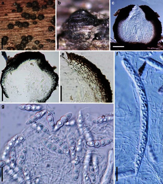
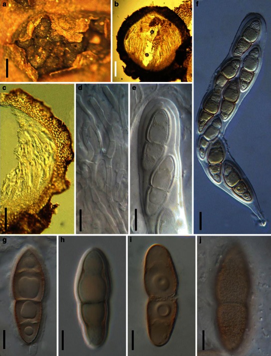
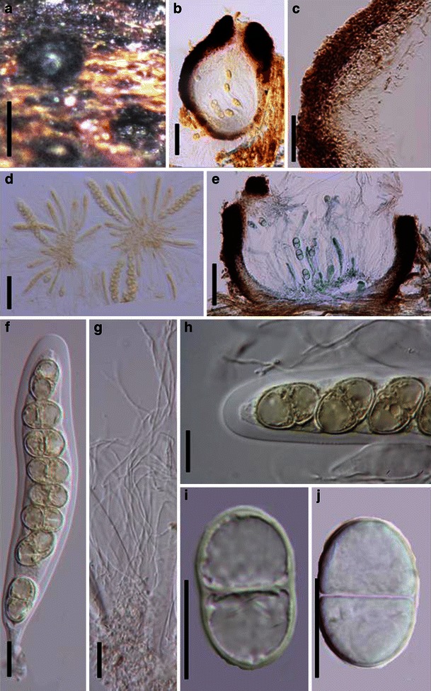
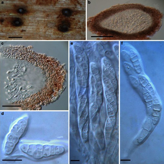
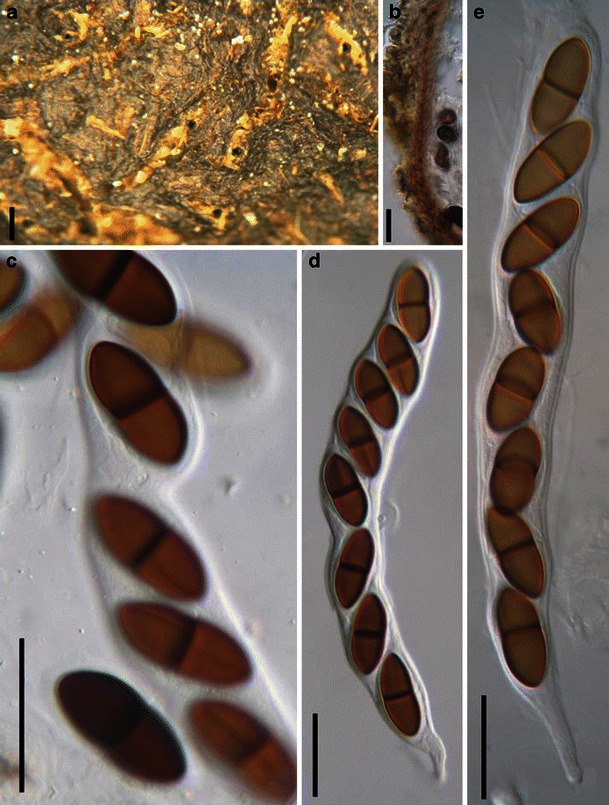
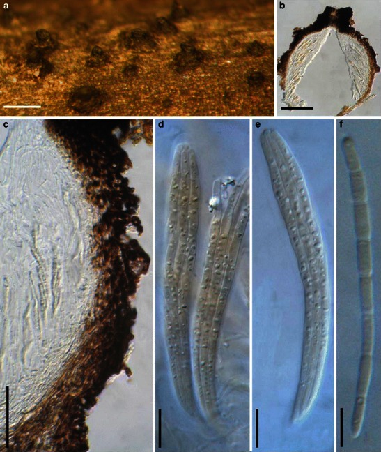
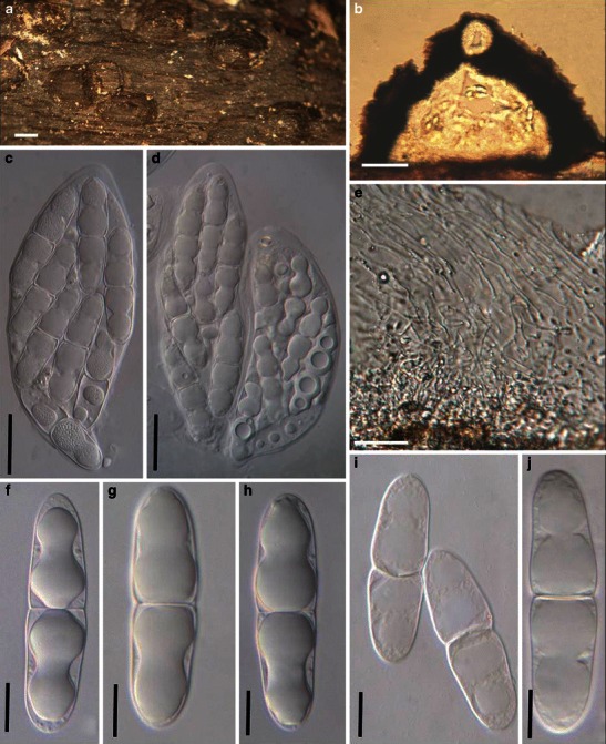
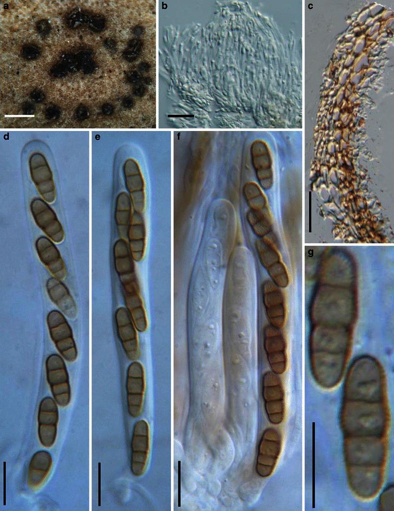
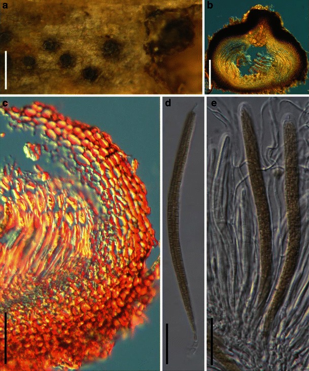
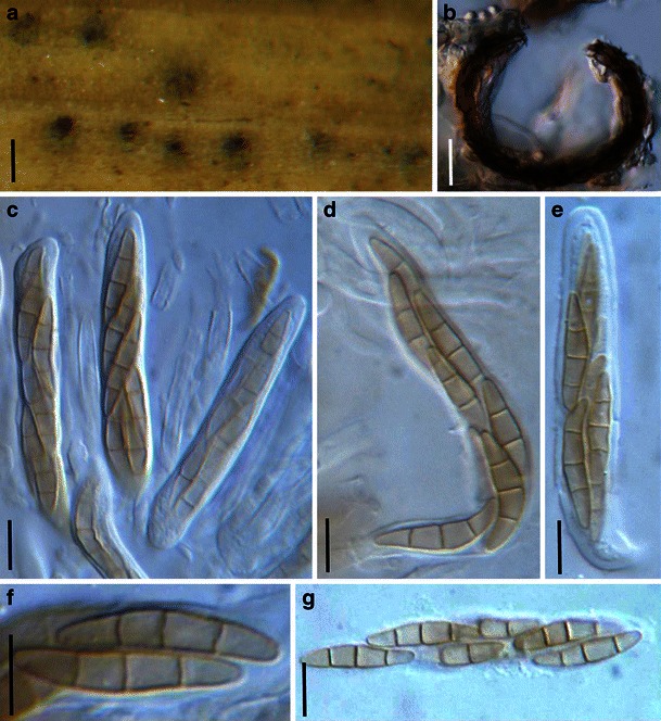
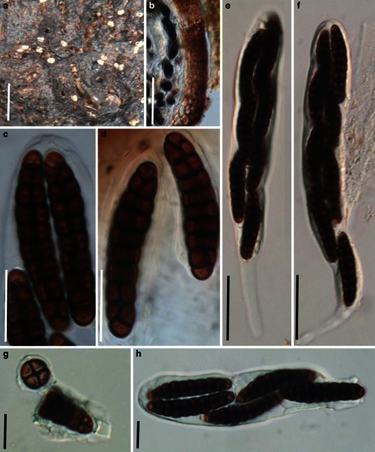
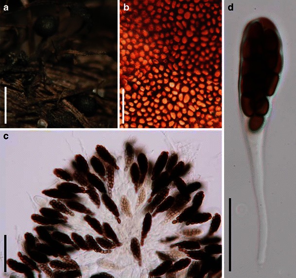
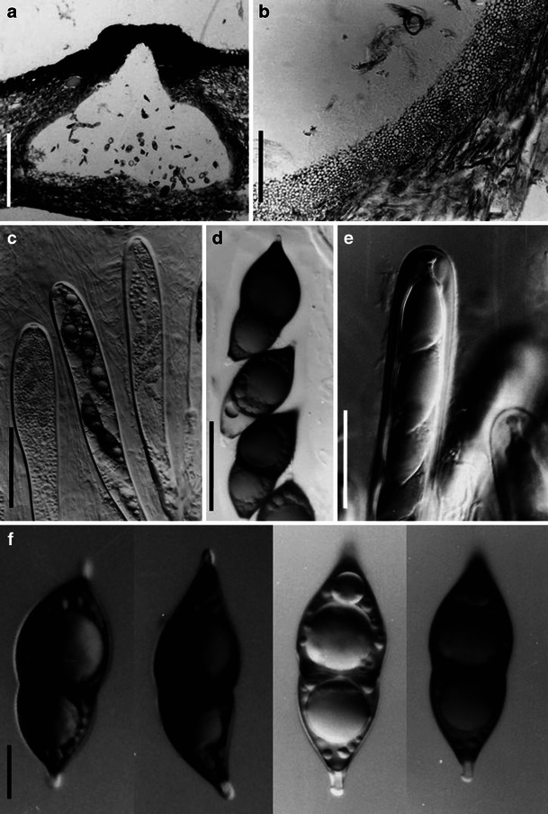
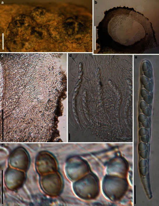
Acrocordiopsis patilii (from IMI 297769, holotype). a Ascomata on the host surface. b Section of an ascoma. c Section of lateral peridium. d Section of the apical peridium. e Section of the basal peridium. Note the paler cells of textura prismatica.
f Cylindrical ascus. g Cylindrical ascus in pseudoparaphyses. h, i One-septate ascospores. Scale bars: a = 3 mm, b = 0.5 mm, c = 200 μm, d, e =50 μm, f, g = 20 μm
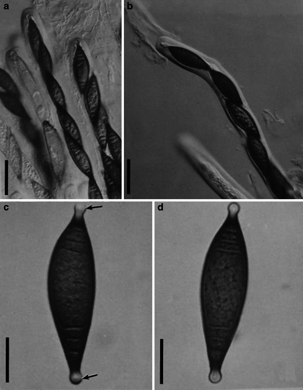
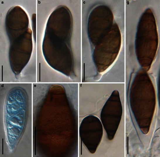
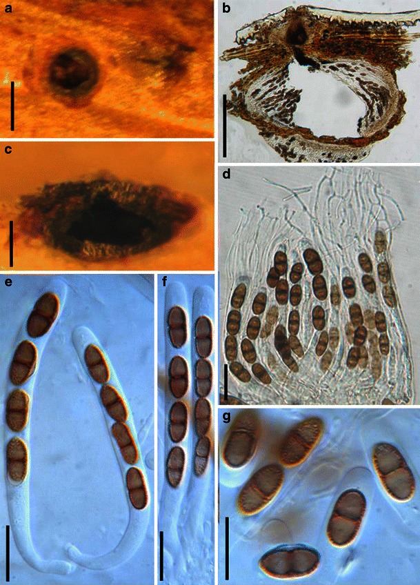
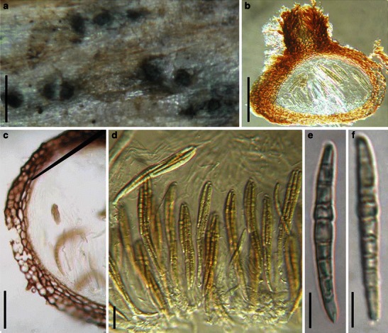
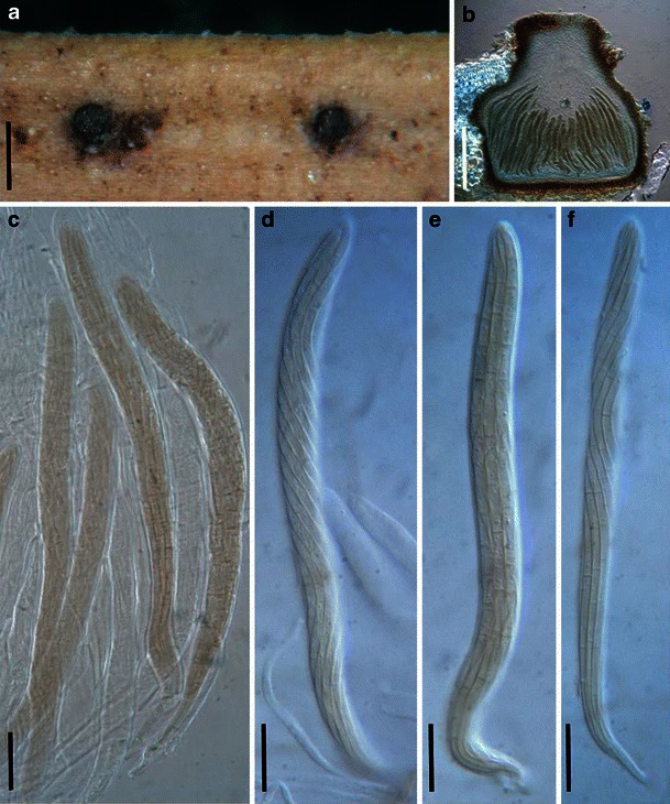
Aigialus grandis (from NY, J.K. 4332b, isotype). a Ascomata on the host surface. Note the longitudinal slit-like furrow which is the ostiole. b Section of the peridium. c, d. Released ascospores. e Ascospores in ascus. Note the conspicuous apical ring. f Cylindrical ascus with a long pedicel. Scale bars: a = 1 mm, b = 200 μm, c–f = 20 μm
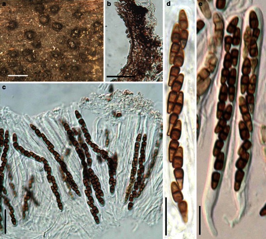
Amniculicola lignicola (from PC 0092661, holotype). a Superficial ascomata gregarious on the host surface. b An erumpent ascoma with elongated papilla and slit-like ostiole. c Habitat section of a superficial ascoma. d, e Section of an ascoma and the partial peridium. f Cylindrical 8-spored ascus with a short pedicel. g Hyaline, 1-septate broadly fusoid ascospores. Scale bars: a = 1 mm, b–d = 100 μm, e = 50 μm, f, g = 20 μm
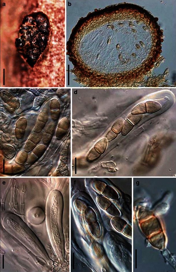
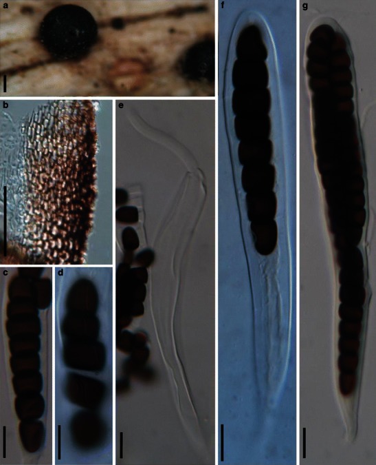
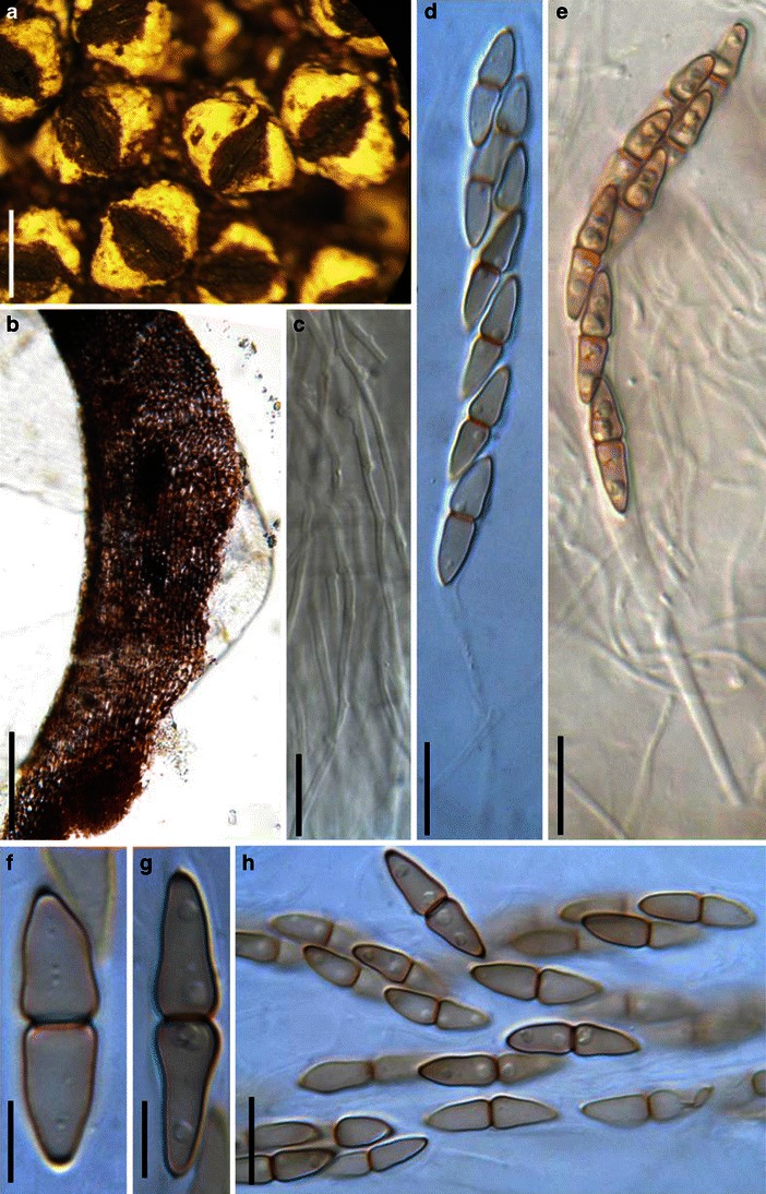
Anomalemma epochnii (K(M):143936, syntype). a Gregarious ascomata on the host surface. b, c Bitunicate asci. Note the wide pseudoparaphyses. d Section of the apical peridium comprising thick-walled cells of textura angularis. e–h Fusoid to broadly fusoid ascospores. Scale bars: a = 0.5 mm, b–h = 20 μm
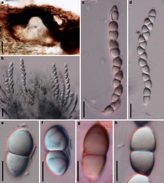
Appendispora frondicola (from BRIP 21354, holotype). a Immersed ascomata on host surface. b Valsoid configuration of the ascomata. c Cylindrical ascus. d Squash showing asci and numerous pseudoparaphyses. e Thin strands of anastomosing pseudoparaphyses. f, g Ascospores with one or two appendages. Scale bars: a = 0.5 mm, b = 100 μm, c–g = 10 μm
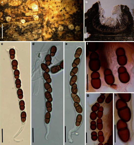
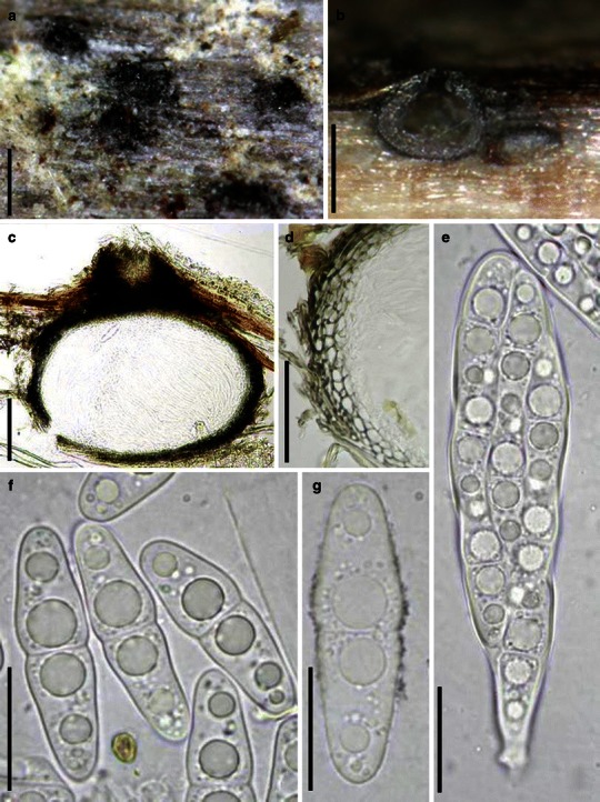
Ascorhombispora aquatica (from HKU(M) 10859, holotype). a Section of an ascoma. b Section of a partial peridium. c Immature ascus. d–f Mature asci with ascospores. Note the deliquescent ascal wall in f. Note the wide, dark band in the medium septum of ascospores in d and e and the mucilaginous sheath and paler end cells in e and f. Scale bars: a = 20 μm, b–f = 10 μm (figures referred to Cai and Hyde 2007)
Asteromassaria macrospora (from L, 1004). a Ascomata clustered in a group breaking through the host surface. b Section of an ascoma. c Section of a partial peridium. Note the cells of textura angularis. d Pseudoparaphyses. Note the branches. e Upper part of the ascus illustrating the ocular chamber. f Ascus with a short pedicel. g–j Ascospores. Note the mucilaginous sheath in G and minutely verruculose ornamentation in J. Scale bars: a = 0.5 mm, b, c = 100 μm, d–j = 10 μm
Astrosphaeriella fusispora (BISH 145726). a Ascomata forming a small group on host surface. Note the remains of the host forming flanges around the ascomata. b Section of the partial peridium. Note the black peridium and wedge of palisade cells between the lateral and basal walls. c Asci in trabeculate pseudoparaphyses. d–f Narrowly fusoid ascospores. Scale bars: a = 1 mm, b = 100 μm, c = 50 μm, d–f = 10 μm
Asymmetricospora calamicola (from HKU(M) 7794, holotype). a Ascomata immersed in the substrate. b Section of the peridium. c Mature and immature asci in pseudoparaphyses (in cotton blue). d Clavate ascus with a small ocular chamber. e–g Ascospores with sheath. Scale bars: a, b = 0.5 mm, c = 50 μm, d–g = 20 μm
Barria piceae (from NY 92003, isotype). a Ascoma on the host surface. Note the wide opening ostiole. b Section of the partial peridium with two types of cells. c, d Asci with ocular chambers and short pedicels. e, f Ellipsoid ascospores which are turning brown with thin sheath around them. Scale bars: a = 0.5 mm, b = 50 μm, c, d = 20 μm, e, f = 10 μm
Belizeana tuberculata (from Herb. J. Kohlmeyer No. 4398, holotype). a Immersed to semi-immersed ascomata. b, e Vertical section of an ascoma. c Section of a partial peridium. d Squash mounts with a large number of asci. f Broadly cylindrical ascus with a large ocular chamber. g Filliform pseudoparaphyses. h Apical part of an ascus. Note the large ocular chamber. i, j One-septate ascospores. Scale bars: a = 0.3 mm, b = 100 μm, c = 20 μm, d, e = 50 μm, f–i = 10 μm
1
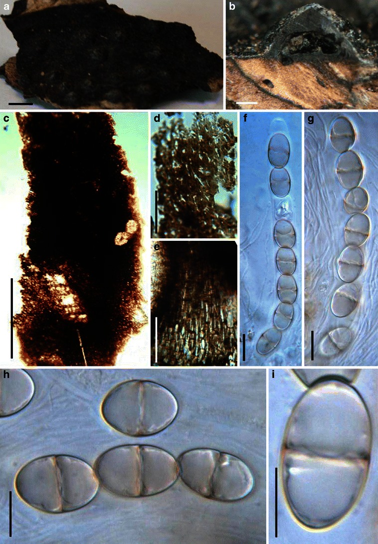
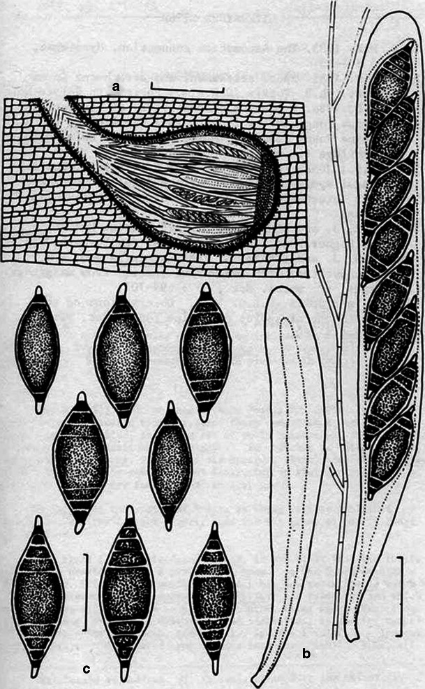
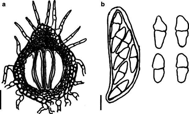
Biatriospora marina (from IMI 297768, holotype). a, b Cylindrical asci. Note the mucilage pseudoparaphyses in (a) and the conspicuous ocular chamber in (b). c, d Ascospores with hyaline end chambers (arrowed). Scale bars: a, b = 50 μm, c, d = 20 μm. 2 Line drawings of Biatriospora marina (based on holotype). a Section through ascocarp showing asci and pseudoparaphyses. b Asci and pseudoparaphyses. c Ascospores. Scale bars: a = 200 μm, b = 40 μm, c = 30 μm (figure with permission from Hyde and Borse 1986)
1
Biatriospora marina (from IMI 297768, holotype). a, b Cylindrical asci. Note the mucilage pseudoparaphyses in (a) and the conspicuous ocular chamber in (b). c, d Ascospores with hyaline end chambers (arrowed). Scale bars: a, b = 50 μm, c, d = 20 μm. 2 Line drawings of Biatriospora marina (based on holotype). a Section through ascocarp showing asci and pseudoparaphyses. b Asci and pseudoparaphyses. c Ascospores. Scale bars: a = 200 μm, b = 40 μm, c = 30 μm (figure with permission from Hyde and Borse 1986)
Bicrouania maritima (from IMI 330806, isotype). a Section of an ascoma. b Section of papilla. Note the periphyses. c–e Eight-spored asci. Note the furcated pedicel. Scale bars: a, b = 100 μm, c–e = 20 μm
Bimuria novae-zelandiae (from CBS 107.79, isotype). a–c Asci with a short pedicel and small ocular chamber. d Immature ascus. e Partial ascospore. Note the convex verrucae on the ascospore surface. f Released ascospores. Note the lighter end cells, germ pore and the longiseptum (arrowed). g Fissitunicate ascus dehiscent. Scale bars: a–g = 20 μm
Bricookea sepalorum (from S, type). a Ascomata on host surface (arrowed). b Section of partial peridium. Note thick-walled out layer and thin-walled inner layer. c–e Cylindrical to slightly obclavate asci with short knob-like pedicels. f–j Hyaline, 3-septate smooth-walled ascospores. Scale bars: a = 0.5 mm, b = 50 μm, c–j = 10 μm
Byssolophis byssiseda (from K(M):164030, isotype). a Ascomata gregarious on the host surface. b Numerous pseudoparaphyses. c Fusoid ascospores with or without terminal appendages. d Clavate ascus with a short furcate pedicel. Scale bars: a = 1 mm, b–d = 10 μm
Byssosphaeria schiedermayriana (from K(M):108784, holotype). a Superficial ascomata on the host surface. b Brown, 1-septate ascospores. c Section of the lateral peridium. Note the outer textura angularis and inner textura epidermoidea cells. d, e Furcate asci with a long pedicel. f Dehiscent ascus. Scale bars: a = 0.5 mm, c = 50 μm, b, d–f = 10 μm
Calyptronectria platensis (from LPS 1209, holotype). a Appearance of ascomata scattered in the substrate (after removing the out layer of the substrate). Note the protruding papilla. b Section of an ascoma. c Section of the partial peridium. Note the lightly pigmented pseudoparenchymatous cells. d Released ascospores with mucilaginous sheath. e Eight-spored asci in hamathecium and embedded in gel matrix. f Ascus with a short pedicel. Scale bars: a = 0.5 mm, b = 100 μm, c = 50 μm, d–f = 10 μm
Carinispora nypae (from BRIP 17106, holotype). a Ascomata on the host surface. b Section of an ascoma. c Section of a partial peridium. d, e, g, h Asci with ocular chambers and short pedicels. f The ocular chamber and apical ring of ascus. i–j Narrowly fusoid ascospores. Scale bars: a = 1 mm, b, c = 100 μm, d, h, i = 50 μm, f = 20 μm, g, e, j =10 μm
Caryosporella rhizophoriae (from NY. Herb. J. Kohlmeyer No. 4532a, holotype). a Gregarious ascomata on host surface. b Section of an ascoma. c, d Section of partial peridium at sides (c) and base (d). Note the three layers. e Asci with long peduncles in pseudoparaphyses. f, g Ascospores. Note the “net”-like ridged ornamentation of spore surface and hyaline germ pores. Scale bars: a = 1 mm, b = 200 μm, c–e = 100 μm, f, g = 10 μm
Chaetomastia hirtula (from H, FFE 825, kleptotype). a Superficial ascomata gregarious on the host surface. b Section of a partial peridium. Note the cells of textura angularis with relatively thick wall. c, d Cylindrical asci with long and furcate pedicels. e, f Brown, 3-septate ascospores. Scale bars: a = 0.5 mm, b = 50 μm, c–f = 10 μm
Chaetoplea calvescens (from FH-81113, isotype). a, b Four-spored and 8-spored asci. c Released ascospores. Scale bars: a–c = 10 μm
Cilioplea coronata (M 175-89-290, lectotype). a Immersed ascomata in small groups on the host surface (the covering host tissue was removed). b Section of a partial ascoma. Note the thin peridium. c Clavate asci within pseudoparaphyses. d Ascus with a small ocular chamber. Scale bars: a = 0.5 mm, b = 100 μm, c = 50 μm, d = 10 μm
Crivellia papareracea (from UBC F14995, epitype). a Gregarious ascomata immersed within the host surface. b Section of an ascoma. c Asci within pseudoparaphyses. d Cylindrical ascus with a short pedicel. Scale bars: a = 1 mm, b = 100 μm, c, d = 20 μm
Decaisnella spectabilis (NY2082, syntype). a Appearance of ascomata on the host surface. b Section of a partial peridium (immersed in the substrate). Note the pseudoparenchymatous out layer. c, d Muriform ascospores. Note the minuitely verrucose ornamentation. e Ascus with a short pedicel. Scale bars: a = 0.5 mm, b = 100 μm, c–e = 20 μm
Delitschia didyma (from L, 1950). a Ascomata on the substrate surface. Note the ostiolar opening. b Section of peridium. Note the small cells of textura angularis. c Released and unreleased ascospores. Note the germ slit in each cell. d, e Asci with ascospores and short pedicels with rounded ends. Scale bars: a = 0.5 mm, b =30 μm, c–e = 50 μm
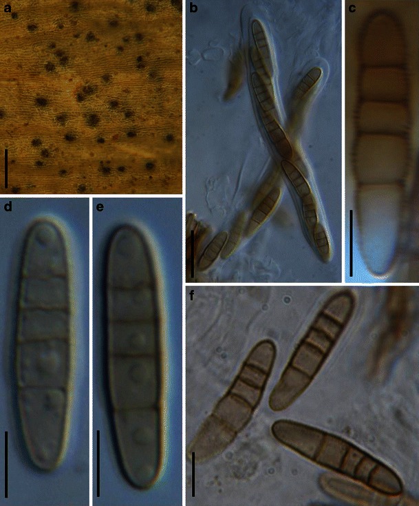
Didymosphaeria futilis (from K(M): 147683, holotype). a Two immersed ascomata on the host surface (one of them is cut horizontally). b Section of an ascoma. Note the thin peridium. c Hand cut portion of ascoma showing habitat in wood. d Asci in pseudoparaphyses. Note the trabeculate pseudonparaphyses anastomosing above the asci. e, f Four-spored asci with long pedicels which are rounded at their bases. g Brown, 1-septate ascospores with spinulose ornamentation. Scale bars: a = 0.3 mm, b, c = 100 μm, d–g = 20 μm
Dothidotthia symphoricarpi (from NY, holotype). a Clustered ascomata on the host stubstrate. b Longitudinal section through an ascoma. c, d Asci with pale brown, 1-septate ascospores. e Immature asci.
f Pale brown, 1-septate ascospores within asci. g Conidia of Thyrostroma anamorph in association with ascomata. Scale bars: a = 0.5 mm, b = 100 μm, c–g = 10 μm. (figure with permission from Phillips et al. 2008)
Dubitatio dubitationum (from NY, isotype; LPS, holotype). a Appearance of ascomata scattered on the host surface. Note the exposed white covering around the ostioles. b, c Section of an ascoma. Note the white covering (see arrow). d–f Cylindrical asci with short furcate pedicels. g–i Asymmetrical, 1-septate reddish-brown ascospores. Scale bars: a = 1 mm, b = 100 μm, c = 50 μm, d–i = 20 μm
Entodesmium rude (from H, Krieger 1070). a Ascomata in groups on the host surface. Note the erumpent papilla which is cylindrical and has an inconspicuous ostiole. b Section of part of an ascoma. Note the arrangement of asci and pseudoparaphyses. c Section of the peridium comprising cells of textura angularis. d Part-spores inside the ascus. e Relatively immature ascus with filliform ascospores and low ocular chamber. f–h Mature and immature asci with pedicels. Scale bars: a = 0.5 mm, b, c = 50 μm, d–h = 10 μm
Eudarluca australis (from LPS 5.415, type). a Ascomata on the host surface. b Section of an ascoma. c Section of a partial peridium. Note the thin peridium with cells of textura angularis. d–g Asci with short pedicels. h Ascospores. Note the 2-septate hyaline ascospore. Scale bars: a, b =100 μm, c = 50 μm, d–h = 10 μm
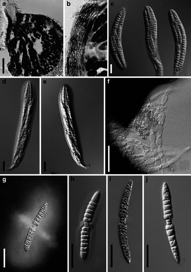
Falciformispora lignatilis (from BRIP 16972, holotype). a Section of a superficial ascoma. The peridium comprises two layers. b, c Squash mounts showing asci with wide pseudoparaphyses. The asci are cylindro-clavate with very short pedicels. d–f Hyaline multiseptate ascospores. Note the elongated appendage at the base (arrow head). Scale bars: a, b =100 μm, c = 50 μm, d–f = 10 μm
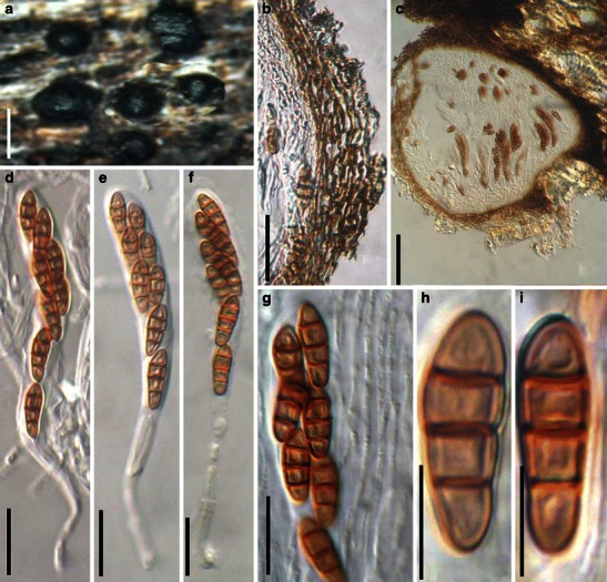
Hadrospora fallax (from BR, Capsa: K 7534, holotype). a Ascomata forming a cluster on the host surface. b Section of an ascoma. Note the peridium structure. c Section of a partial peridium. Note the pseudoparenchymatous cells. d Asci in pseudoparaphyses. e–i Reddish brown multiseptate ascospores. Scale bars: a = 0.5 mm, b = 100 μm, c, d = 50 μm, e–i = 20 μm
Halotthia posidoniae (from S, isotype of Sphaeria posidoniae). a Ascomata gregarious on the host surface. b–d Mature or immature cylindrical asci. e–h Ellipsoidal, dark-brown, 1-septate ascospores. Scale bars: a = 1 mm, b–d = 50 μm, e–h = 5 μm
Helicascus kanaloanus (from Herb. J. Kohlmeyer No. 2566, holotype). a Section of ascostroma immersed in the host tissue. Note the torsellioid ostiole. b One-septate, brown, asymmetrical ascospores within the asci. c, d Released thick-walled ascospores. Note the germ pore at the lower end of the ascospores. Scale bars: a = 0.5 mm, b–d = 20 μm
Herpotrichia rubi (from g, f. rh. 2171, type). a Numerous ascomata gregariously immersed in the host tissue. b Section of an ascoma. Note the central ostiole and peridium structure and also note the arrangement of asci and pseudoparaphyses. c Section of partial lateral peridium which comprises cells of textura angularis. d Part of a mature squashed ascus. e Relatively wide, septate pseudoparaphyses. f Immature ascus. Note the furcate pedicel. g–h One-septate ascospores. Note the verruculose ornamentation which is visible at the sides. Scale bars: a = 0.5 mm, b = 100 μm, c = 50 μm, d = 20 μm, e–h = 10 μm
Immotthia hypoxylon (from holotype of Amphisphaeria hypoxylon). a Ascomata gregarious on host surface. b–d Bitunicate asci. e–h Released 1-septate ascospores. Scale bars: a = 0.5 mm; b–h = 10 μm
Isthmosporella pulchra (from ILLS 53086, holotype). a Section of an ascoma. b Section of a partial peridium. c–e Broadly clavate asci with short pedicels. f Pseudoparaphyses. g–j Ascospores. Note the 2-celled isthmus in J and mucilaginous sheath in G and H. Scale bars: a = 50 μm, b–j = 20 μm
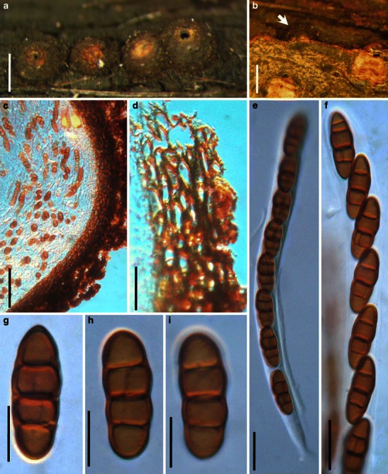
Kalmusia ebuli (from BR 101525–63, holotype). a Immersed to erumpent ascomata scattered on the host surface. b Section of a partial peridium. Note the compressed peridium cells. c Section of an ascoma. d–f Eight-spored asci with long pedicels. g Partial ascus in pseudoparaphyses. h, i Ascospores with 3 thick-walled septa. Scale bars: a = 0.5 mm, b = 50 μm, c = 100 μm, d–g = 20 μm, h, i = 10 μm
Karstenula rhodostoma (from PH 01048835, type). a Line of ascomata on host surface (after remove the decaying cover). Note the wide ostiolar opening and light colored region around the ostiole. b Immersed ascoma under the decaying cover (see arrow). c, d Section of the peridium. The peridium comprises small thick-walled cells in the outer layer. The outside comprises defuse hyphae which is probably part of the subiculum. e Ascus with a short furcate pedicel. f Partial ascus showing arrangement of ascospores. g–i Released ascospores. Note the transverse and rarely vertical septa. Scale bars: a, b = 0.5 mm, c = 50 μm, d–f = 20 μm, g–i = 10 μm
Katumotoa bambusicola (from HHUF 28663, holotype). a Ascomata scattered on the host surface. b Asci in pseudoparaphyses. c Hyaline ascospore with long terminal appendages. d Clavate ascus with a short pedicel. Scale bars: a = 0.5 mm. b–d = 20 μm
Keissleriella sambucina (from FH, holotype of Otthiella aesculi). a Section of an ascoma. b Pseudoparaphyses which are narrow (less than 1.5 μm) and branch and anastomosing as trabeculate. c, d Hyaline ascospores with distinct constrictions at the septa. e Asci amongst narrow pseudoparaphyses. F. Ascus with a pedicel and ocular chamber. Scale bars: a = 100 μm, b–f = 10 μm
Lentithecium fluviatile (from IFRD 2039). a Erumpent ascomata scattering on the host surface. b Habitat section of the immersed ascomata. c, d Section of an ascoma and a partical peridium. Note the peridium cells of textura angularis.
e Clavate 8-spored ascus with a short pedicel. f, g Hyaline, 1-septate broadly fusoid ascospores. Scale bars: a, b = 0.5 mm, c = 100 μm, d = 50 μm, e–g = 20 μm
Leptosphaeria doliolum (from L, lectotype). a Ascomata on the host surface. Note the shiny black surface. b Section of the partial peridium. Note the uneven thickness. c–e Asci with a short pedicel. f Three ascospores in ascus. Scale bars: a = 0.5 mm, b = 100 μm, c–f = 20 μm
Leptosphaerulina australis (from NY, C.T. Rogerson 3836). A. Compressed ascoma. Note the obpyriform asci within the ascoma and the thin peridium. B, C. Eight-spored asci released from the ascomata. Note the apical apparatus (arrowed). D. Ascospores with thin sheath. E. An old pale brown ascospore. Scale bars: A-C = 50 μm, D, E = 10 μm
Lewia scrophulariae (from FH, slide from lectotype). a Cylindrical ascus with a short pedicel. b Ascospores in asci. c–f Released muriform brown ascospores. Scale bars: a = 20 μm, b–f = 10 μm
Lichenopyrenis galligena (from MA-Lichen 12715, holotype). a, b Ascomata forming in the host tissues. c, d Sections of ascomata. e Section of a partial peridium. f–h, k Broadly clavate asci. Note the short rounded pedicel. i, j, l Ascospores. Note the small swellings at the septa. Scale bars: a, b = 0.5 mm, c, d = 100 μm, e = 50 μm, f–h, k = 20 μm, i, j, l = 10 μm
Lineolata rhizophorae (from Herb. J. Kolmeyer No. 2390b, isotype of Didymosphaeria rhizophorae). a Ascomata immersed in the host substrate with protruding papilla. b Ascospores within an ascus. Note the ascospore arrangement. c–f One-septate ascospores. Note the striate ornamentation in (c). Scale bars: a–b = 20 μm, c–f = 10 μm
Loculohypoxylon grandineum (from NY). a Appearance of ascomata on the host surface. b Habitat section of ascomata. c Section of an ascoma. Note the pale brown thin-walled peridium cells. d, e Uniseriate ascospores in asci. f–f Cylindro-clavate asci with ascospores. Note the ocular chamber in (g). Scale bars: a = 100 μm, b = 200 μm, c = 50 μm, d–h = 10 μm
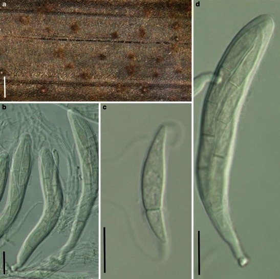
Lophionema vermisporum (from NY-643, holotype). a Appearance of ascomata on the host surface. Note the form of the neck. b Section of the peridium. c Peridium comprising two types of cells which merge in the middle; outer cells small heavily pigmented thick-walled cells of textura angularis, inner cells less pigmented, and comprising thin-walled compressed cells. d, e Cylindro-clavate, 8-spored asci. f A 7-septate filliform ascospore. Scale bars: a = 0.5 mm, b = 100 μm, c = 50 μm, d–f = 10 μm
Lophiostoma macrostomum (a–h, j from UPS, leptotype; i from IFRD 2005). a Appearance of ascomata on the host surface. Note the raised crest-like areas and full length germ slits. b Section of the peridium. c–e Cylindro-clavate asci with ascospores arranged in a 2-3-seriate manner. f Hamathecium comprising branching and septate pseudoparaphyses. g–j Released or unreleased ascospores. Note the smooth young ascospores with terminal sheath, and the verrucose senescent ascospores. Scale bars: a = 0.5 mm, b = 200 μm, c–j = 10 μm
Lophiotrema nucula (from UPS, lectotype). a Ascomata on the host surface. b Section of a partial ascoma. c Peridium structure near the apex. d, h Cylindrical asci in the pseudoparaphyses. e, f Upper part of the asci, showing the small ocular chamber near the apex. h Mature ascospores. i Pseudoparaphyses. Scale bars: a = 0.5 mm, b = 100 μm, c, d = 30 μm, e–i = 10 μm
Macroventuria wenti. a Ascomata. Note the setae. b Ascus and ascospores. Scale bars: a = 50 μm, b = 10 μm (figures referred to van der Aa 1971)
Mamillisphaeria dimorphospora (from HKU(M) 7425, paratype?). a Ascomata scattered on the host surface. Note the small papilla. b Section of an ascoma. c, d Asci (TYPE 1). e Trabeculate pseudoparaphyses in a gelatinous matrix. f–j Ascospores. Scale bars: a = 0.5 mm, b–d = 100 μm, e = 10 μm, f–j = 20 μm
Massarina eburnea (from IFRD 2006). a Ascomata on the host surface. b Section of an ascoma. c Ascus with a short pedicel. d Cellular pseudoparaphyses. e Section of the peridium comprising a few layers of compressed cells. f Asci in pseudoparaphyses. g Three-septate ascospores. Scale bars: a = 0.5 mm, b = 100 μm, c–g = 20 μm
Massariosphaeria phaeospora (ZT, holotype). a Ascomata scattering on the host surface. Note the immersed to erumpent ascomata. b Section of a partial peridium. Note the peridium structure. c, d Released ascospores. Scale bars: a = 200 μm, b–d = 20 μm
Mauritiana rhizophorae (from HKU(M)10219, holotype). a Vertical section of an ascoma. Note the thin layer of fungal tissue (pseudostroma?) on the host surface. b Section of a partial peridium. c Pseudoparaphyses and immature ascus. d Fissitunicate asci. e Asci showing thickening of the apical wall. f–i Ascospores with transverse septa and paler polar cells. Scale bars: a = 40 μm, b, d–i = 10 μm, c = 20 μm
Melanomma pulvis-pyrius (a–b, d–e, h–j from UPS, holotype; c, g, k, l from epitype). a Ascomata gregarious on the host surface. b Vertical section of an ascoma. c–f Asci with pedicels. g Dehiscent ascus. h–l Ascospores. Scale bars: a = 0.5 mm, b = 200 μm, c–l = 10 μm
Metameris japonica (from S, F7166, type). a Ascostroma arrangement on the host surface. b Section of two ascomata from one ascostroma. c Immature asci within pseudoparaphyses. d, e Hyaline ascospores. Scale bars: a = 0.5 mm. b = 100 μm, c–e = 20 μm
Mixtura saginata (from S reg. nr F8934, type). a, b Leaf spots in leaves of Chusquea serrulatae. Note the erumpent ascomata surrounded by white material in (b). c Section of an ascoma. Note the peridium structure which comprises cells of textura angularis. The arrangement of the asci and pseudoparaphyses can also be seen. d Immature asci in pseudoparaphyses. Note the stumpy pedicel and thickened apex with flattened ocular chamber. e, f Mature ascospores. Note the hyaline ends and distosepta. Scale bars: a = 10 mm, b, c = 100 μm, d = 50 μm, e–f = 20 μm
Montagnula infernalis (from M 1183, holotype). a Appearance of ascomata immersed in host tissue. b Section of an immersed ascoma. Note the hyaline closely adhering cells in the ostiole region. c Section of the peridium comprising a few layers of cells. d An immature ascus with a long pedicel. e, g Mature muriform ascospores in asci. f Cellular pseudoparaphyses. Scale bars: a = 0.5 mm, b, c = 100 μm, d–g = 20 μm
Moristroma polysporum (from BAFC 32036, holotype). a Two multiculate ascostroma on the host surface. b Section of an ascostroma. Note the multilocula. c Section of the peridium. Note the thick walled cells. d, e Broadly cylindrical to fusoid asci containing numerous part spores. f Released part spores. Scale bars: a = 0.5 mm, b = 200 μm, c = 50 μm, d–f = 10 μm
Morosphaeria velataspora (from IMI 297770, type). a Section of an ascoma. b Cylindrical asci embedded in pseudoparaphyses. c–e Hyaline, 1-3-septate, ascospores with mucilaginous sheath. Scale bars: a = 100 μm, b = 50 μm, c–e = 20 μm
Murispora rubicunda (from IFRD 2017). a Habitat section of the immersed ascomata. b Section of an ascoma. Note the thin peridium and cells of textura angularis.
c Mature and immature asci. d Muriform ascospores. Scale bars: a, b = 100 μm, c, d = 20 μm
Neomassariosphaeria typhicola (from IFRD 2018). a Immersed ascomata gregarious in the host substrate. b–d Cylindro-clavate asci embedded in pseudoparaphyses. Note the phragmosporous ascospores. Scale bars: a, b = 200 μm, c, d = 20 μm
Neophaeosphaeria filamentosa (from NY, holotype). a Ascomata as a circular cluster on the host surface. b Hamathecium of wide psuedoparaphyses. c Section of peridium comprising cells of textura angularis. d–f Cylindrical asci with thickened apex. Note the short furcate pedicel. g Pale brown, 3-septate ascospores. Note the verruculose ornamentation. Scale bars: a = 200 μm, b, c = 20 μm, d–g = 10 μm
Nodulosphaeria hirta (from BR 101945–95, holotype). a Appearance of ascomata on the host surface. b Vertical section of an ascoma. Note the setae at the apex and in the ostiole. c Section of a partial peridium. Note the outer layer cells of textura angularis and inner layer compressed cells. d Squash mount showing asci in pseudoparaphyses. e, f. The light brown filiform ascospores. Scale bars: a = 0.5 mm, b = 100 μm, c = 50 μm, d = 20 μm, e, f = 10 μm
Ohleria modesta (from g: f. rh. 2173, isotype). a Ascomata scattering on host surface. b Section of a partial peridium. c Asci embedded in pseudoparaphyses. d, e Cylindrical asci with short pedicels. Scale bars: a = 1 mm, b, c = 50 μm, d, e = 20 μm
Ohleriella neomexicana (NY, holotype). a Ascoma scattering on the host surface. b Section of a partial peridium. Note the small cells of textura angularis. cAscospore in ascus. d Ascospore breaking into part spores. Note the sigmoid germ slit. e Dehiscent ascus. f, g Asci with short pedicels. Scale bars: a = 100 μm, b = 50 μm, c–g = 10 μm
Ophiobolus disseminans (from BPI-629021, type). a Immersed ascomata scattered on the host surface. Note the erumpent papilla. b Section of an ascoma. c. Section of a partial peridium. Note the thick-walled outer layer and thin-walled inner layer (orange colour due to DIC). d Ascus with a short furcate pedicel. e Squash mount showing asci in pseudoparaphyses. Scale bars: a = 0.5 mm, b = 100 μm, c = 50 μm, d, e = 20 μm
Ophiosphaerella graminicola (from LPS 858, holotype). a Ascomata on the host surface. Note the protruding disk-like papilla. b Section of an ascoma. c Asci in pseudoparaphyses with short pedicels. d–f Cylindrical asci with short pedicels. Scale bars: a = 0.5 mm, b = 100 μm, c–f =10 μm
Ostropella albocincta (K(M): 143941, syntype). a Ascomata gregarious on host surface. b Section of the partial peridium. Note the peridium comprising two cell types and the whitening tissue (arrowed). c Pseudoparaphyses. d, e Asci with long pedicel. f–h Ascospores, which are strongly constricted at the central septum. Scale bars: a = 1 mm, b = 100 μm, d, e, h = 20 μm, c, f, g = 10 μm
Paraliomyces lentifer (from Herb. J. Kohlmeyer No. 1720). a Section of an immersed ascoma. b Eight-spored cylindrical asci embedded in pseudoparaphyses. c, d Cylindrical asci with short pedicels. e–h One-septate hyaline ascospores. Scale bars: a = 100 μm, b–d = 20 μm, e–h = 10 μm
Phaeosphaeria oryzae (from S nr F9572, F9573, lectotype). a Appearance of ascomata on the host surface. b Section of an ascoma. c Squash mount showing asci in pseudoparaphyses. Note that asci with short pedicels. d, e Asci with short pedicels. F, G. Light brown 3-septate ascospores. Scale bars: a = 100 μm, b–g = 10 μm
Phaeosphaeriopsis glauco-punctata (from Cooke M.C. 166). a Ascomata immersed in the substrate. b Eight-spored cylindrical asci. c–f. Pale brown baculate ascospores which are released from asci. Scale bars: a = 200 μm, b = 20 μm, c, d–f = 10 μm
Platysporoides chartarum (from G NASSAU: 210558, type). a, b Ascomata scattered among fibers. Note the central ostioles. c Asci in numerous cellular pseudoparaphyses. d, e Cylindro-clavate asci with short pedicels. f–h. Muriform ascospores. Scale bars: a, b = 200 μm, c–e = 20 μm, f–h = 10 μm
1
Pleomassaria siparia (from BR, type). a Ascomata on the host surface. b Section of a partial peridium. c, d Asci with short pedicels. e–g Ascospores with thin sheath. Scale bars: a = 0.5 mm, b–d = 50 μm, e–g = 20 μm. 2
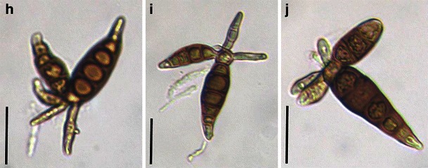
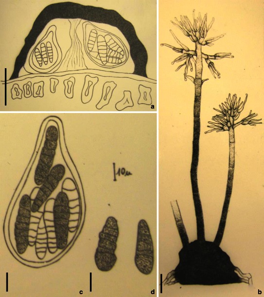
Prosthemium betulinum (from BR, type). h–i Conidia with arms. Scale bars: h–j = 20 μm
1
Pleomassaria siparia (from BR, type). a Ascomata on the host surface. b Section of a partial peridium. c, d Asci with short pedicels. e–g Ascospores with thin sheath. Scale bars: a = 0.5 mm, b–d = 50 μm, e–g = 20 μm. 2
Prosthemium betulinum (from BR, type). h–i Conidia with arms. Scale bars: h–j = 20 μm
Pleophragmia leporum (from G. Fungi rhenani n2272, type). a Appearance of ascomata on the substrate surface. Note the ostiolar pore. b Section of a partial peridium. c, h Apical part of an ascus. Note the apical apparatus in (c). d Released ascospores. e, f Clavate Asci with pedicels. g Part of a broken ascospore. Note the crossing septa. Scale bars: a = 0.5 mm, B = 50 μm, c–f = 20 μm, g, h = 10 μm
Pleoseptum yuccaesedum (from BPI 802381, holotype). a Appearance of ascomata scattered on the host surface. Only the upper region is visible. b Squash mount of asci in pseudoparaphyses. c Section of an ascoma. Note the peridium comprising cells of textura angularis. d, e Asci with short furcate pedicels. f, g Muriform dark-brown ascospores. Scale bars: a = 0.5 mm, b = 40 μm, c = 100 μm, d, e = 20 μm, f, g = 10 μm
Pleospora herbarium (from E, Krieger 683). a Immersed ascomata scattering on host surface. b Ascomata in small groups. Note: the surface layer of the host is removed. c Section of an ascoma. Note the peridium cells of textura angularis. D, E. Asci with short pedicels. Scale bars: a, b = 0.5 mm, c = 100 μm, d, e = 30 μm, f–k = 20 μm
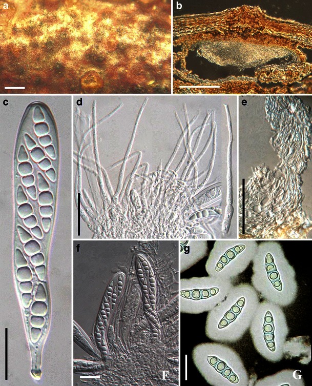
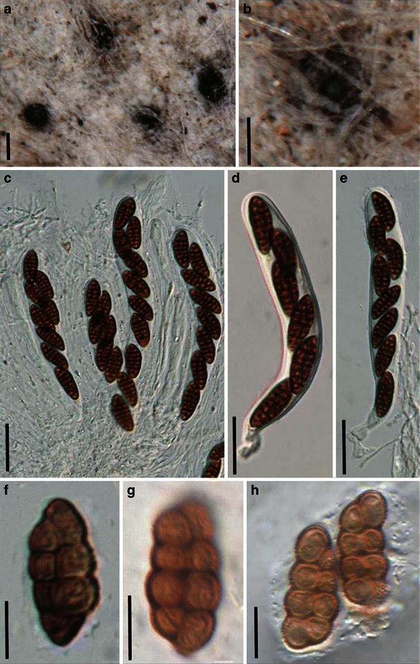
Preussia funiculata (from TRTC 46985). a Superficial cleistothecoid ascomata. b Part of peridium from front view. c Squash mounts showing a large number of asci. d A clavate ascus with a long and thin pedicel. Scale bars: a = 0.5 mm, b = 20 μm, c, d = 100 μm
Quintaria lignitalis (from J. Kohlmeyer No. 4365a, holotype). a Ascomata immersed in substrate. b Section of an ascoma. Note the thin peridium and elongated papilla. c, e Asci embedded in pseudoparaphyses. d Five septate fusoid hyaline ascospores. Scale bars: a = 0.5 mm, b = 200 μm, c, e = 50 μm, d =20 μm
Roussoëlla nitidula (from PAD Paol. 2484, holotype). a Appearance of the stroma on host surface. b Asci and pseudoparaphyses. c, d Long cylindrical furcate asci. E-H. Ascospores. Note the striate ornamentation. Scale bars: a = 0.5 mm, b–d = 20 μm, e–h = 10 μm
Saccharicola bicolor (from IMI 215888, holotype). a Section of an ascomata immersed in the host tissue. b Section of a partial pycnidia. Note the phragmosporous conidia. c Clavate ascus with ocular chamber and short pedicel. d Ascospores. Note the pigmented central cell(s). Scale bars: a, b = 50 μm, c = 20 μm, d = 10 μm
Salsuginea ramicola (from BRIP 17102, holotype). a Habitat section of an ascoma. b Section of the partial peridium. c Clavate mature and immature asci. d Ascospores within ascus. e Apical part of immature asci. f Ascospores with an apical chamber at each end. Scale bars: a = 0.5 mm, b–e = 50 μm, f = 10 μm
Semidelitschia agasmatica (from TRTC 40697, holotype). a Immersed ascomata scattered on the surface of the substrate. b Squash of ascoma. Note the numerous released asci. c Apical ring of cylindrical asci. d One-celled ascospores. Note the germ slits (see arrow). e Cylindrical ascus. Note the tapering pedicel. Scale bars: a = 0.5 mm, b–e = 100 μm
Setomelanomma holmii (from UPS F-117969 (slide), isotype). a, b Asci with short pedicels in pseudoparaphyses. c Partial view of ascus. d Branching and septate pseudoparaphyses. a Three-septate lightly pigmented ascospores in ascus. Scale bars: a–e = 10 μm
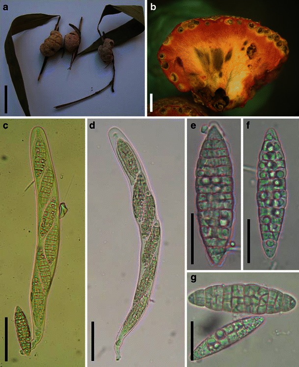
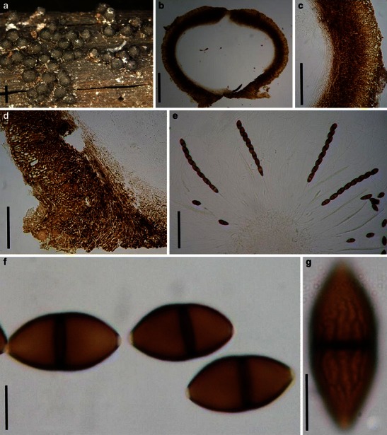
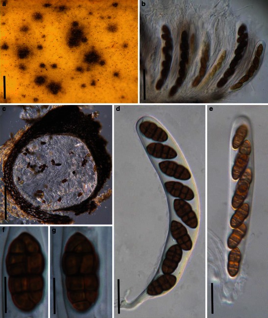
Shiraia bambusium (from IFRD 2040). a Ascostroma form a nubby structures on the twigs of host. b Vertical section of an ascostroma. Note the reddish staining of the inner tissue. c, d Cylindrical asci with a short pedicel. e–g Muriform fusoid hyaline ascospores. Scale bars: a = 1 cm, b = 1 mm, c, d = 50 μm, e–g = 20 μm
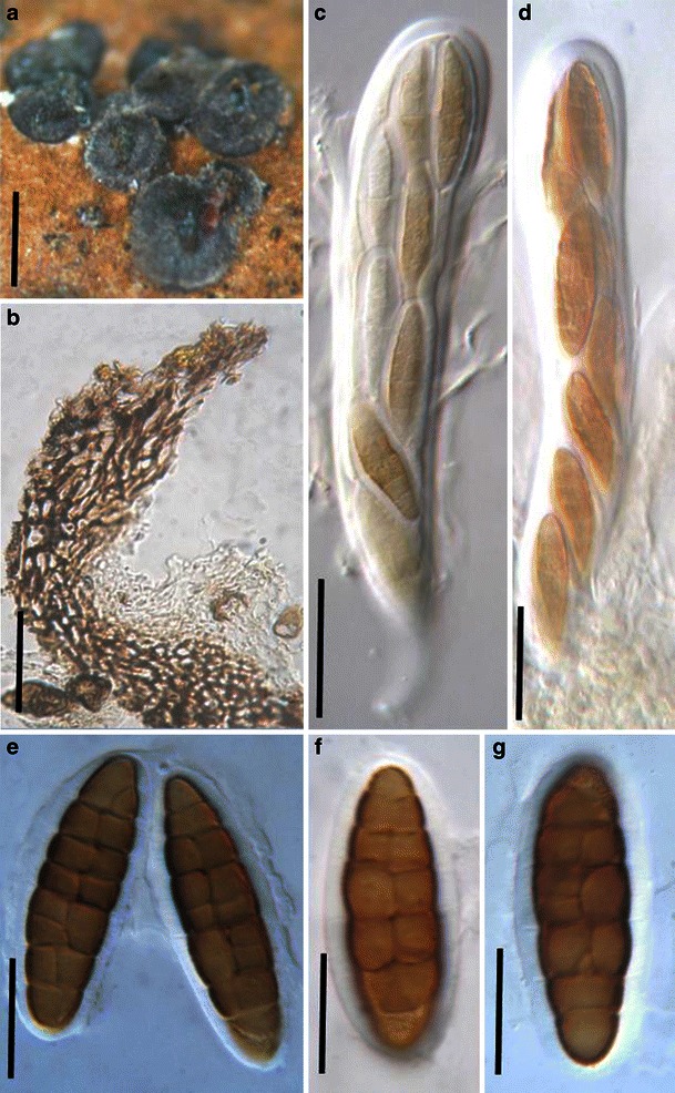
Sinodidymella verrucosa (from W 16366, type). a Ascomata on the host surface. Note the radial ridges around the pseudostiolar region. b Section of an ascoma. c Section of peridium. Note the hyaline small cells and interwoven hyphae. d Cylindrical asci in pseudoparaphyses. e Eight-spored ascus with short pedicel. f Hyaline, 1-septate ascospores which turn pale brown when mature. Scale bars: A = 1 mm, B = 100 μm, c = 50 μm, d–f = 20 μm

Splanchnonema pustulatum (from L, No. 910.251–352, No. 910.251–371). a Appearnce of ascomata on the host surface beneath a slightly raised area with minute ostiolar opening. b Section of the partial peridium. Note the compressed cells. c Dehiscent ascus. d Cluster of three asci joined in hymenium and pseudoparaphyses. e, f Asymmetric ascospores. Note the conspicuous sheath. Scale bars: a = 1 mm, b–d = 50 μm, e, f = 20 μm
Sporormia fimetaria (from RO, type). a Appearance of ascomata on the host surface. Note the scattered distribution. b–d Broad cylindrical asci with a short and thick pedicel. e Released filiform ascospores which may break up into part spores. Scale bars: a = 0.5 mm, b–d = 20 μm, e = 10 μm
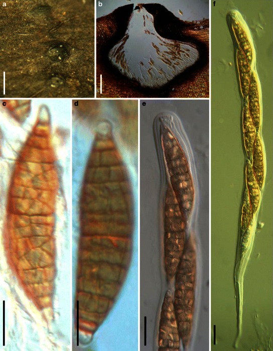
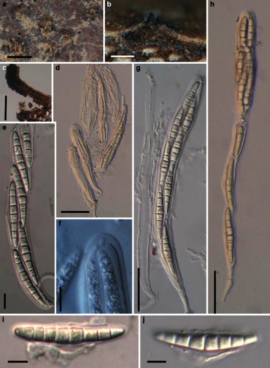
Trematosphaeria pertusa (a, d, f–i from epitype, b, c, e, j from neotype). a Ascomata on the host surface. b Section of an ascoma. c, h Section of the peridium. c shows the peridium structure at sides, and h indicates the basal peridium structure. Note the hyaline and thin-walled cells in (h). d Asci amongst pseudoparaphyses. e Ascus with pedicle. f, g Dehiscent ascus. i Upper part of the ascus, showing the ocular chamber and the mucilage covering the apex. j, k Ascospores. Scale bars: a = 0.5 mm, b, c = 100 μm, d–h = 20 μm, i–k = 10 μm
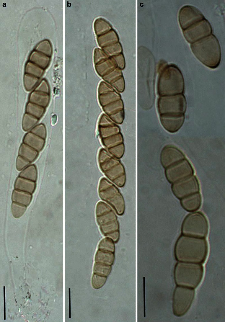
Verruculina enalia (from KDH 2137, slide). a Cylindrical asci with short pedicels. b One-septate verruculose ascospores. Scale bars: a = 20 μm, b = 10 μm
Westerdykella ornata (from CBS 379.55 holotype). a Appearance of the ascomata on culture substrate surface. b–f Mature and immature asci as well as the released ascospores. Note the spiral bands around the ascospores. Scale bars: a = 1 mm, b–f = 10 μm
Wettsteinina gigantospora (from S, holotype of Massarina gigantospora). a Ascomata with protruding papilla scattered on the host surface. b Obpyriform thick-walled ascus with small apical apparatus. c Fissitunicate ascus. d Released hyaline ascospores. Note the distinct primary septum and less distinct secondary septa. e Ascospore with sheath. Scale bars: a = 0.5 mm, b–d = 100 μm, e = 50 μm
Wilmia brasiliensis (from UB Col. Microl 8438, holotype). a Section of an ascoma. Note the setae in the ostiole. b Conidioma of the coelomycetous anamorphic stage. c, d Clavate asci with short furcate pedicels. e, f Released 1-septate pale brown ascospores. Scale bars: a, b = 100 μm, c, d = 20 μm, e, f = 10 μm
Xenolophium applanatum (from IFRD 2038). a Gregarious ascomata on the host surface. Note protruding papilla and slit-like ostiole. b Vertical section of the papilla and ostiole. c Section of the partial peridium. Note the two layers of the peridium. d Eight-spored asci in trabeculate pseudoparaphyses. Note the long pedicels. e–g Pale brown ascospores. Scale bars: a = 2 mm, b = 200 μm, c = 50 μm, d = 20 μm, e–g = 10 μm
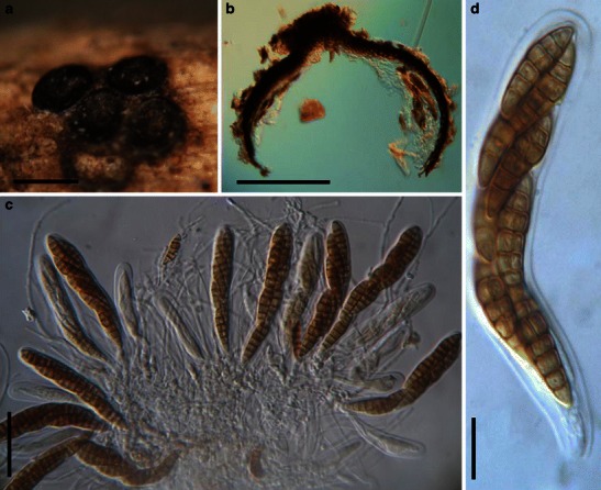
Javaria samuelsii (from isotype). a Ascoma on the host surface. Note reflexed pieces of the ruptured host tissue. b, c Cylindro-clavate asci within narrow pseudoparaphyses in gelatinous matrix. d Released ascospore with sheath. Scale bars: a = 1 mm, b = 50 μm, c, d = 20 μm
Pycnidiophora dispersa (A from CBS 297.56; B-D from MSC 133.118, type). a Ascomata scattering on the surface of the substrate. b Crashed ascoma. Note the numerous released asci. c Globose asci and released ascospores. d One-celled ascospores. Scale bars: a = 200 μm, b–d = 20 μm
Sporormiella nigropurpurea (from NY, holotype). a Section of an ascoma. b Section of the papilla. Note the dense pseudoparaphyses. c Section of a partial peridium. d, e Eight-spored cylindro-clavate asci with furcate pedicels. f, g Four-celled, brown ascospores. Note the sigmoid germ slit in each cell. Scale bars: a = 200 μm, b, c = 50 μm, d, e = 20 μm, f, g = 10 μm
Spororminula tenerifae (from HCBS 9812, holotype). a Appearance of ascomata on the host surface. b, c Sections of the partial peridium. Note the elongate cells of textura angularis. d, e Asci with thin pedicels. f, g Ascospores, which may break into part spores. Scale bars: a = 0.5 mm, b = 100 μm, c = 50 μm, d–g = 20 μm
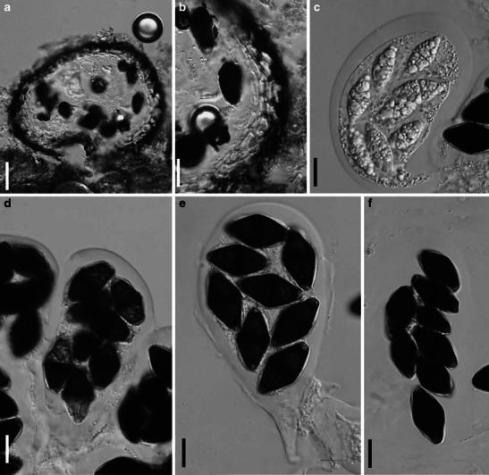
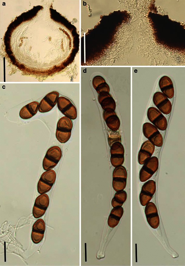
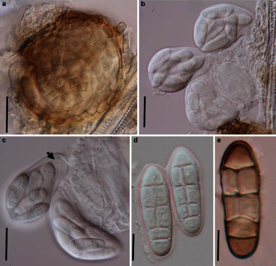
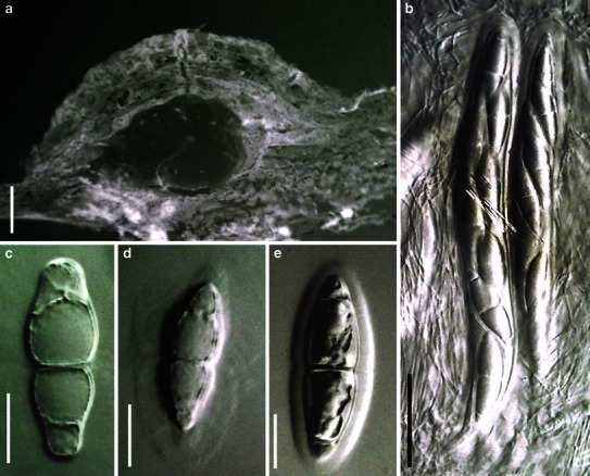
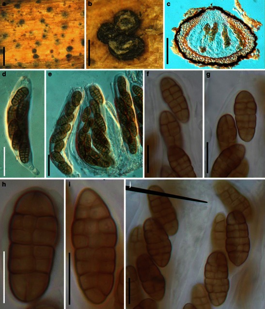
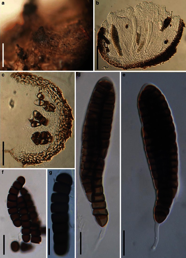
Kriegeriella mirabilis (from S reg. nr F12638, isolectotype). a Section of a superficial ascoma. b Anamorphic stage. c Obpyriform ascus. Note the pigmented ascospores and hyaline ascospores coexisted in a single ascus. d Ascospores. Scale bars: a = 50 μm, b–d = 10 μm. e Ascomata on the host surface. f, g Crashed ascoma. Note the peridium structure. h, i Hyaline asymmetric ascospores. Scale bars: e, f =100 μm, c = 50 μm, h, i = 10 μm
Kriegeriella mirabilis (from S reg. nr F12638, isolectotype). a Section of a superficial ascoma. b Anamorphic stage. c Obpyriform ascus. Note the pigmented ascospores and hyaline ascospores coexisted in a single ascus. d Ascospores. Scale bars: a = 50 μm, b–d = 10 μm. e Ascomata on the host surface. f, g Crashed ascoma. Note the peridium structure. h, i Hyaline asymmetric ascospores. Scale bars: e, f =100 μm, c = 50 μm, h, i = 10 μm
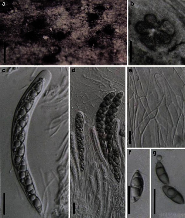
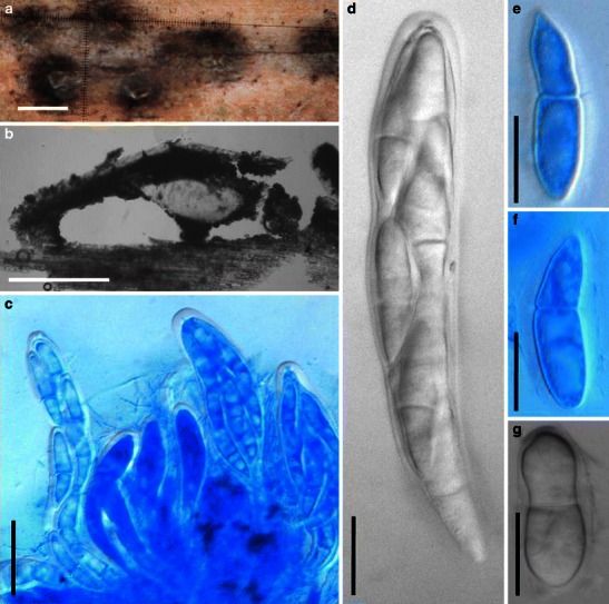
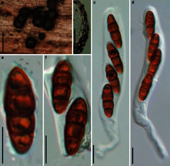
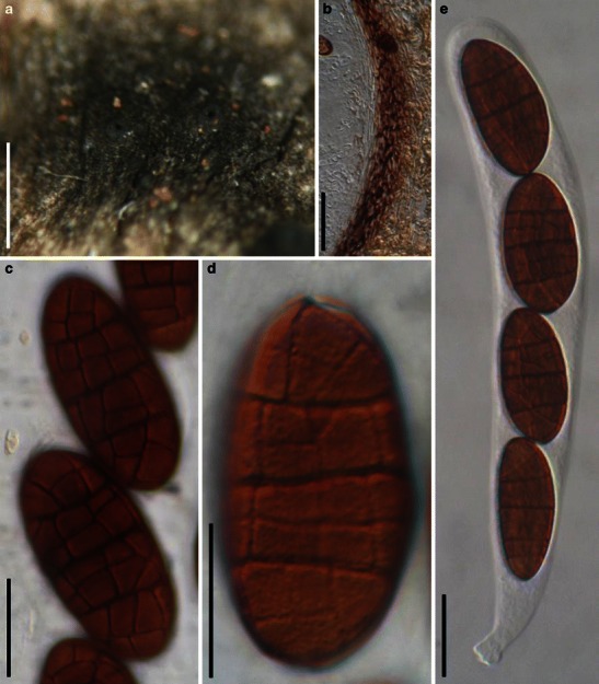
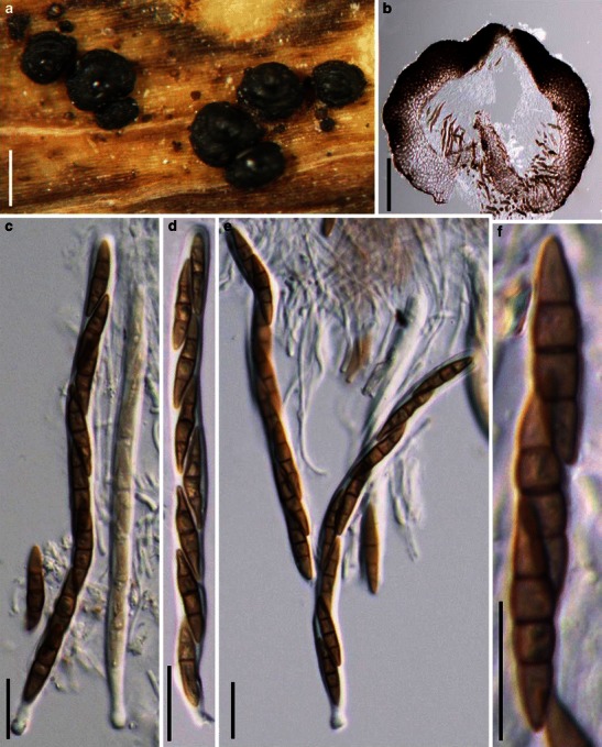
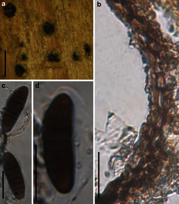
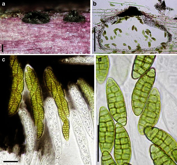
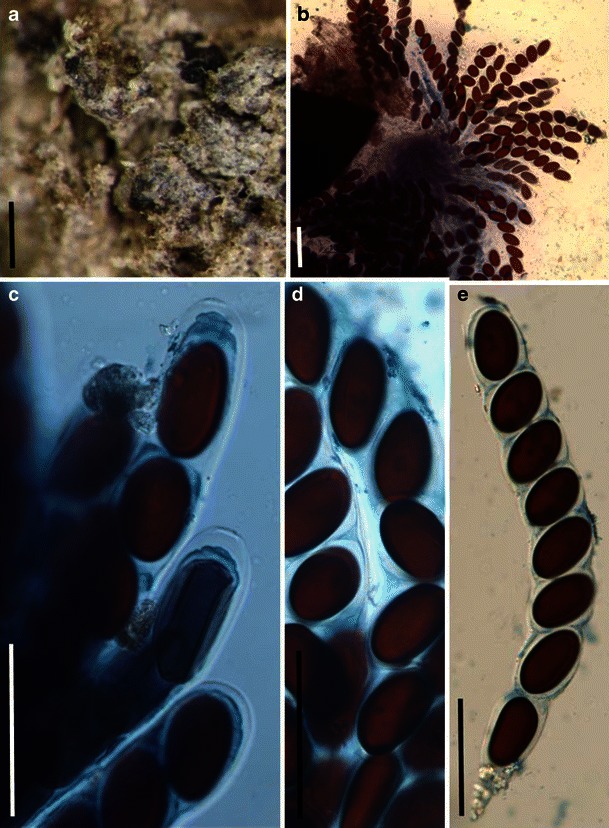
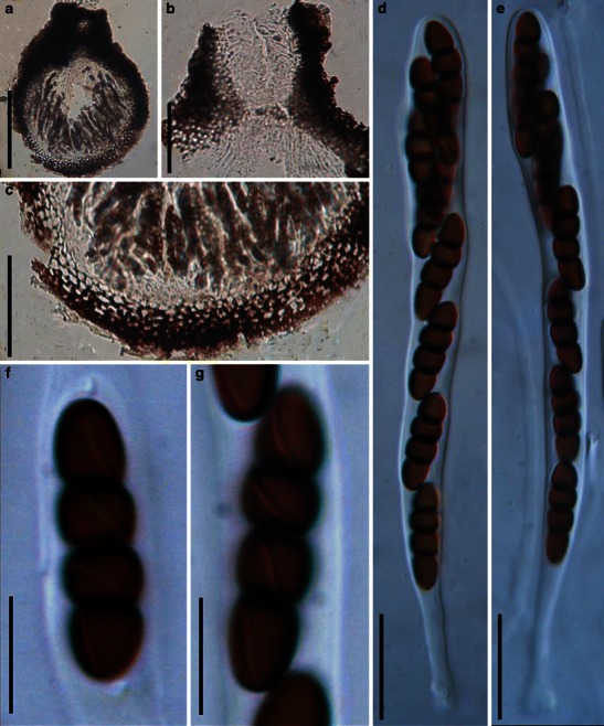
Phaeotrichum hystricinum (from TRTC 4361, holotype). a Superficial ascomata on host surface. Note the long and black appendages. b Part of peridium. Note the large cells in surface view. c–f Released reddish brown ascospores with hyaline end cells. Note the strongly constricted middle septum. Scale bars: a = 0.5 mm, b–f = 20 μm

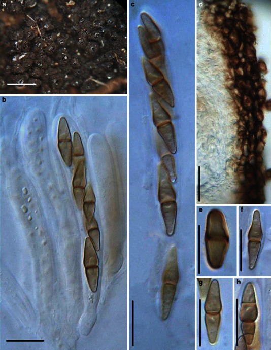
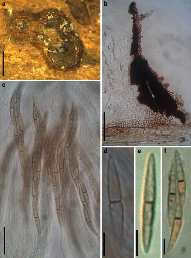
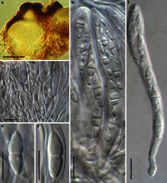
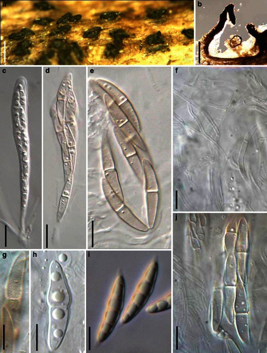
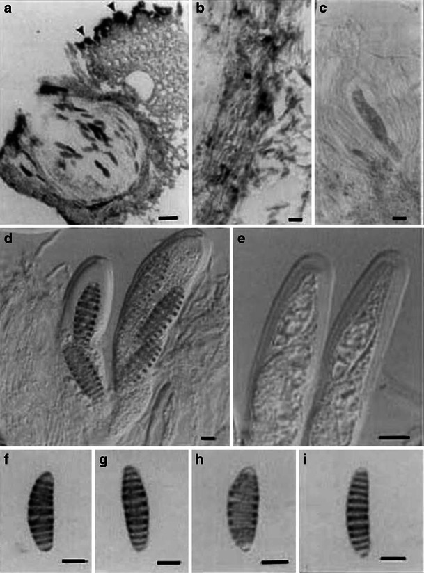
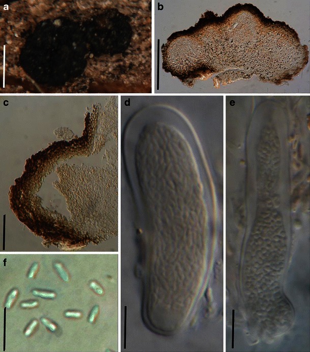
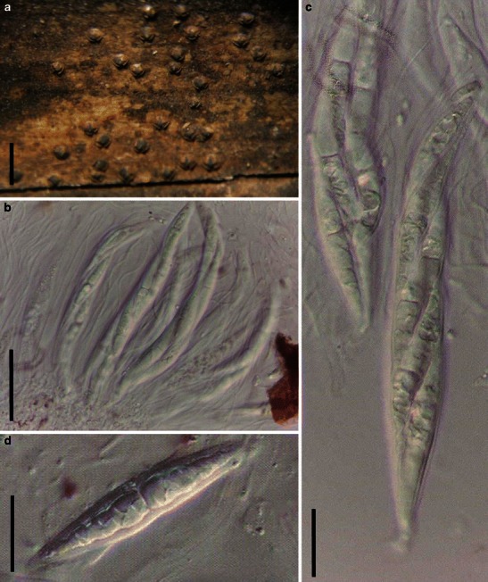
Zeuctomorpha arecae (from IMI 246067, holotype). a Gregarious ascomata on host surface. Note the numerous setae on the surface of ascomata. b Asci with ocular chamber and short peduncles. c, d Ascus with ocular chamber and knob-like pedicel. e–i One septate ascospores which are slightly asymmetrical. Scale bars: a = 0.5 mm, b–i = 20 μm
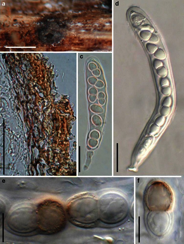
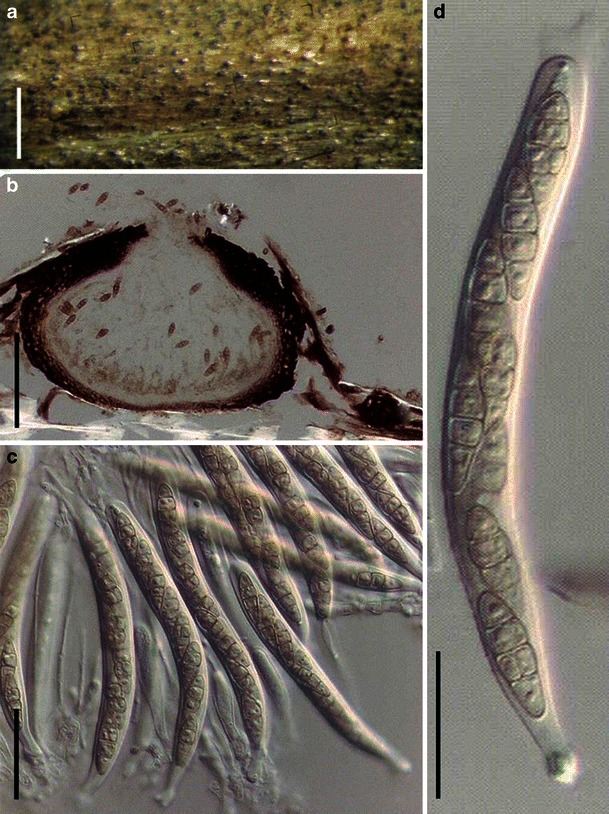
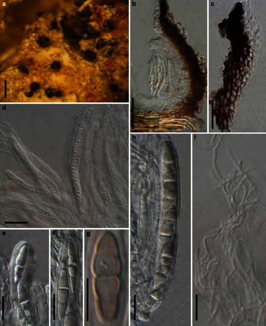
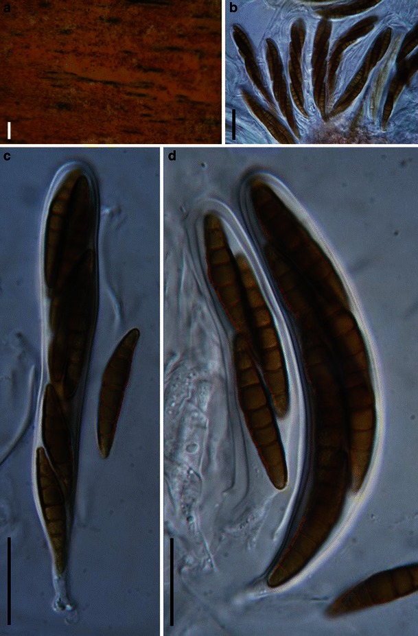
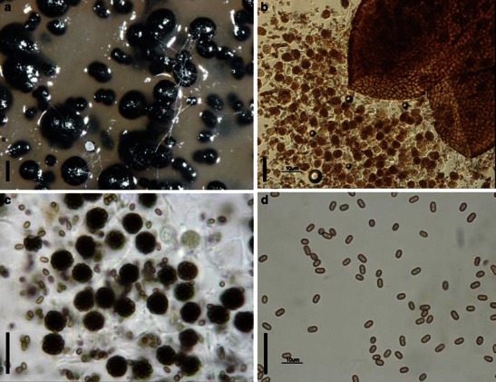
Muroia nipponica (TNS-F-230252, isotype). a Linear ascostroma parallel to the host fibers. b Crashed ascus with ascospores released. c–e Released hyaline ascospores. Scale bars: a = 5 mm, b–e = 20 μm
References
-
- Adams GC, Wingfield MJ, Common R, Roux J (2005) Phylogenetic relationships and morphology of Cytospora species from Eucalyptus. Stud Mycol 52:1–146
-
- Aguirre-Hudson B. A taxonomic study of the species referred to the ascomycete genus Leptorhaphis. Bull Br Mus Nat Hist (Bot) 1991;21:85–192.
-
- Ahmed SI, Asad F. Sporormia fimicola sp. nov. and Sporormiella inaequalis sp. nov. from West Pakistan. Sydowia. 1968;21:290–294.
-
- Ahmed SI, Cain RF. Revision of the genera Sporormia and Sporormiella. Can J Bot. 1972;50:419–478.
-
- Alias SA, Jones EBG, Torres J. Intertidal fungi from the Philippines, with a description of Acrocordiopsis sphaerica sp. nov. (Ascomycota) Fungal Divers. 1999;2:35–41.
Grants and funding
LinkOut - more resources
Full Text Sources