. 2012 Oct 5:3:184.
doi: 10.3389/fgene.2012.00184. eCollection 2012.

Molecules affecting hypothalamic control of core body temperature in response to calorie intake

Affiliations

Molecules affecting hypothalamic control of core body temperature in response to calorie intake

Tamas Bartfai et al. Front Genet. .

Abstract

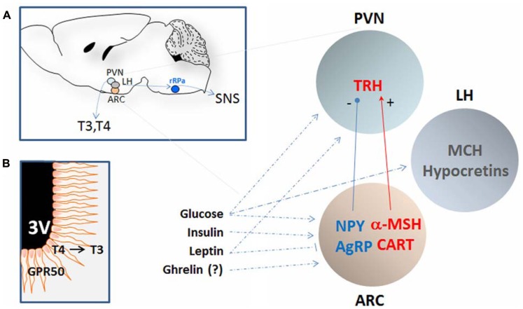
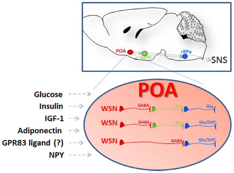
Core body temperature (CBT) and calorie intake are main components of energy homeostasis and two important regulators of health, longevity, and aging. In homeotherms, CBT can be influenced by calorie intake as food deprivation or calorie restriction (CR) lowers CBT whereas feeding has hyperthermic effects. The finding that in mice CBT prolonged lifespan independently of CR, suggested that the mechanisms modulating CBT may represent important regulators of aging. Here we summarize the current knowledge on the signaling molecules and their receptors that participate in the regulation of CBT responses to calorie intake. These include hypothalamic neuropeptides regulating feeding but also energy expenditure via modulation of thermogenesis. We also report studies indicating that nutrient signals can contribute to regulation of CBT by direct action on hypothalamic preoptic warm-sensitive neurons that in turn regulate adaptive thermogenesis and hence CBT. Finally, we show the role played by two orphans G protein-coupled receptor: GPR50 and GPR83, that were recently demonstrated to regulate temperature-dependent energy expenditure.

Keywords: GPCR; calorie restriction; core body temperature; homeostasis; hypothalamus; neuropeptides; warm-sensitive neurons.


Figures

FIGURE 1
FIGURE 1
Schematic representation of the hypothalamic nuclei and the neuropeptides that regulates calorie intake and energy expenditure by affecting core body temperature. (A) The approximate location of the periventricular nucleus (PVN), the arcuate nucleus (ARC), and the lateral hypothalamus (LH) are shown in a sagittal section of the mouse brain (left). These regions are in polysynaptic contact with the BAT (not shown) via the raphe pallidus (RPa) and can regulate CBT by affecting the sympathetic nervous system (SNS). In addition, CBT can also be affected by thyrotropin-releasing hormones (TRH) that determines the levels of circulating thyroid hormones T3 and T4. The PVN and the ARC contain neurons that can respond to nutritional state by modulating the level of specific orexigenic or anorexigenic neuropeptides. The orexigenic neuropeptide Y (NPY) and agouti-related protein (AgRP) are elevated during calorie restriction or food deprivation and are strong stimulator of appetite and reduce CBT by acting on the SNS as well as by reducing the level of TRH in the PVN. In contrast, the anorexigenic peptide α-melanocyte-stimulating hormone (α-MSH), and possibly the cocaine-and amphetamine-regulated transcript (CART), stimulates thermogenesis. The LH is also an area important in the regulation of energy homeostasis and the melanin-concentrating hormone (MCH) and the hypocretins (Hcrt) can contribute to CBT elevation at least indirectly by increasing the locomotor activity associated with food seeking behavior and possibly via BAT activation. Neurons in these nuclei are sensitive to changes in the level of glucose, leptin, insulin, and possibly ghrelin all positively correlated to CBT. (B) Schematic representation of tanycytes cells lining the third ventricle (3V) and projecting into the hypothalamus. These cells can contribute to the regulation of CBT in response to nutrient signals in at least two ways: (1) by producing of T3 from T4 thus increasing its local concentration and in doing so inhibiting the synthesis of TRH even when circulating T3 levels are low; (2) by expressing GPR50, an orphan G-coupled protein receptor proposed to serve as a regulator of adaptive thermogenesis in response to nutrient signals. See text for details and references.
FIGURE 2
FIGURE 2
Schematic representation of the localization and the organization of the nuclei and the cells participating to central thermoregulation. Lesions studies indicate that the hypothalamic preoptic area (POA) exerts the function of a bona fide thermostat allowing sensing and proper thermoregulatory responses to local as well as peripheral temperature changes (afferent pathways are not shown). In addition to respond to changes in temperature the POA can also sense nutrient signals: POA injection of insulin, IGF-1, and adiponectin were followed by hyperthermia via BAT activation while treatment with NPY or downregulation of the G-protein coupled receptor 83 (GPR83) induced hypothermia. The receptors for some of these ligands, as well as GPR83, were demonstrated in the POA warm-sensitive neurons (WSN). These specialized GABAergic cells exert a tonic inhibition on the raphe pallidus (aRP) either directly or through neurons in the dorsomedial hypothalamus (DMH) and control thermogenesis by activation of brown adipose tissue (BAT), muscular shivering, or the regulation of vasodilation (scheme adapted from Morrison and Nakamura, 2011). WSN are one component of the POA thermoregulatory neurocircuitry that comprises also temperature insensitive and cold-sensitive neurons that might also participate in the POA responses to nutrients (not shown in this scheme).

References

    1. Adachi A., Shimizu N., Oomura Y., Kobashi M. (1984). Convergence of hepatoportal glucose-sensitive afferent signals to glucose-sensitive units within the nucleus of the solitary tract. Neurosci. Lett. 46 215–218 - PubMed
    1. Adamantidis A, de Lecea L. (2009). The hypocretins as sensors for metabolism and arousal. J. Physiol. 587 33–40 - PMC - PubMed
    1. Adams F., Grassie M., Shahid M., Hill D. R., Henry B. (2003). Acute oral dexamethasone administration reduces levels of orphan GPCR glucocorticoid-induced receptor (GIR) mRNA in rodent brain: potential role in HPA-axis function. Brain Res. Mol. Brain Res. 117 39–46 - PubMed
    1. Ahima R. S. (2000). Leptin and the neuroendocrinology of fasting. Front. Horm. Res. 26 42–56 - PubMed
    1. Ahima R. S., Prabakaran D., Mantzoros C., Qu D., Lowell B., Maratos-Flier E., et al. (1996). Role of leptin in the neuroendocrine response to fasting. Nature 382 250–252 - PubMed

LinkOut - more resources