. 2012 Sep;9(3):258-68.
doi: 10.3724/SP.J.1263.2012.02292.

Salubrinal protects against tunicamycin and hypoxia induced cardiomyocyte apoptosis via the PERK-eIF2α signaling pathway

Affiliations

Salubrinal protects against tunicamycin and hypoxia induced cardiomyocyte apoptosis via the PERK-eIF2α signaling pathway

Chun-Lei Liu et al. J Geriatr Cardiol. 2012 Sep.

Abstract

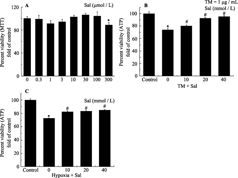
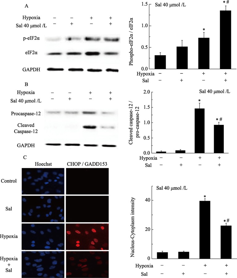
Objectives: This study examined the protective effect of salubrinal and the mechanism underlying this protection against tunicamycin (TM)- and hypoxia-induced apoptosis in rat cardiomyocytes.

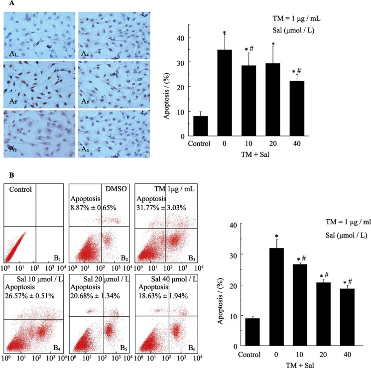
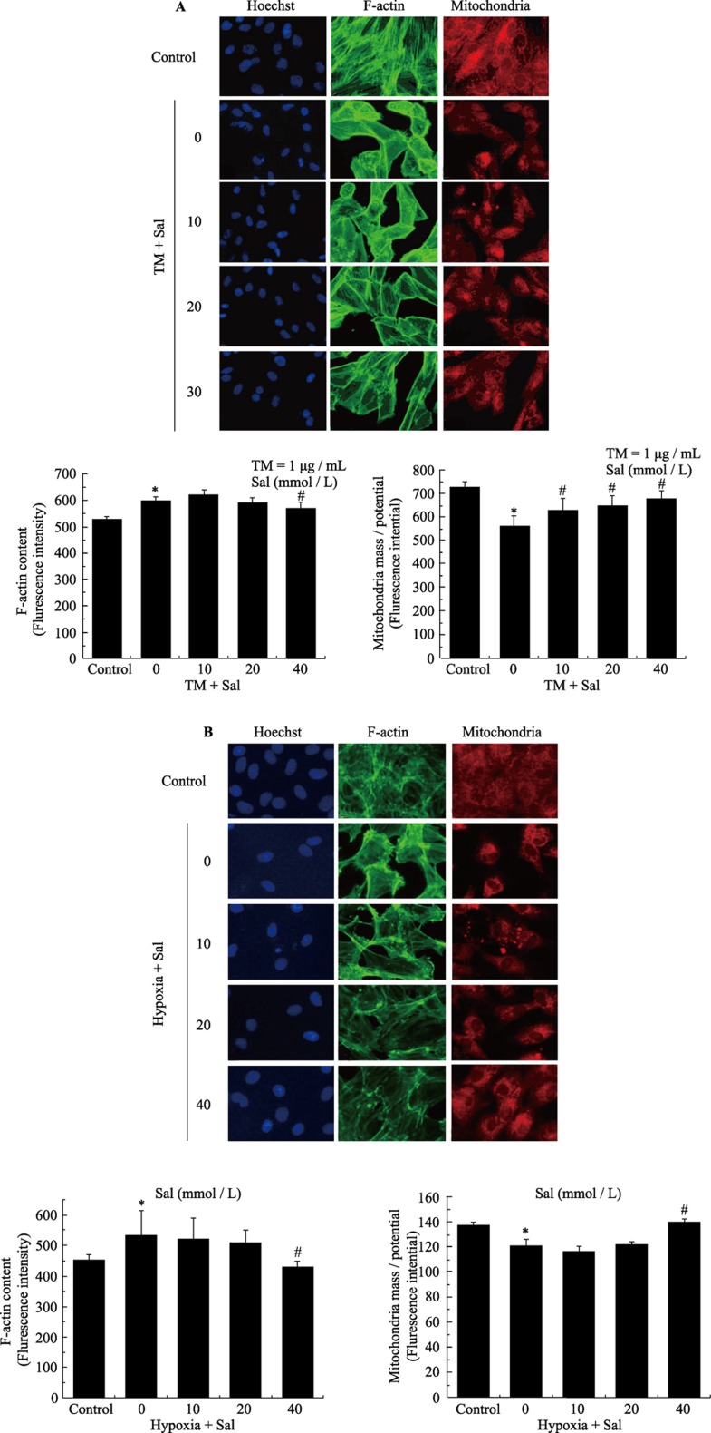
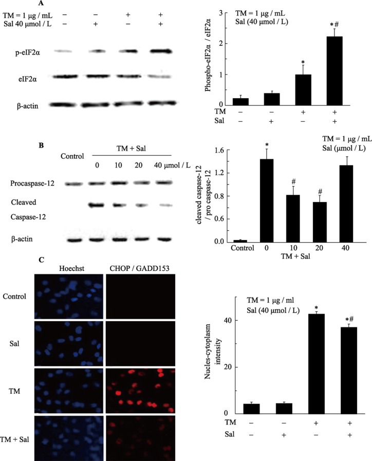
Methods: Neonatal rat cardiomyocytes were cultured from the ventricles of 1-day-old Wistar rats. Cells were exposed to different concentrations of salubrinal (10, 20, and 40 µmol/L) for 30 min followed by TM treatment or hypoxia for 36 h. Apoptosis was measured by a multiparameter HCS (high content screening) apoptosis assay, TUNEL assay and flow cytometry. The phosphorylation of eukaryotic translation initiation factor 2 subunit alpha (eIF2α) and the expression of cleaved caspase-12 were determined by Western blotting. C/EBP homologous protein (CHOP) was detected by immunocytochemistry.

Results: HCS, TUNEL assays and flow cytometry showed that salubrinal protected cardiomyocytes against apoptosis induced by TM or hypoxia. Western blotting showed that salubrinal protected cardiomyocytes against apoptosis by inducing eIF2α phosphorylation and down-regulating the expression of the endoplasmic reticulum stress-mediated apoptotic proteins, CHOP and cleaved caspase-12.

Conclusions: Our study suggests that salubrinal protects rat cardiomyocytes against TM- or hypoxia-associated apoptosis via a mechanism involving the inhibition of ER stress-mediated apoptosis.

Keywords: Apoptosis; Cell protection; Endoplasmic reticulum stress; Rat cardiomyocytes; Salubrinal.


Figures

Figure 1.
Figure 1.. Salubrinal mediates concentration-dependent protection against cytotoxicity in cardiomyocytes.
(A): Cultured neonatal rat cardiomyocytes were exposed to the indicated concentrations of salubrinal alone for 36 h. *P < 0.05 vs. control. (B): Cultured neonatal rat cardiomyocytes were exposed to TM (1 µg/mL) for 36 h in the presence or absence of the indicated concentrations of salubrinal for 30 min. The cell viability was then measured as described in Methods. *P < 0.001 vs. control; #P < 0.05 vs. TM. (C): Cultured neonatal rat cardiomyocytes were exposed to hypoxia for 36 h in the presence or absence of the indicated concentrations of salubrinal for 30 min. The cell viability was then measured as described in Materials and Methods. *P < 0.05 vs. control. #P < 0.05 vs. hypoxia. ATP: adenosine triphosphate; MTT: methylthiazoletetrazolium; Sal: salubrinal; TM: tunicamycin.
Figure 2.
Figure 2.. Salubrinal reduces TM-induced apoptosis in cardiomyocytes as demonstrated by TUNEL and flow cytometry assays.
(A): Cardiomyocytes were treated with tunicamycin (TM) (1 µg/mL) in the presence (A4: 10 µmol/L, A5: 20 µmol/L, A6: 40 µmol/L) or absence (A3) of salubrinal for 36 h. The negative control (A1) and positive control (A2) cells were analyzed by TUNEL assay. The results are presented as the means ± SE of three experiments. *P < 0.001 vs. control. #P < 0.05 vs. TM. (B): Cardiomyocytes treated with TM (1 µg/mL) in the presence (B4: 10 µmol/L, B5: 20 µmol/L, B6: 40 µmol/L) or absence (B3) of different concentrations of salubrinal for 24 h. Cells were not treated with either TM or salubrinal in (B1, B2). Apoptosis was assessed by flow cytometry. The results are the means ± SE of three experiments. *P < 0.001 vs. control; *P < 0.05 vs. TM. DMSO: Dimethylsulfoxide; Sal: salubrinal; TM: tunicamycin.
Figure 3.
Figure 3.. Salubrinal reduces TM- and hypoxia-induced apoptosis in cardiomyocytes as demonstrated by a multi-parameter HCS apoptosis assay.
(A): Analysis of apoptosis in response to TM-treatment of cardiomyocytes using the ArrayScan HCS Reader. Cardiomyocytes were exposed to TM (1 µg/mL) in the presence or absence of salubrinal. The cells were then fixed and stained with Hoechst 33342 to analyze nuclear morphology, Alexa fluor 488-phalloidin to analyze F-actin content and MitoTracker Red to analyze mitochondrial mass/potential. Apoptotic parameters were quantified by analyzing the stained cells on a Thermo Scientific ArrayScan HCS Reader. Dose-response plots for F-actin content and mitochondrial mass/potential. *P < 0.05 vs. control; #P < 0.05 vs. TM. (B): Measurement of apoptosis in response to hypoxia in cardiomyocytes on the ArrayScan HCS Reader. The cardiomyocytes were exposed to hypoxia in the presence or absence of salubrinal. The cells were then fixed and stained with Hoechst 33342 to analyze nuclear morphology, Alexa fluor 488-phalloidin to analyze F-actin content and MitoTracker Red to analyze mitochondrial mass/potential. Apoptotic parameters were quantified by analyzing the stained cells on a Thermo Scientific ArrayScan HSC Reader. Dose-response plots for F-actin content and mitochondrial mass/potential. *P < 0.05 vs. control. #P < 0.05 vs. hypoxia. HCS: high content screening; Sal: salubrinal; TM: tunicamycin.
Figure 4.
Figure 4.. Salubrinal activates eIF-2 expression and attenuates TM induced CHOP and caspase-12 expression in cardiomyocytes.
(A): Salubrinal maintains eIF-2α phosphorylation. Cardiomyocytes were exposed to tunicamycin (TM) (1 µg/mL) in the presence or absence of salubrinal for four hours. The expression of eIF2α and p-eIF2α was analyzed by Western Blotting using anti-eIF2α and anti-p-eIF2α antibodies. *P < 0.001 vs. control; #P < 0.01 vs. TM. (B): Salubrinal regulates the expression of caspase-12. Cardiomyocytes were exposed to TM (1 µg/mL) in the presence or absence of salubrinal for eight hours. Caspase-12 protein expression was analyzed by western blotting with anti-caspase-12 antibodies. *P < 0.001 vs. control. #P < 0.01 vs. TM. (C): Measurement of CHOP expression in cardiomyocytes with the ArrayScan HSC Reader. The cardiomyocytes were exposed to TM (1 µg/mL) in the presence or absence of salubrinal and then fixed. The cells were then stained with Hoechst 33342 to analyze nuclear morphology, and with CHOP antibody followed by a DyLight 488 secondary antibody to quantify CHOP expression. *P < 0.05 vs. control; #P < 0.05 vs. hypoxia. CHOP: C/EBP homologous protein; Sal: salubrinal; TM: tunicamycin.
Figure 5.
Figure 5.. Salubrinal activates eIF-2α expression and attenuates hypoxia induced CHOP and caspase-12 expression in cardiomyocytes.
(A): Salubrinal maintains eIF-2α phosphorylation. Cardiomyocytes were exposed to hypoxia in the presence or absence of salubrinal for four hours. The expression of eIF2α and p-eIF2α was analyzed by Western Blotting using anti-eIF2α, anti-p-eIF2α antibodies. *P < 0.001 vs. control; #P < 0.01 vs. hypoxia. (B): Salubrinal regulates the expression of caspase-12. Cardiomyocytes were exposed to hypoxia in the presence or absence of salubrinal for eight hours. Caspase-12 protein expression was analyzed by Western Blotting using anti-caspase-12 antibodies. *P < 0.001 vs. control. #P < 0.01 vs. hypoxia. (C): Measurement of CHOP expression in cardiomyocytes with the ArrayScan HCS Reader. The cardiomyocytes were exposed to hypoxia in the presence or absence of salubrinal. The cells were then fixed and stained with Hoechst 33342 to analyze nuclear morphology, and with CHOP antibody followed by a DyLight 488 secondary antibody to quantify CHOP expression. *P < 0.05 vs. control; #P < 0.05 vs. hypoxia. CHOP: C/EBP homologous protein; HCS: high content screening; Sal: salubrinal; TM: tunicamycin.

References

    1. Chiu CZ, Wang BW, Chung TH, et al. Angiotensin II and the ERK pathway mediate the induction of myocardin by hypoxia in cultured rat neonatal cardiomyocytes. Clin Sci (Lond) 2010;119:273–282. - PMC - PubMed
    1. Calmettes G, Deschodt-Arsac V, Gouspillou G, et al. Improved energy supply regulation in chronic hypoxic mouse counteracts hypoxia-induced altered cardiac energetics. PLoS One. 2010;5:e9306. - PMC - PubMed
    1. Tajiri S, Oyadomari S, Yano S, et al. Ischemia-induced neuronal cell death is mediated by the endoplasmic reticulum stress pathway involving CHOP. Cell Death Differ. 2004;11:403–415. - PubMed
    1. Terai K, Hiramoto Y, Masaki M, et al. AMP-activated protein kinase protects cardiomyocytes against hypoxic injury through attenuation of endoplasmic reticulum stress. Mol Cell Biol. 2005;25:9554–9575. - PMC - PubMed
    1. Kaufman RJ. Stress signaling from the lumen of the endoplasmic reticulum: coordination of gene transcriptional and translational controls. Genes Dev. 1999;13:1211–1233. - PubMed

LinkOut - more resources