. 2012 Oct 26:9:246.
doi: 10.1186/1743-422X-9-246.

Whole genome sequencing of phage resistant Bacillus anthracis mutants reveals an essential role for cell surface anchoring protein CsaB in phage AP50c adsorption

Affiliations

Whole genome sequencing of phage resistant Bacillus anthracis mutants reveals an essential role for cell surface anchoring protein CsaB in phage AP50c adsorption

Kimberly A Bishop-Lilly et al. Virol J. .

Abstract

Background: Spontaneous Bacillus anthracis mutants resistant to infection by phage AP50c (AP50R) exhibit a mucoid colony phenotype and secrete an extracellular matrix.

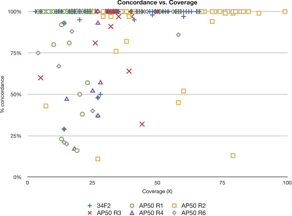
Methods: Here we utilized a Roche/454-based whole genome sequencing approach to identify mutations that are candidates for conferring AP50c phage resistance, followed by genetic deletion and complementation studies to validate the whole genome sequence data and demonstrate that the implicated gene is necessary for AP50c phage infection.

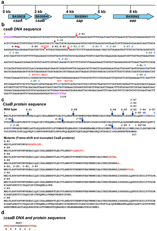
Results: Using whole genome sequence data, we mapped the relevant mutations in six AP50R strains to csaB. Eleven additional spontaneous mutants, isolated in two different genetic backgrounds, were screened by PCR followed by Sanger sequencing of the csaB gene. In each spontaneous mutant, we found either a non-synonymous substitution, a nonsense mutation, or a frame-shift mutation caused by single nucleotide polymorphisms or a 5 base pair insertion in csaB. All together, 5 and 12 of the 17 spontaneous mutations are predicted to yield altered full length and truncated CsaB proteins respectively. As expected from these results, a targeted deletion or frame-shift mutations introduced into csaB in a different genetic background, in a strain not exposed to AP50c, resulted in a phage resistant phenotype. Also, substitution of a highly conserved histidine residue with an alanine residue (H270A) in CsaB resulted in phage resistance, suggesting that a functional CsaB is necessary for phage sensitivity. Conversely, introduction of the wild type allele of csaB in cis into the csaB deletion mutant by homologous recombination or supplying the wild type CsaB protein in trans from a plasmid restored phage sensitivity. The csaB mutants accumulated cell wall material and appeared to have a defective S-layer, whereas these phenotypes were reverted in the complemented strains.

Conclusions: Taken together, these data suggest an essential role for csaB in AP50c phage infection, most likely in phage adsorption. (The whole genome sequences generated from this study have been submitted to GenBank under SRA project ID: SRA023659.1 and sample IDs: AP50 R1: SRS113675.1, AP50 R2: SRS113676.1, AP50 R3: SRS113728.1, AP50 R4: SRS113733.1, AP50 R6: SRS113734.1, JB220 Parent: SRS150209.1, JB220 Mutant: SRS150211.1).


Figures

Figure 1
Figure 1
Filtration of true positive variations from false positive variations by percent concordance and coverage. The graph displays 260 high quality (HQ) variations in the genome sequences (34F2 and its derivatives) produced by GS Reference Mapper software as a function of percentage concordance. The graph indicates all the high-quality variations in the six genomic sequences obtained in this work. The variations from the six strains are indicated by different symbols. AP50 R1, AP50 R2, AP50 R3, AP50 R4 and AP50 R6 are S-R1, S-R2, S-R3, S-R4 and S-R6 respectively.
Figure 2
Figure 2
Genetic map of csa operon, DNA and protein sequences of csaB.a) Genetic map of the csaB region on B. anthracis chromosome. The block arrows indicate the genes and the direction of transcription of the different genes. b) Nucleotide sequence of the wild type csaB gene and the location and nucleotide changes in the AP50R mutants. Insertions and deletions are indicated by the red arrowheads and inverted arrowheads respectively. The various strains are indicated next to the mutated sites: derivatives of Sterne 34F2 and JB220 are indicated by the prefixes S and J respectively. The mutant loci labeled in black were discovered by WGS and those in green by Sanger sequencing of csaB PCR fragments. The altered bases are indicated in blue with the changed base above the wild type. A693C and H270A mutations were genetically engineered mutants. The sequences at the beginning and end of the gene marked in pink are the junctions the csaB deletion mutant. c) Amino acid sequence of wild type and mutant CsaB proteins. The sites of truncation of CsaB protein due to frame-shift and nonsense mutations are indicated by blue downward arrows. The amino acid changes in non-synonymous mutants are indicated in blue above the wild type residue. The amino acid sequences of the frame-shift and truncated proteins are shown. The residues in red are the additional residues added due to the frame-shift before truncation of the mutant proteins. d) DNA and protein sequence of the csaB gene in csaB deletion mutant.
Figure 3
Figure 3
Phage sensitivity spot test on Sterne 7702 and various derivatives. For each row the left spot was spotted with medium (M), the middle spot received AP50c (A) and the right spot was spotted with γ (G). The clearing in the bacterial spot indicates infection and killing of bacteria. In rows 3 through 6, the γ spots completely cleared the bacterial spots. The various strains are as follows: 1) and 2) BA663 (wild type); 3) BAP350 (ΔcsaB); 4) BAP356 (csaB ∇G234); 5) BAP366 (csaB ∇G46); 6) BAP411 (csaB ∇GCTTA219); 7) BAP435 (csaB ΔA203); 8) BAP503 (csaB H270A); 9) BAP533 (csaB A693C); 10) BAP553 (ΔBAS3946); 11) BAP560 (BAS3946 G1024A); 12) BGBN 001 (ΔcsaB/pBGBN1003)
Figure 4
Figure 4
Amino acid sequence alignment of csaB orthologs. BAS0840 was aligned with the Pfam PF04230 seed alignment, and percent conservation was determined as described in Methods. Only the alignment of the amino acids in the vicinity of histidine 270 of CsaB protein is shown. The full-length alignment is provided in Additional file 1: Figure S1.
Figure 5
Figure 5
Complementation of csaB mutation.a) PCR verification of XmaI mutant csaB. The wild-type sequence of csaB was restored in the ΔcsaB strain with the addition of a silent mutation creating an XmaI site (see Methods). The region was amplified by PCR, and PCR products were purified and digested with XmaI. The lane labeled BA663 indicates the csaB PCR product from wild type strain (7702) after digestion with XmaI, whereas the lane labeled BAP533 is the PCR product from the mutant showing the two bands after digestion with XmaI. b) Genetic map of csaB complementing plasmid pNBGD1003. pNBGD1003 is a shuttle vector derived from pSW4. The locations of the Gram positive and Gram negative replicons (from pUB110 and ColE1, respectively), antibiotic resistance genes aadD and bla, origin of conjugational transfer oriT, constitutively expressed B. anthracis pagA promoter (labeled PA promoter) and the csaB gene downstream of the promoter are indicated.
Figure 6
Figure 6
Transmission electron microscopic images of wt and ∆csaB mutants and the complemented strains.a) BA663 (csaB+); b) BA663+AP50c; c) BAP350 (∆csaB); d) BAP350 (∆csaB)+AP50c; e) BAP533 (csaB+XmaI); f) BAP533+AP50c; g) BAP350 (∆csaB)+ pBGBN 1003 (csaB+); h) BAP350 (∆csaB)+ pBGBN 1003 (csaB+)+AP50c. The scale bar is in the bottom right corner of each figure.

References

    1. Labrie SJ, Samson JE, Moineau S. Bacteriophage resistance mechanisms. Nat Rev Microbiol. 2010;8(5):317–327. doi: 10.1038/nrmicro2315. - DOI - PubMed
    1. Stern A, Sorek R. The phage-host arms race: Shaping the evolution of microbes. Bioessays. 2011;33(1):43–51. doi: 10.1002/bies.201000071. - DOI - PMC - PubMed
    1. Hagens S, Loessner MJ. Application of bacteriophages for detection and control of foodborne pathogens. Appl Microbiol Biotechnol. 2007;76(3):513–519. doi: 10.1007/s00253-007-1031-8. - DOI - PubMed
    1. McAuliffe ORR, Fitzgerald GF. The new phage biology: from genomics to applications. Norfolk, UK: Caister Academic Press; 2007.
    1. McKinstry MER. Phages: their role in bacterial pathogenesis and biotechnology Use of phages in therapy and bacterial detection. Washington DC: ASM press; 2005.

MeSH terms

Substances