. 2012 Oct 26;16(5):R209.
doi: 10.1186/cc11841.

Mechanisms underlying ICU muscle wasting and effects of passive mechanical loading

Mechanisms underlying ICU muscle wasting and effects of passive mechanical loading

Monica Llano-Diez et al. Crit Care. .

Abstract

Introduction: Critically ill ICU patients commonly develop severe muscle wasting and impaired muscle function, leading to delayed recovery, with subsequent increased morbidity and financial costs, and decreased quality of life for survivors. Critical illness myopathy (CIM) is a frequently observed neuromuscular disorder in ICU patients. Sepsis, systemic corticosteroid hormone treatment and post-synaptic neuromuscular blockade have been forwarded as the dominating triggering factors. Recent experimental results from our group using a unique experimental rat ICU model show that the mechanical silencing associated with CIM is the primary triggering factor. This study aims to unravel the mechanisms underlying CIM, and to evaluate the effects of a specific intervention aiming at reducing mechanical silencing in sedated and mechanically ventilated ICU patients.

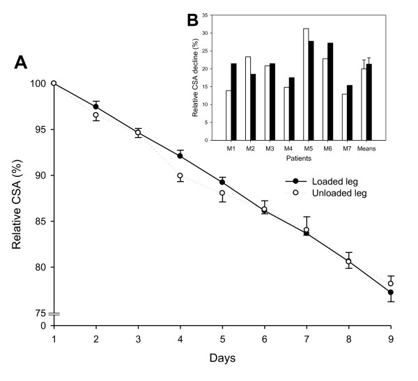
Methods: Muscle gene/protein expression, post-translational modifications (PTMs), muscle membrane excitability, muscle mass measurements, and contractile properties at the single muscle fiber level were explored in seven deeply sedated and mechanically ventilated ICU patients (not exposed to systemic corticosteroid hormone treatment, post-synaptic neuromuscular blockade or sepsis) subjected to unilateral passive mechanical loading for 10 hours per day (2.5 hours, four times) for 9 ± 1 days.

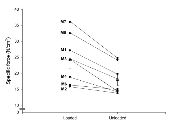
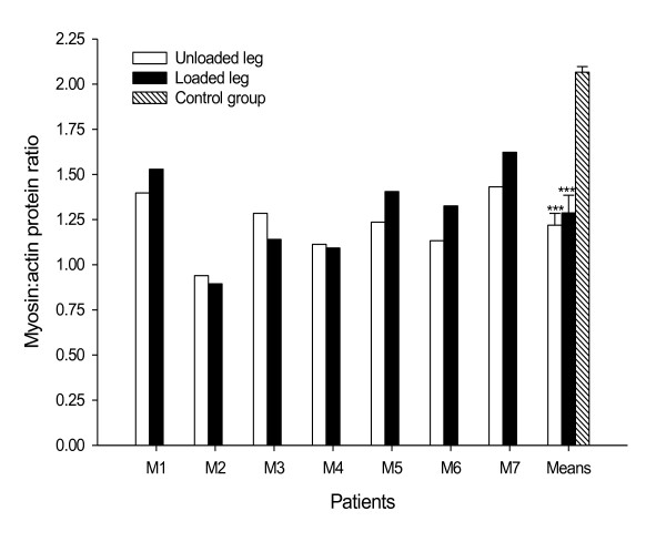
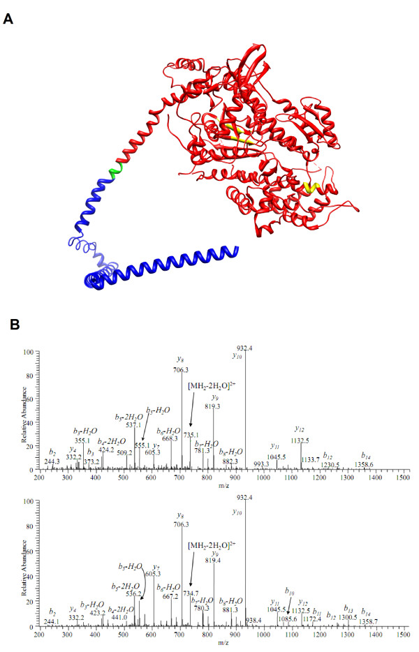
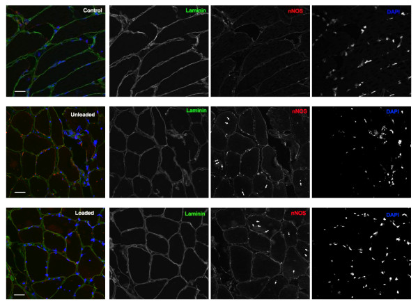
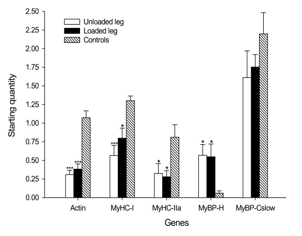
Results: These patients developed a phenotype considered pathognomonic of CIM; that is, severe muscle wasting and a preferential myosin loss (P < 0.001). In addition, myosin PTMs specific to the ICU condition were observed in parallel with an increased sarcolemmal expression and cytoplasmic translocation of neuronal nitric oxide synthase. Passive mechanical loading for 9 ± 1 days resulted in a 35% higher specific force (P < 0.001) compared with the unloaded leg, although it was not sufficient to prevent the loss of muscle mass.

Conclusion: Mechanical silencing is suggested to be a primary mechanism underlying CIM; that is, triggering the myosin loss, muscle wasting and myosin PTMs. The higher neuronal nitric oxide synthase expression found in the ICU patients and its cytoplasmic translocation are forwarded as a probable mechanism underlying these modifications. The positive effect of passive loading on muscle fiber function strongly supports the importance of early physical therapy and mobilization in deeply sedated and mechanically ventilated ICU patients.


Figures

Figure 1
Figure 1
Ultrasound measurements of tibialis anterior cross-sectional area. (A) Relative cross-sectional area (CSA) during the intervention period (9 ± 1 days). Solid line, loaded side; dashed line, unloaded side. Values are mean ± standard error of the mean (SEM). The value at day 1 is equivalent to 100%. (B) Relative CSA decline in the loaded leg (black bars) and the unloaded leg (white bars) for each ICU patient on the final day of the intervention, and mean ± SEM for all patients.
Figure 2
Figure 2
Specific force in single muscle fibers. Specific force in single muscle fibers expressing the type I myosin heavy chain isoform in the loaded and unloaded legs from patients exposed to unilateral loading and mechanical ventilation for 9 ± 1 days. Black circles, individual means; open triangles, average for all patients pooled together ± standard error of the mean.
Figure 3
Figure 3
Myosin:actin protein ratios in unloaded and loaded legs. Myosin:actin protein ratios in tibialis anterior muscle cross-sections from each mechanically ventilated ICU patient in the unloaded leg (white bars) and the loaded leg (black bars), and from healthy controls (hashed bar). Mean ± standard error of the mean given for patients in the loaded and unloaded legs as well as in healthy controls. ***Statistically significant differences versus the healthy control group (P < 0.001).
Figure 4
Figure 4
Post-translational modifications of the myosin motor domain. (A) Ribbon diagram of the myosin motor domain (red) and part of the tail region (blue). Three ICU-condition-specific post-translational modifications (PTMs) were observed in the motor domain of the protein (yellow) and one lost modification was in the tail region (green). Since only part of the myosin protein is modeled, PTMs located further down on the tail region are not shown. (B) Upper panel, spectrum of deamidated peptide ENQSILITGESGAGK; bottom panel, spectrum of intact peptide ENQSILITGESGAGK. The deamidation is safely determined by high-resolution mass spectrometry (MS) in MS mode (the mass difference between the molecular ion masses is 0.984 Da, corresponding to N+H-O) and in MS/MS mode as well. In the MS/MS spectrum one can see the mass shift of +1 Da for all b-ion series, including those that correspond to neutral losses from b-ions, except b2. Since b2 ion has the same mass for both peptides and contains asparagine, the deamidation is assigned to glutamine Gln3 and not Asn2.
Figure 5
Figure 5
Cross-sections of tibialis anterior muscle stained for neuronal nitric oxide synthase, Laminin and 4',6-diamidino-2-phenylindol. Cross-sections of tibialis anterior muscle stained for neuronal nitric oxide synthase (nNOS; red), Laminin (green) and 4',6-diamidino-2-phenylindol (DAPI; blue). In control, basal expression of nNOS is low and localized to the sarcolemma. ICU conditions induce strong expression of nNOS and dislocation of nNOS to the cytoplasm (arrowheads). Scale bar = 50 μm. The overlay image for each row is shown to the left.
Figure 6
Figure 6
Myofibrillar mRNA expression. Actin, myosin heavy chain (MyHC; types I and IIa), myosin binding protein (MyBP)-H, and MyBP-Cslow mRNA expression in the unloaded leg (white bar) and the loaded leg (black bar) from ICU patients, and in the tibialis anterior muscles from healthy controls (hashed bar). Values are starting quantity ± standard error of the mean. Statistical significance versus healthy control group: *P < 0.05 and ***P < 0.001.

References

    1. Cheung AM, Tansey CM, Tomlinson G, Diaz-Granados N, Matte A, Barr A, Mehta S, Mazer CD, Guest CB, Stewart TE, Al-Saidi F, Cooper AB, Cook D, Slutsky AS, Herridge MS. Two-year outcomes, health care use and costs in survivors of ARDS. Am J Respir Crit Care Med. 2006;16:538–544. - PubMed
    1. Hudson LD, Lee CM. Neuromuscular sequelae of critical illness. N Engl J Med. 2003;16:745–747. doi: 10.1056/NEJMe020180. - DOI - PubMed
    1. Herridge MS, Cheung AM, Tansey CM, Matte-Martyn A, Diaz-Granados N, Al-Saidi F, Cooper AB, Guest CB, Mazer CD, Mehta S, Stewart TE, Barr A, Cook D, Slutsky AS. One-year outcomes in survivors of the acute respiratory distress syndrome. N Engl J Med. 2003;16:683–693. doi: 10.1056/NEJMoa022450. - DOI - PubMed
    1. Herridge MS, Tansey CM, Matte A, Tomlinson G, Diaz-Granados N, Cooper A, Guest CB, Mazer CD, Mehta S, Stewart TE, Kudlow P, Cook D, Slutsky AS, Cheung AM. Functional disability 5 years after acute respiratory distress syndrome. N Engl J Med. 2011;16:1293–1304. doi: 10.1056/NEJMoa1011802. - DOI - PubMed
    1. Norman H, Zackrisson H, Hedstrom Y, Andersson P, Nordquist J, Eriksson LI, Libelius R, Larsson L. Myofibrillar protein and gene expression in acute quadriplegic myopathy. J Neurol Sci. 2009;16:28–38. doi: 10.1016/j.jns.2009.04.041. - DOI - PMC - PubMed

Publication types

MeSH terms