. 2012 Nov;7(11):2056-66.
doi: 10.1038/nprot.2012.127. Epub 2012 Oct 25.

Stiffness-controlled three-dimensional extracellular matrices for high-resolution imaging of cell behavior

Affiliations

Stiffness-controlled three-dimensional extracellular matrices for high-resolution imaging of cell behavior

Robert S Fischer et al. Nat Protoc. 2012 Nov.

Abstract

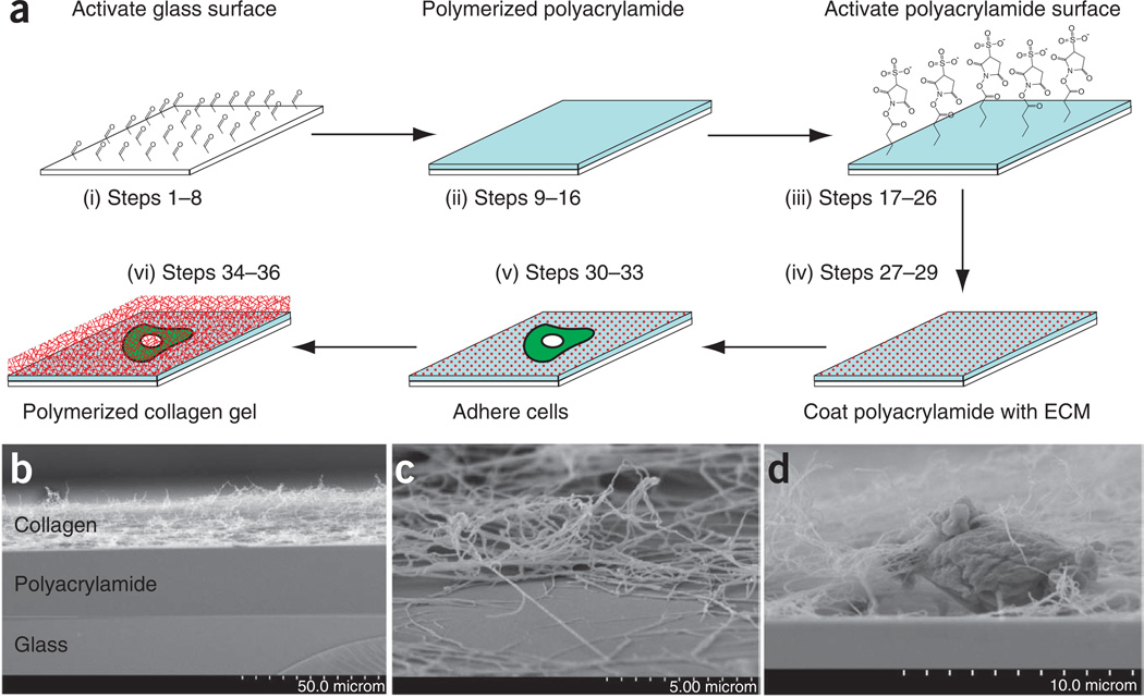
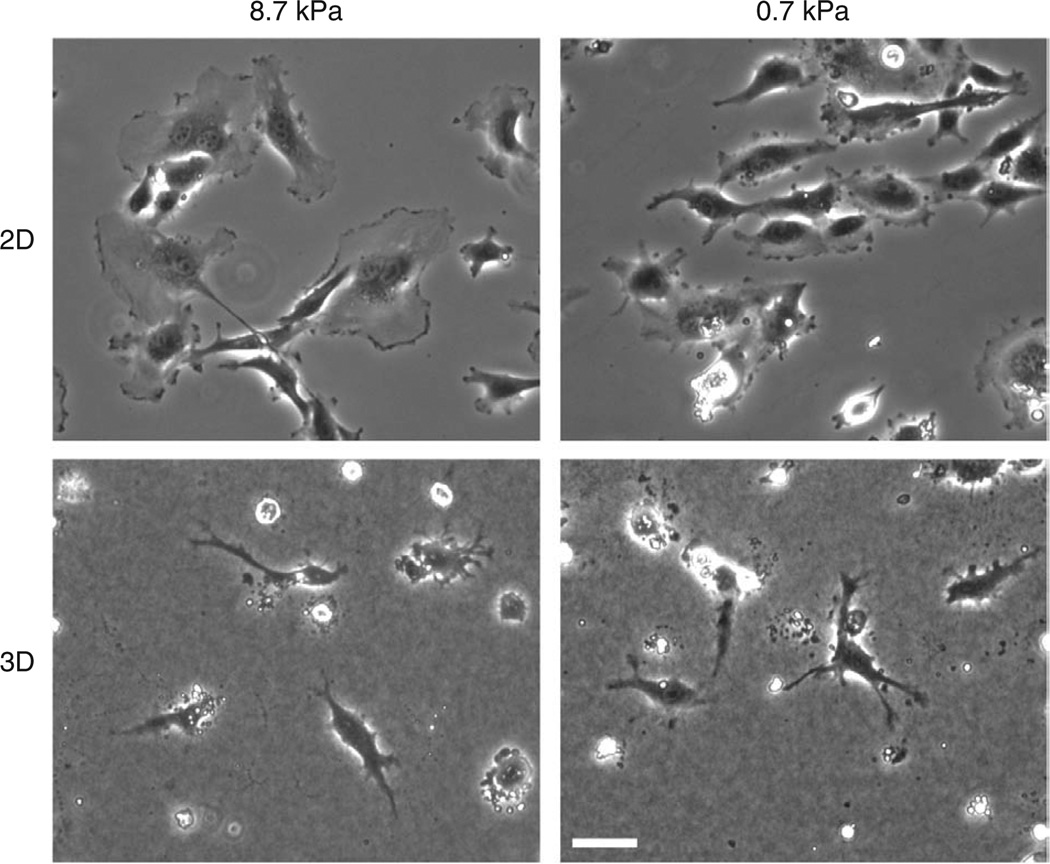
Regulation of cell functions by the physical properties of the extracellular matrix (ECM) has emerged as a crucial contributor to development and disease. Two specific physical properties of the ECM, stiffness and dimensionality, each influence cell signaling and function. As these ECM physical properties are linked to other properties that also regulate cell behavior, e.g., integrin ligand density, parsing the specific contributions of ECM stiffness and dimensionality has proven difficult. Here we detail a simple protocol, which can be completed in 1-2 d, for combining three-dimensional (3D) ECM engagement with controlled underlying ECM stiffness. In these 'sandwich gels', cells are sandwiched between a 3D fibrillar ECM and an ECM-coupled polyacrylamide gel of defined compliance, allowing the study of the specific effects of ECM compliance on cell function in physiologically relevant 3D ECMs. This type of system enables high-resolution time-lapse imaging and is suitable for a wide range of cell types and molecular perturbations.


Figures

Figure 1
Figure 1. Assembly of sandwich gels
(a) Schematic of sandwich gels. (i) Silanized glass cover slips are activated with glutaraldehyde. (ii) A 20–25-µm-thick polyacrylamide gel (light blue) is then polymerized on top of the activated glass. (iii) The surface of the polyacrylamide gel is activated with sulfo-SANPAH, wherein the photoactivatable nitrophenyl azide group covalently bonds the polyacrylamide, whereas the NHS ester group is available to bond to free amines in the ECM proteins (red dots). Cells (green) are then allowed to adhere as for normal culture, the supernatant medium is removed and a collagen gel (red cross hatch) is polymerized on top of the cells and polyacrylamide. (b) Scanning electron microscopy (SEM) of cross-section of sandwich gels. Sandwich gels were fixed as described and critical point dried for SEM, and then the back of the cover slip was etched with a diamond pen and broken to reveal the cross-sectional area of the sandwich gel. (c) SEM of collagen fibers from the polymerized gel and attached to the polyacrylamide surface. Shown is an area where the bulk of the collagen gel has retracted during fixation, illustrating that a portion of the collagen gel remains closely associated with the polyacrylamide. (d) Cross-sectional area of a sandwich gel in SEM showing the posterior side of the cell embedded within the collagen and interacting with the polyacrylamide. Note small set of collagen fibers being condensed by dorsal surface of cell.
Figure 2
Figure 2. Anticipated results with sandwich gel culture setup
Phase contrast images of HUVECs on 0.7-kPa or 8.7-kPa polyacrylamide gels without (2D) and with (3D) collagen gel on top. Note that when adhered to 2D collagen–coated polyacrylamide gels, cells spread to a larger area on stiffer ECM. In contrast, in 3D sandwich gels, cells are less spread and more branched, and softer ECM (0.7 kPa) induces more cell branches. This illustrates the different effects of stiffness in 2D and 3D ECMs. Scale bar, 50 µm.

Similar articles

Cited by

References

    1. Ghosh K, Thodeti CK, Dudley AC, Mammoto A, Klagsbrun M, et al. Tumor-derived endothelial cells exhibit aberrant Rho-mediated mechanosensing and abnormal angiogenesis in vitro. Proc. Natl. Acad. Sci. USA. 2008;105:11305–11310. - PMC - PubMed
    1. Ingber DE. Mechanical signaling and the cellular response to extracellular matrix in angiogenesis and cardiovascular physiology. Circ. Res. 2002;91:877–887. - PubMed
    1. Mammoto A, Connor KM, Mammoto T, Yung CW, Huh D, et al. A mechanosensitive transcriptional mechanism that controls angiogenesis. Nature. 2009;457:1103–1108. - PMC - PubMed
    1. Discher DE, Janmey P, Wang YL. Tissue cells feel and respond to the stiffness of their substrate. Science. 2005;310:1139. - PubMed
    1. Pelham RJ, Jr, Wang YL. Cell locomotion and focal adhesions are regulated by the mechanical properties of the substrate. Biol. Bull. 1998;194:348–349. - PubMed

Publication types

MeSH terms