

Pitfalls in the interpretation of structural changes in mutant proteins from crystal structures
- PMID: 23099666
- PMCID: PMC4109977
- DOI: 10.1007/s10969-012-9147-1
Pitfalls in the interpretation of structural changes in mutant proteins from crystal structures
Abstract
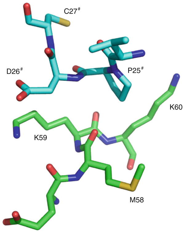
PpcA is a small protein with 71 residues that contains three covalently bound hemes. The structures of single mutants at residue 58 have shown larger deviations in another part of the protein molecule than at the site of the mutation. Closer examination of the crystal packing has revealed the origin of this unexpected structural change. The site of mutation is within Van der Waals distance from another protein molecule related by a crystallographic twofold axis within the crystal. The structural changes occurred at or near the mutation site have led to a slight adjustment of the surface residues in contact. The observed deviations between the native and the mutant molecular structures are derived from the new crystal packing even though the two crystals are essentially isomorphous. Without careful consideration of the crystal lattice a non-expert looking at only the coordinates deposited in the Protein Data Bank could draw erroneous conclusion that mutation in one part of the molecule affected the structure of the protein in a distant part of the molecule.
Figures


References
-
- Pokkuluri PR, Londer YY, Duke NEC, Long WC, Schiffer M. Family of cytochrome c7-type proteins from Geobacter sulfurreducens. The structure of one cytochrome c7 at 1.45 Å resolution. Biochemistry. 2004;43:849–859. - PubMed
-
- Pokkuluri PR, Londer YY, Yang X, Duke NE, Erickson J, Orshonsky V, Johnson G, Schiffer M. Structural characterization of a family of cytochromes c7 involved in Fe(III) respiration by Geobacter sulfurreducens. Biochim Biophys Acta. 2010;1797:222–232. - PubMed
-
- Morgado L, Paixao VB, Schiffer M, Pokkuluri PR, Bruix M, Salgueiro CA. Revealing the structural origin of the redox-Bohr effect: the first solution structure of a cytochrome from Geobacter sulfurreducens. Biochem J. 2012;441:179–187. - PubMed
-
- Lovley DR, Holmes DE, Nevin KP. Dissimilatory Fe(III) and Mn(IV) reduction. Adv Microb Physiol. 2004;49:219–286. - PubMed
Publication types
MeSH terms
Substances
Grants and funding
LinkOut - more resources
Full Text Sources
