Review
. 2013 Mar;41(3):446-68.
doi: 10.1007/s10439-012-0678-1. Epub 2012 Oct 26.

Physical non-viral gene delivery methods for tissue engineering

Affiliations
Review

Physical non-viral gene delivery methods for tissue engineering

Adam J Mellott et al. Ann Biomed Eng. 2013 Mar.

Abstract

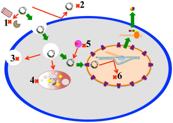
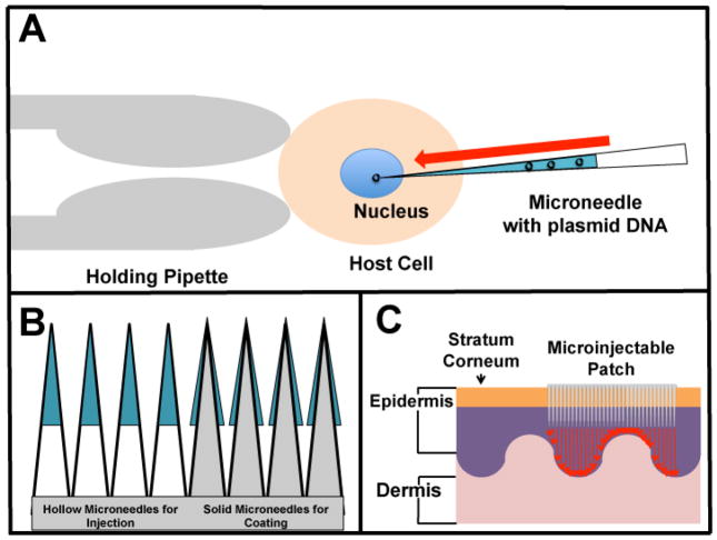
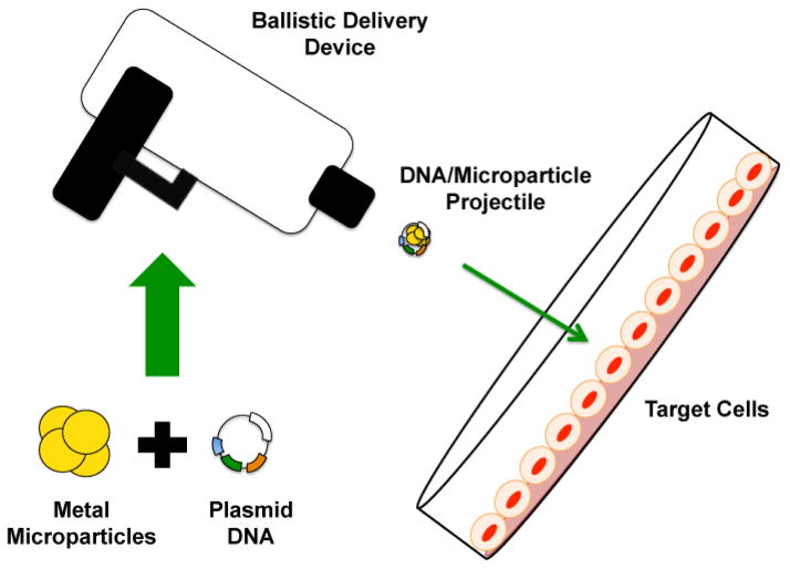
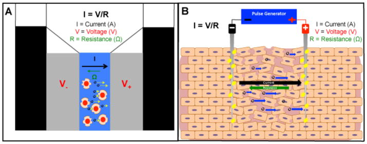
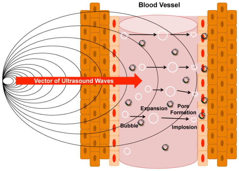
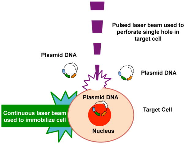
The integration of gene therapy into tissue engineering to control differentiation and direct tissue formation is not a new concept; however, successful delivery of nucleic acids into primary cells, progenitor cells, and stem cells has proven exceptionally challenging. Viral vectors are generally highly effective at delivering nucleic acids to a variety of cell populations, both dividing and non-dividing, yet these viral vectors are marred by significant safety concerns. Non-viral vectors are preferred for gene therapy, despite lower transfection efficiencies, and possess many customizable attributes that are desirable for tissue engineering applications. However, there is no single non-viral gene delivery strategy that "fits-all" cell types and tissues. Thus, there is a compelling opportunity to examine different non-viral vectors, especially physical vectors, and compare their relative degrees of success. This review examines the advantages and disadvantages of physical non-viral methods (i.e., microinjection, ballistic gene delivery, electroporation, sonoporation, laser irradiation, magnetofection, and electric field-induced molecular vibration), with particular attention given to electroporation because of its versatility, with further special emphasis on Nucleofection™. In addition, attributes of cellular character that can be used to improve differentiation strategies are examined for tissue engineering applications. Ultimately, electroporation exhibits a high transfection efficiency in many cell types, which is highly desirable for tissue engineering applications, but electroporation and other physical non-viral gene delivery methods are still limited by poor cell viability. Overcoming the challenge of poor cell viability in highly efficient physical non-viral techniques is the key to using gene delivery to enhance tissue engineering applications.


Figures

Figure 1
Figure 1. Gene Delivery Barriers
DNA must overcome several barriers during the delivery process to successfully produce desired gene expression. The green arrows are the pathway DNA must follow to induce gene expression, while the red arrows indicate potential barriers that prevent gene delivery. 1) DNA must avoid extracellular nucleases and 2) DNA must associate with the cellular membrane in some form to gain access to the cell via penetration, electrostatic interactions, adsorption, or ligand mediated receptor binding. DNA that enters through endocytosis must escape the endosome before the endosome 3) is recycled back to the cell membrane or 4) before the endosome matures into a lysosome, and DNA is degraded. In the cytoplasmic compartment, DNA must traffic toward the nuclear envelope and 5) avoid degradation by intracellular nucleases. Finally, to produce gene expression, 6) DNA must cross the nuclear envelope by transport through a nuclear pore (non-dividing cells) or passively re-locate into the nucleus between the disassembly and reformation of the nuclear envelope during mitosis (dividing cells). Gene expression is produced when enough intact DNA is transcribed in the nucleus into mRNA, and then translated into a protein, composed of amino acids, in the cytoplasm.
Figure 2
Figure 2. Microinjection
Microinjection strategies utilize microneedles to deliver DNA directly to cell nuclei. A) In traditional microinjection, an individual cell is held in place by the tip of a pipette while a technician uses a microscope to pierce the cell membrane and nuclear envelope with a microneedle to deliver genetic material to the cell nucleus. B) Microneedles can be fabricated so that the shaft is hollow and able to carry a suspension of genetic material for injection, or microneedles can be fabricated so that the shaft is solid and the tip is dipped in a suspension of genetic material for application to tissues via coating or scratching. C) Microneedles can be arranged in arrays on patches that can be applied directly to the skin. The microneedle patches are capable of penetrating the stratum corneum and delivering drugs or genetic material to the epidermal tissues.
Figure 3
Figure 3. Ballistic Gene Delivery
Plasmid DNA is mixed with gold or tungsten particles ranging in size from nanometers to micrometers. An electric or plasma discharge is used to propel the DNA/particle complexes into tissues or cell cultures.
Figure 4
Figure 4. Electroporation
Electroporation strategies apply a current across cells or tissues to make cell membranes more permeable to exogenous DNA. A) Traditional electroporators have a pulse generator and a pair of electrodes that can be applied directly to tissues or cells. A cuvette utilizes plate electrodes to apply a voltage potential across cells in suspension. Since resistance is constant, the current is proportional to the voltage potential. As voltage reaches a critical threshold, hydrophilic pores form in the cell membrane make it permeable to plasmid DNA. The negatively charged DNA is mobile in the electrical field (toward the positive electrode) so DNA transport into permeabilized cells is greater than by diffusion alone. B) Needle electrodes have been used for in vivo applications where needles are inserted directly into primary tissues such as skin or skeletal muscle fibers after DNA has been injected. A current is applied across a very small area of tissue to facilitate the same process as in a cuvette.
Figure 5
Figure 5. Sonoporation
Ultrasonic frequencies are used to induce the cavitation of microbubbles for creating pores in cells contained in culture or tissue. The acoustic waves cause microbubbles to expand and then collapse. When the microbubbles collapse, a microshockwave is emitted that can rupture a cell membrane if the collapsing microbubble is in close proximity to the cell membrane. The ruptured cell membrane forms a pore, which allows cells to be temporarily more permeable to plasmid DNA.
Figure 6
Figure 6. Laser Induced Pore Formation
Pulsed lasers have been shown to perforate cell membranes similar to microinjection strategies, but without the use of a needle. Investigators have shown a variety of laser beams of varying wavelengths are capable of making precise “holes” in cell membranes when beam energy, pulse frequency, and exposure duration are manipulated. Investigators can precisely target individual cells in culture or in tissue with aid of a microscope to target specific sites on cells for perforation to allow DNA to enter cells. A second laser with an uninterrupted beam can be used to immobilize individual cells in suspension while a pulsed laser is used to perforate cells.

References

    1. Adler AF, Leong KW. Emerging links between surface nanotechnology and endocytosis: impact on nonviral gene delivery. Nano Today. 2010;5:553–569. - PMC - PubMed
    1. Adler AF, Speidel AT, Christoforou N, Kolind K, Foss M, Leong KW. High-throughput screening of microscale pitted substrate topographies for enhanced nonviral transfection efficiency in primary human fibroblasts. Biomaterials. 2011;32:3611–9. - PMC - PubMed
    1. Aihara H, Miyazaki J. Gene transfer into muscle by electroporation in vivo. Nat Biotechnol. 1998;16:867–70. - PubMed
    1. Akinc A, Thomas M, Klibanov AM, Langer R. Exploring polyethylenimine-mediated DNA transfection and the proton sponge hypothesis. J Gene Med. 2005;7:657–63. - PubMed
    1. Alberts B, Bray D, Johnson A, Lewis J, Raff M, Roberts K, Walter P, Campbell A. Essential cell biology. Garland Science; New York: 2004.

LinkOut - more resources