. 2013 Feb;62(2):510-8.
doi: 10.2337/db12-0294. Epub 2012 Oct 25.

Ectopic expression of GIP in pancreatic β-cells maintains enhanced insulin secretion in mice with complete absence of proglucagon-derived peptides

Affiliations

Ectopic expression of GIP in pancreatic β-cells maintains enhanced insulin secretion in mice with complete absence of proglucagon-derived peptides

Ayako Fukami et al. Diabetes. 2013 Feb.

Abstract

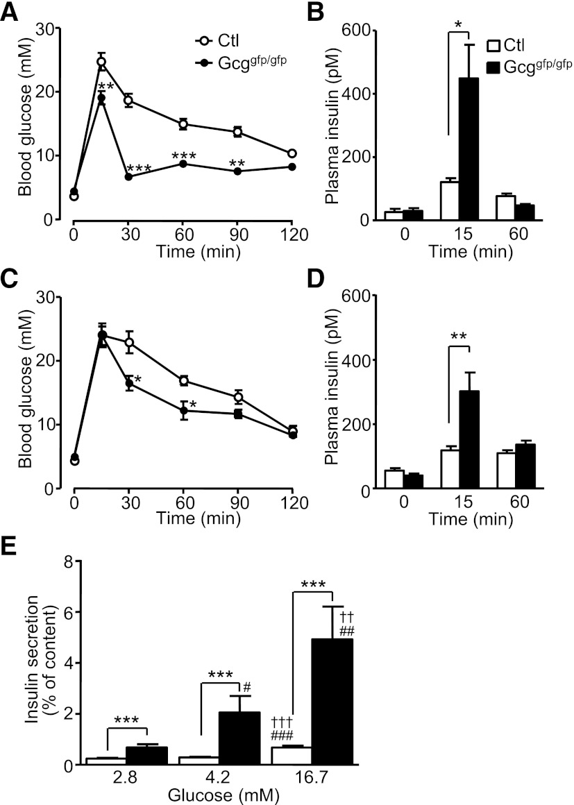
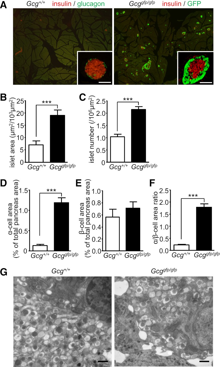
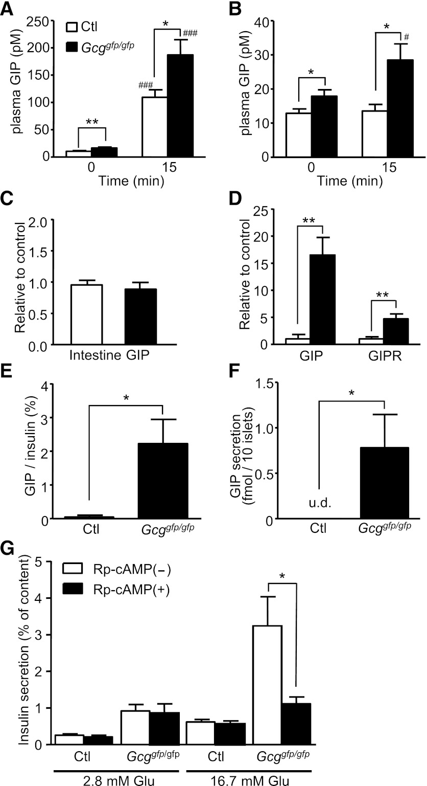
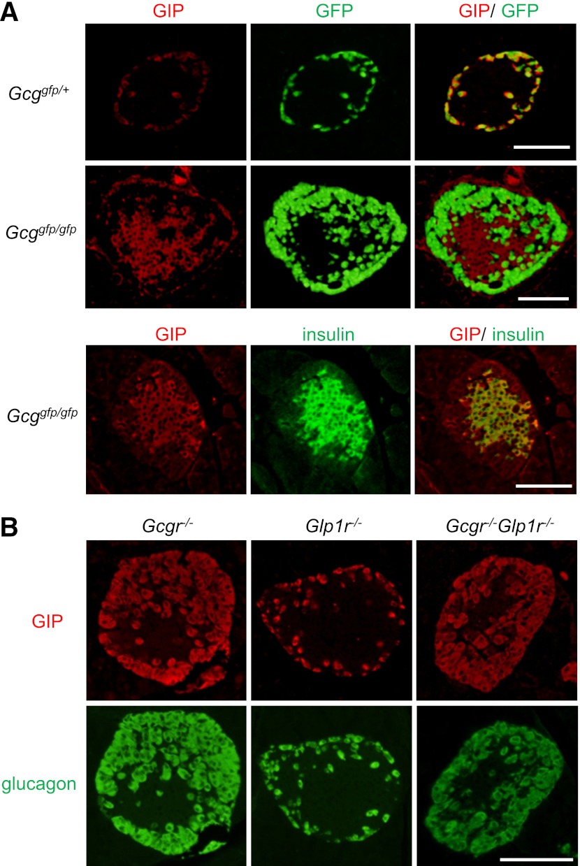
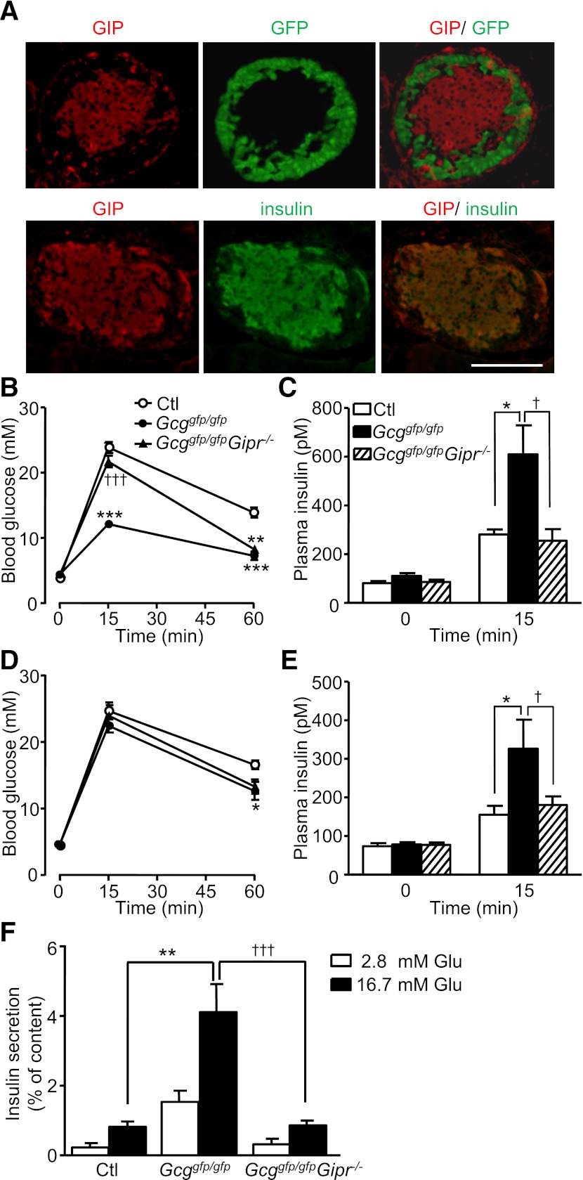
Glucagon and glucagon-like peptide-1 (GLP-1) are produced in pancreatic α-cells and enteroendocrine L-cells, respectively, in a tissue-specific manner from the same precursor, proglucagon, that is encoded by glucagon gene (Gcg), and play critical roles in glucose homeostasis. Here, we studied glucose homeostasis and β-cell function of Gcg-deficient mice that are homozygous for a Gcg-GFP knock-in allele (Gcg(gfp/gfp)). The Gcg(gfp/gfp) mice displayed improved glucose tolerance and enhanced insulin secretion, as assessed by both oral glucose tolerance test (OGTT) and intraperitoneal glucose tolerance test (IPGTT). Responses of glucose-dependent insulinotropic polypeptide (GIP) to both oral and intraperitoneal glucose loads were unexpectedly enhanced in Gcg(gfp/gfp) mice, and immunohistochemistry localized GIP to pancreatic β-cells of Gcg(gfp/gfp) mice. Furthermore, secretion of GIP in response to glucose was detected in isolated islets of Gcg(gfp/gfp) mice. Blockade of GIP action in vitro and in vivo by cAMP antagonism and genetic deletion of the GIP receptor, respectively, almost completely abrogated enhanced insulin secretion in Gcg(gfp/gfp) mice. These results indicate that ectopic GIP expression in β-cells maintains insulin secretion in the absence of proglucagon-derived peptides (PGDPs), revealing a novel compensatory mechanism for sustaining incretin hormone action in islets.


Figures

FIG. 1.
FIG. 1.
Disruption of PGDPs improves glucose tolerance and enhances insulin secretion. Blood glucose levels (A) and plasma insulin levels (B) in the OGTT in 12- to 15-week-old control (Ctl, Gcg+/+ and Gcggfp/+) and Gcggfp/gfp mice (n = 5–6 mice per group). Blood glucose levels (C) and plasma insulin levels (D) in the IPGTT in 11- to 22-week-old mice (n = 9–10 mice per group). E: Glucose-induced insulin secretion from isolated islets. Pancreatic islets were isolated from 5- to 7-month-old control (Gcg+/+ and Gcggfp/+, n = 13–18 in each group) and Gcggfp/gfp (n = 8–9 in each group) mice and incubated with the indicated concentration of glucose for 30 min. Insulin secretion is expressed as the ratio of insulin released into the medium relative to insulin content. Values are expressed as means ± SEM. *P < 0.05; **P < 0.01; ***P < 0.001 vs. Ctl. ††P < 0.01. †††P < 0.001 vs. 4.2 mmol/L glucose. #P < 0.05; ##P < 0.01; ###P < 0.001 vs. 2.8 mmol/L glucose.
FIG. 2.
FIG. 2.
Gcggfp/gfp mice exhibit α-cell hyperplasia. A: Representative pancreatic sections from 9-week-old Gcg+/+ (left) and Gcggfp/gfp (right) mice. In the left panel, sections were immunostained for glucagon (green) and insulin (red). In the right panel, sections were immunostained for insulin (red) and shown with autofluorescence of GFP. Scale bar, 100 µm. B–F: Morphometric analysis of islets. B: Islet area shown relative to a 103-µm2 sectional area. C: Islet number shown relative to a 106-µm2 sectional area. D: The α-cell area shown as glucagon-positive or GFP-positive area relative to total pancreas area. E: The β-cell area shown as insulin-positive area relative to total pancreas area. F: The α-cell/β-cell area ratio shown as a ratio of glucagon-positive or GFP-positive area relative to insulin-positive area. G: Electron microscopy of islets. Scale bar, 500 nm. Values are expressed as means ± SEM. ***P < 0.001. (A high-quality digital representation of this figure is available in the online issue.)
FIG. 3.
FIG. 3.
Gcggfp/gfp mice display enhanced GIP secretion and GIP expression in pancreatic islets, and enhancement of glucose-induced insulin secretion is blocked by Rp-cAMP in Gcggfp/gfp mice. Plasma GIP levels at 0 and 15 min after oral (A) or intraperitoneal (B) glucose administration in 12- to 17-week-old control (Ctl, Gcg+/+, and Gcggfp/+) and Gcggfp/gfp mice (n = 6–11 mice per group). C: mRNA expression of GIP in the intestine (n = 6–8). The expression level of GIP mRNA was normalized to that of glyceraldehyde-3-phosphate dehydrogenase mRNA. D: mRNA expression of GIP and GIP receptor (GIPR) in isolated islets (n = 4). The expression levels normalized to that of insulin 1 are shown. E: GIP content in isolated islets (n = 5–7). GIP content was normalized to insulin content. F: GIP secretion from islets (n = 5–8). Isolated islets were incubated with 16.7 mmol/L glucose for 16 h. G: Effect of Rp-cAMP on glucose-induced insulin secretion. Isolated islets from control (n = 14–22 in each group) and Gcggfp/gfp (n = 13–22 in each group) mice were incubated with the indicated concentration of glucose and 500 μmol/L Rp-cAMP for 30 min. Insulin secretion is expressed as the ratio of insulin released into the medium relative to insulin content. Values are expressed as means ± SEM. *P < 0.05; **P < 0.01. #P < 0.05; ###P < 0.001 vs. 0 min. u.d., undetectable.
FIG. 4.
FIG. 4.
GIP is expressed in pancreatic β-cells in Gcggfp/gfp mice, but not in Gcgr−/−, Glp1r−/−, or Gcgr−/−Glp1r−/− mice. A: Representative immunostaining for GIP (red) and insulin (green), and GFP fluorescence (green) of islets from Gcggfp/gfp mice and Gcggfp/+ mice. B: Representative immunostaining for GIP (red) and glucagon (green) of islets from Gcgr−/−, Glp1r−/−, and Gcgr−/−Glp1r−/− mice. Scale bar, 100 µm. (A high-quality digital representation of this figure is available in the online issue.)
FIG. 5.
FIG. 5.
Lack of GIP receptor signaling abrogates enhanced insulin secretion in Gcggfp/gfp mice. A: Representative immunostaining for GIP (red) and insulin (green), and GFP fluorescence (green) of islets from Gcggfp/gfpGipr−/− mice. Scale bar, 100 µm. B: Blood glucose levels during the OGTT in 15- to 24-week-old control (Ctl), Gcggfp/gfp, and Gcggfp/gfpGipr−/− mice (n = 5–7 mice per group). C: Plasma insulin levels at 0 and 15 min after oral glucose loading. D: Blood glucose levels during the IPGTT in 13- to 22-week-old mice (n = 6–8 mice per group). E: Plasma insulin levels 0 and 15 min after intraperitoneal glucose administration. F: Glucose-induced insulin secretion from isolated islets. Pancreatic islets were isolated from 5- to 7-month-old control (Ctl), Gcggfp/gfp, and Gcggfp/gfpGipr−/− mice, and then incubated with the indicated concentration of glucose for 30 min. Insulin secretion is expressed as the ratio of insulin released into the medium relative to insulin content. Values are expressed as means ± SEM. *P < 0.05; **P < 0.01; ***P < 0.001 vs. Ctl. †P < 0.05; †††P < 0.001 vs. Gcggfp/gfp, Ctl, Gcggfp/+Gipr+/−; Gcggfp/gfp, Gcggfp/gfpGipr+/+, and Gcggfp/gfpGipr+/−. (A high-quality digital representation of this figure is available in the online issue.)

References

    1. Baggio LL, Drucker DJ. Biology of incretins: GLP-1 and GIP. Gastroenterology 2007;132:2131–2157 - PubMed
    1. Hayashi Y. Metabolic impact of glucagon deficiency. Diabetes Obes Metab 2011;13(Suppl 1):151–157 - PubMed
    1. Unger RH. Glucagon physiology and pathophysiology in the light of new advances. Diabetologia 1985;28:574–578 - PubMed
    1. Jiang G, Zhang BB. Glucagon and regulation of glucose metabolism. Am J Physiol Endocrinol Metab 2003;284:E671–E678 - PubMed
    1. Cherrington AD, Chiasson JL, Liljenquist JE, Lacy WW, Park CR. Control of hepatic glucose output by glucagon and insulin in the intact dog. Biochem Soc Symp 1978;43:31–45 - PubMed

Publication types

MeSH terms