. 2013 Jan 4;288(1):285-93.
doi: 10.1074/jbc.M112.385625. Epub 2012 Oct 25.

Site-specific phosphorylation of the p65 protein subunit mediates selective gene expression by differential NF-κB and RNA polymerase II promoter recruitment

Affiliations

Site-specific phosphorylation of the p65 protein subunit mediates selective gene expression by differential NF-κB and RNA polymerase II promoter recruitment

Karin Hochrainer et al. J Biol Chem. .

Abstract

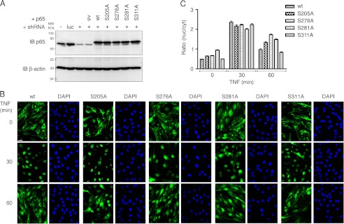
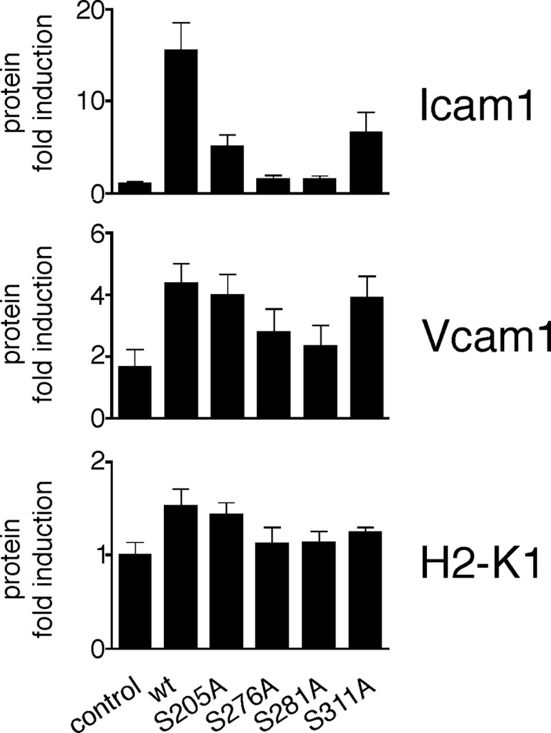
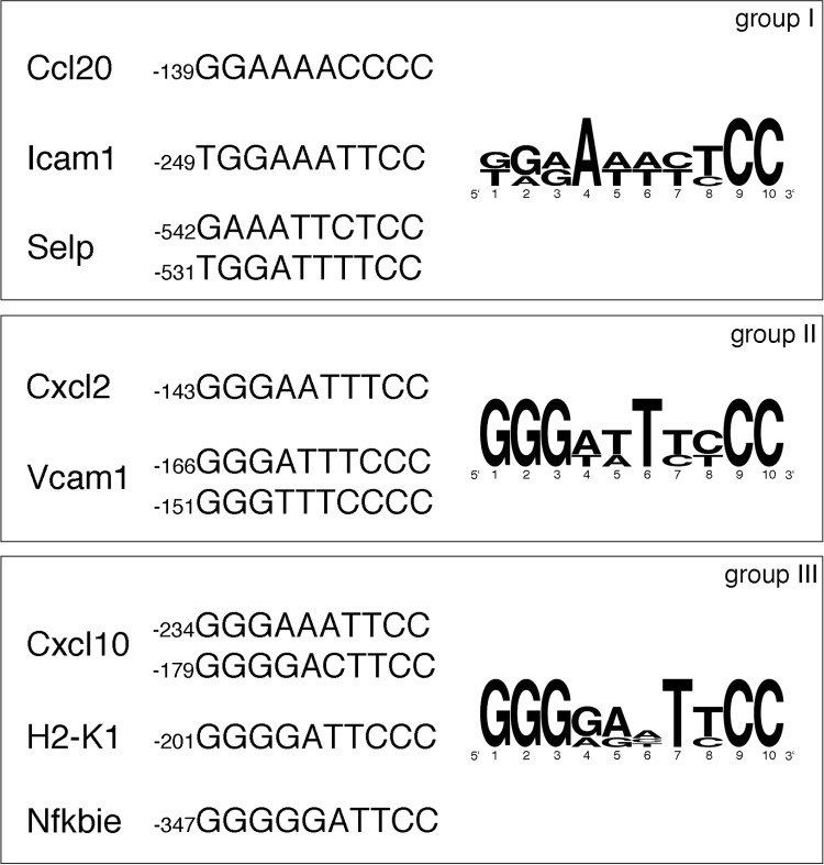
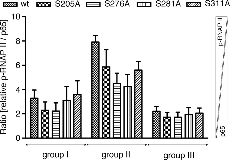
Phosphorylation of NF-κB plays an important role in modulating transcriptional activity of NF-κB independently of inhibitor of κB (IκB) proteins. For the p65 subunit, multiple phosphorylation sites have been mapped in and adjacent to both the N-terminal Rel homology domain and the C-terminal transactivation domain. Their impact on NF-κB-dependent transcription, however, has never been assessed at a broader level. In this study, we evaluate the importance of differential p65 phosphorylation on four serine acceptor sites in the Rel homology domain for the expression of an array of NF-κB-dependent genes in endothelial cells. We find that inhibition of p65 phosphorylation on these serine residues targets NF-κB activity to distinctive gene subsets in a κB enhancer element-specific context. We show that the phosphorylation-dependent alterations in gene and protein expression are reflective of the amount of p65 and phosphorylated RNA polymerase II (p-RNAP II) bound to respective gene promoter regions. Depending on the gene subset, impaired gene expression was either a result of decreased p65 promoter recruitment or of a failure of bound p65 to recruit p-RNAP II. In conclusion, our findings demonstrate that site-specific p65 phosphorylation targets NF-κB activity to particular gene subsets on a global level by influencing p65 and p-RNAP II promoter recruitment.


Figures

FIGURE 1.
FIGURE 1.
Generation and characterization of wild type and phosphorylation-deficient p65-expressing bEND.3 cells. A, Western blot analysis of p65 expression in bEND.3 cells transduced with luciferase (luc) control and p65-targeting shRNAs (+). c-Myc-tagged human p65 wild type (wt) and amino acids 205, 276, 281, 311 serine to alanine (SA) mutants were stably introduced into successful p65 knockdown cells. The fusion of p65 with N-terminal c-Myc leads to a shift in molecular weight of 10 amino acids reflected by slower migration of human c-Myc-p65 in SDS-PAGE. IB, immunoblot. B, subcellular localization of p65 variants in bEND.3 in the absence or presence of 10 ng/ml TNF for 30 and 60 min. DAPI staining is included as nuclear reference. Bar, 20 μm. C, semi-automated quantification of cytosolic and nuclear p65 immunofluorescence (n = 128–248 cells, derived from three experiments). Nuclear/cytosolic values of >1 indicate predominantly nuclear p65.
FIGURE 2.
FIGURE 2.
Comparison of expression profiles of 37 genes in p65 WT, S205A-, S276A-, S281A-, and S311A-expressing bEND.3 cells after 3 h of TNF stimulation. Stably transduced p65 WT and mutant bEND.3 cells were either left untreated or stimulated with 10 ng/ml TNF for 3 h; RNA was isolated, and the induction of NF-κB-dependent genes was tested by quantitative RT-PCR. Depending on the mRNA expression patterns obtained in p65 WT and mutant cells, the tested genes were subdivided into three groups as outlined in the main text. Arrows indicate the allowable range of variation for a given mRNA/p65 variant combination to assign the gene to the respective groups. Double arrowheads signal that the variation is permitted beyond the indicated levels. Asterisk, either p65 S276A- or S281A-derived mRNA expression levels remain below half of p65 WT mRNA levels. Data are represented as mean ± S.E. (n = 3). All datasets were calculated relative to the mean of unstimulated, pooled controls.
FIGURE 3.
FIGURE 3.
Protein expression levels of three selected genes as observed by FACS analysis after 3 h of TNF treatment. Untreated or 3-h TNF-treated retrovirally transduced p65 WT and mutant bEND.3 cells were processed for FACS analysis as described under “Experimental Procedures.” Data represent mean ± S.E. (n = 3), calculated relative to the mean of unstimulated, pooled controls.
FIGURE 4.
FIGURE 4.
Relation of mRNA expression of two selected genes from each group to their promoter κB consensus sequences. The sequences of decameric κB sites for the tested genes (Ccl20 (38), Icam1 (39), Selp (40), Cxcl2 (41), Vcam1 (42), Cxcl10 (43), H2-k1 (44), and Nfkbie (45)) and their position relative to the translational start site are listed. Sequences for each group were subjected to a sequence conservation analysis using the tool WebLogo (46), where the relative height of letters corresponds to the relative frequency of bases at each position.
FIGURE 5.
FIGURE 5.
NF-κB p65 and p-RNAP II promoter binding. ChIP analysis was carried out by RT-PCR in p65 WT and mutant-expressing bEND.3 cells left untreated or treated with 10 ng/ml TNF for 0.5 and 3 h. Chromatin was isolated, and DNA was immunoprecipitated with p65 and p-RNAP II antibodies. Data are represented as mean ± S.E. (n = 3).
FIGURE 6.
FIGURE 6.
p-RNAP II promoter binding expressed as ratio to p65 promoter binding. ChIP data obtained from Ccl20, Icam1, Selp, Cxcl2, Vcam1, Cxcl10, H2-k1, and Nfkbie promoter sites were used to determine the ratio of promoter-bound p-RNAP II to p65 in the three regulatory groups. Higher y values signify relatively more bound p-RNAP II than p65.

References

    1. Barkett M., Gilmore T. D. (1999) Control of apoptosis by Rel/NF-κB transcription factors. Oncogene 18, 6910–6924 - PubMed
    1. Hayden M. S., West A. P., Ghosh S. (2006) NF-κB and the immune response. Oncogene 25, 6758–6780 - PubMed
    1. Karin M., Lin A. (2002) NF-κB at the crossroads of life and death. Nat. Immunol. 3, 221–227 - PubMed
    1. Baeuerle P. A., Baltimore D. (1988) IκB. A specific inhibitor of the NF-κB transcription factor. Science 242, 540–546 - PubMed
    1. Henkel T., Machleidt T., Alkalay I., Krönke M., Ben-Neriah Y., Baeuerle P. A. (1993) Rapid proteolysis of IκB-α is necessary for activation of transcription factor NF-κB. Nature 365, 182–185 - PubMed

Publication types

LinkOut - more resources