

A single residue within the V5 region of HIV-1 envelope facilitates viral escape from the broadly neutralizing monoclonal antibody VRC01
- PMID: 23100255
- PMCID: PMC3522310
- DOI: 10.1074/jbc.M112.399402
A single residue within the V5 region of HIV-1 envelope facilitates viral escape from the broadly neutralizing monoclonal antibody VRC01
Abstract
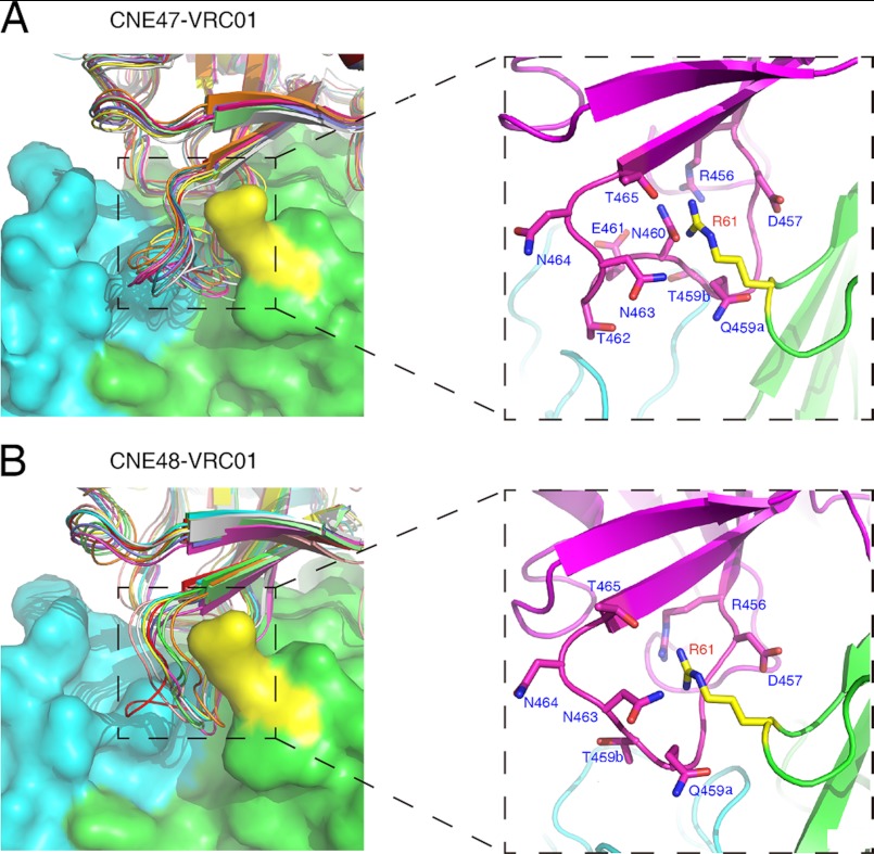
VRC01, a broadly neutralizing monoclonal antibody, is capable of neutralizing a diverse array of HIV-1 isolates by mimicking CD4 binding with the envelope glycoprotein gp120. Nonetheless, resistant strains have been identified. Here, we examined two genetically related and two unrelated envelope clones, derived from CRF08_BC-infected patients, with distinct VRC01 neutralization profiles. A total of 22 chimeric envelope clones was generated by interchanging the loop D and/or V5 regions between the original envelopes or by single alanine substitutions within each region. Analysis of pseudoviruses built from these mutant envelopes showed that interchanging the V5 region between the genetically related or unrelated clones completely swapped their VRC01 sensitivity profiles. Mutagenesis analysis revealed that the asparagine residue at position 460 (Asn-460), a potential N-linked glycosylation site in the V5 region, is a key factor for observed resistance in these strains, which is further supported by our structural modeling. Moreover, changes in resistance were found to positively correlate with deviations in VRC01 binding affinity. Overall, our study indicates that Asn-460 in the V5 region is a critical determinant of sensitivity to VRC01 specifically in these viral strains. The long side chain of Asn-460, and potential glycosylation, may create steric hindrance that lowers binding affinity, thereby increasing resistance to VRC01 neutralization.
Figures
Similar articles
-
Resistance mutations that distinguish HIV-1 envelopes with discordant VRC01 phenotypes from multi-lineage infections in the HVTN703/HPTN081 trial: implications for cross-resistance.J Virol. 2025 Feb 25;99(2):e0173024. doi: 10.1128/jvi.01730-24. Epub 2025 Jan 16. J Virol. 2025. PMID: 39817771 Free PMC article.
-
A 2-4-Amino Acid Deletion in the V5 Region of HIV-1 Env gp120 Confers Viral Resistance to the Broadly Neutralizing Human Monoclonal Antibody, VRC01.AIDS Res Hum Retroviruses. 2017 Dec;33(12):1248-1257. doi: 10.1089/aid.2017.0063. Epub 2017 Oct 17. AIDS Res Hum Retroviruses. 2017. PMID: 28903577
-
HIV-1 fitness cost associated with escape from the VRC01 class of CD4 binding site neutralizing antibodies.J Virol. 2015 Apr;89(8):4201-13. doi: 10.1128/JVI.03608-14. Epub 2015 Jan 28. J Virol. 2015. PMID: 25631091 Free PMC article.
-
Human antibodies that neutralize HIV-1: identification, structures, and B cell ontogenies.Immunity. 2012 Sep 21;37(3):412-25. doi: 10.1016/j.immuni.2012.08.012. Immunity. 2012. PMID: 22999947 Free PMC article. Review.
-
A matrix of structure-based designs yields improved VRC01-class antibodies for HIV-1 therapy and prevention.MAbs. 2021 Jan-Dec;13(1):1946918. doi: 10.1080/19420862.2021.1946918. MAbs. 2021. PMID: 34328065 Free PMC article. Review.
Cited by
-
Detailed study of the interaction between human herpesvirus 6B glycoprotein complex and its cellular receptor, human CD134.J Virol. 2014 Sep;88(18):10875-82. doi: 10.1128/JVI.01447-14. Epub 2014 Jul 9. J Virol. 2014. PMID: 25008928 Free PMC article.
-
Development of Protein- and Peptide-Based HIV Entry Inhibitors Targeting gp120 or gp41.Viruses. 2019 Aug 1;11(8):705. doi: 10.3390/v11080705. Viruses. 2019. PMID: 31374953 Free PMC article. Review.
-
HIV-1 envelope facilitates the development of protease inhibitor resistance through acquiring mutations associated with viral entry and immune escape.Front Microbiol. 2024 Apr 18;15:1388729. doi: 10.3389/fmicb.2024.1388729. eCollection 2024. Front Microbiol. 2024. PMID: 38699474 Free PMC article.
-
Resistance mutations that distinguish HIV-1 envelopes with discordant VRC01 phenotypes from multi-lineage infections in the HVTN703/HPTN081 trial: implications for cross-resistance.J Virol. 2025 Feb 25;99(2):e0173024. doi: 10.1128/jvi.01730-24. Epub 2025 Jan 16. J Virol. 2025. PMID: 39817771 Free PMC article.
-
Diverse pathways of escape from all well-characterized VRC01-class broadly neutralizing HIV-1 antibodies.PLoS Pathog. 2018 Aug 20;14(8):e1007238. doi: 10.1371/journal.ppat.1007238. eCollection 2018 Aug. PLoS Pathog. 2018. PMID: 30125330 Free PMC article.
References
-
- Chan D. C., Fass D., Berger J. M., Kim P. S. (1997) Core structure of gp41 from the HIV envelope glycoprotein. Cell 89, 263–273 - PubMed
-
- Weissenhorn W., Dessen A., Harrison S. C., Skehel J. J., Wiley D. C. (1997) Atomic structure of the ectodomain from HIV-1 gp41. Nature 387, 426–430 - PubMed
-
- Zhu P., Liu J., Bess J., Jr., Chertova E., Lifson J. D., Grisé H., Ofek G. A., Taylor K. A., Roux K. H. (2006) Distribution and three-dimensional structure of AIDS virus envelope spikes. Nature 441, 847–852 - PubMed
-
- Dalgleish A. G., Beverley P. C., Clapham P. R., Crawford D. H., Greaves M. F., Weiss R. A. (1984) The CD4 (T4) antigen is an essential component of the receptor for the AIDS retrovirus. Nature 312, 763–767 - PubMed
Publication types
MeSH terms
Substances
LinkOut - more resources
Full Text Sources
Other Literature Sources
Research Materials
