

Somatic gain-of-function mutations in PIK3CA in patients with macrodactyly
- PMID: 23100325
- PMCID: PMC3542862
- DOI: 10.1093/hmg/dds440
Somatic gain-of-function mutations in PIK3CA in patients with macrodactyly
Abstract
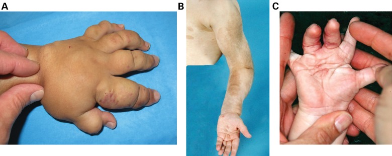
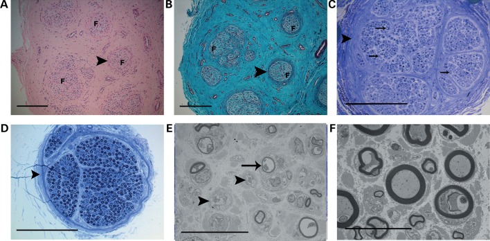
Macrodactyly is a discrete congenital anomaly consisting of enlargement of all tissues localized to the terminal portions of a limb, typically within a 'nerve territory'. The classic terminology for this condition is 'lipofibromatous hamartoma of nerve' or Type I macrodactyly. The peripheral nerve, itself, is enlarged both in circumference and in length. It is not related to neurofibromatosis (NF1), nor is it associated with vascular malformations, such as in the recently reported CLOVES syndrome. The specific nerve pathophysiology in this form of macrodactyly has not been well described and a genetic etiology for this specific form of enlargement is unknown. To identify the genetic cause of macrodactyly, we used whole-exome sequencing to identify somatic mutations present in the affected nerve of a single patient. We confirmed a novel mutation in PIK3CA (R115P) present in the patient's affected nerve tissue but not in blood DNA. Sequencing PIK3CA exons identified gain-of-function mutations (E542K, H1047L or H1047R) in the affected tissue of five additional unrelated patients; mutations were absent in blood DNA available from three patients. Immunocytochemistry confirmed AKT activation in cultured cells from the nerve of a macrodactyly patient. Additionally, we found that the most abnormal structure within the involved nerve in a macrodactylous digit is the perineurium, with additional secondary effects on the axon number and size. Thus, isolated congenital macrodactyly is caused by somatic activation of the PI3K/AKT cell-signaling pathway and is genetically and biochemically related to other overgrowth syndromes.
Figures


References
-
- Flatt A. E. The Care of Congenital Hand Anomalies. St. Louis, Missouri: Quality Medical Publishing, Inc; 1994.
-
- Ho C.A., Herring J.A., Ezaki M. Long-term follow-up of progressive macrodystrophia lipomatosa. A report of two cases. J. Bone Joint Surg. Am. 2007;89:1097–1102. doi:10.2106/JBJS.F.01378. - DOI - PubMed
-
- Lindhurst M.J., Parker V.E., Payne F., Sapp J.C., Rudge S., Harris J., Witkowski A.M., Zhang Q., Groeneveld M.P., Scott C.E., et al. Mosaic overgrowth with fibroadipose hyperplasia is caused by somatic activating mutations in PIK3CA. Nat. Genet. 2012;44:928–933. doi:10.1038/ng.2332. - DOI - PMC - PubMed
-
- Muzaffar A.R., Rafols F., Masson J., Ezaki M., Carter P.R. Keloid formation after syndactyly reconstruction: associated conditions, prevalence, and preliminary report of a treatment method. J. Hand Surg. [Am.] 2004;29:201–208. doi:10.1016/j.jhsa.2003.10.017. - DOI - PubMed
-
- Bamshad M.J., Ng S.B., Bigham A.W., Tabor H.K., Emond M.J., Nickerson D.A., Shendure J. Exome sequencing as a tool for Mendelian disease gene discovery. Nat. Rev. Genet. 2011;12:745–755. doi:10.1038/nrg3031. - DOI - PubMed
Publication types
MeSH terms
Substances
Supplementary concepts
LinkOut - more resources
Full Text Sources
Other Literature Sources
Molecular Biology Databases
Research Materials
Miscellaneous
