. 2013 Jan 15;22(2):372-83.
doi: 10.1093/hmg/dds434. Epub 2012 Oct 25.

A critical role for Telethonin in regulating t-tubule structure and function in the mammalian heart

Affiliations

A critical role for Telethonin in regulating t-tubule structure and function in the mammalian heart

Michael Ibrahim et al. Hum Mol Genet. .

Abstract

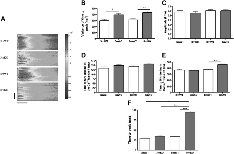
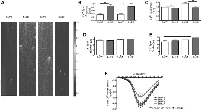
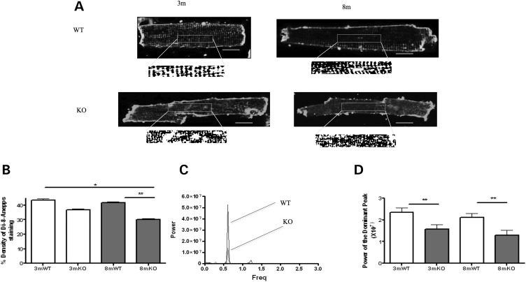
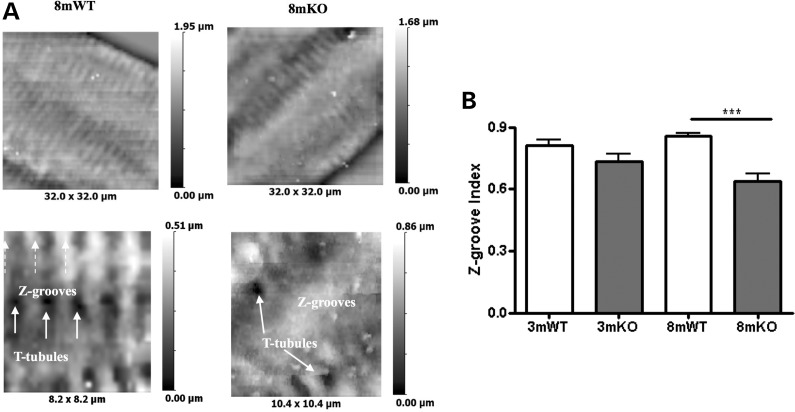
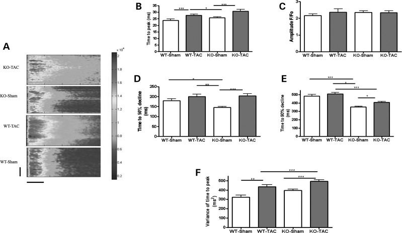
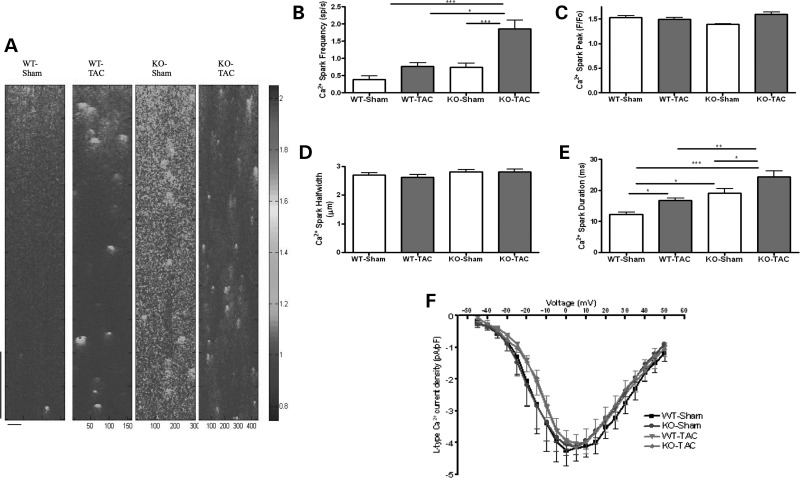
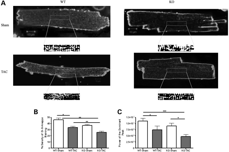
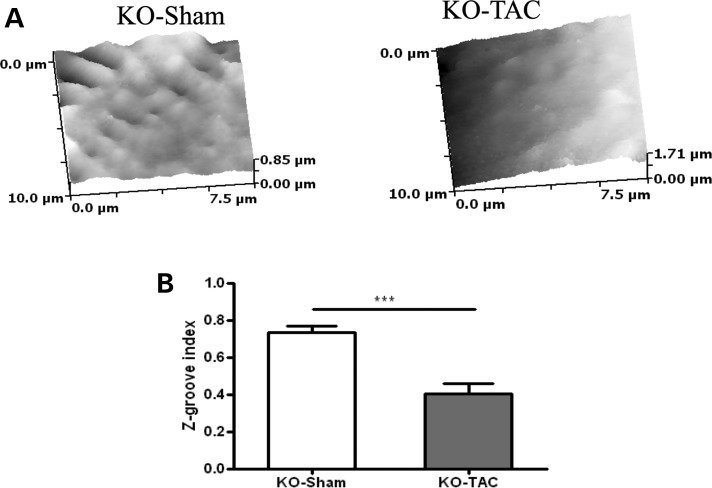
The transverse (t)-tubule system plays an essential role in healthy and diseased heart muscle, particularly in Ca(2+)-induced Ca(2+) release (CICR), and its structural disruption is an early event in heart failure. Both mechanical overload and unloading alter t-tubule structure, but the mechanisms mediating the normally tight regulation of the t-tubules in response to load variation are poorly understood. Telethonin (Tcap) is a stretch-sensitive Z-disc protein that binds to proteins in the t-tubule membrane. To assess its role in regulating t-tubule structure and function, we used Tcap knockout (KO) mice and investigated cardiomyocyte t-tubule and cell structure and CICR over time and following mechanical overload. In cardiomyocytes from 3-month-old KO (3mKO), there were isolated t-tubule defects and Ca(2+) transient dysynchrony without whole heart and cellular dysfunction. Ca(2+) spark frequency more than doubled in 3mKO. At 8 months of age (8mKO), cardiomyocytes showed progressive loss of t-tubules and remodelling of the cell surface, with prolonged and dysynchronous Ca(2+) transients. Ca(2+) spark frequency was elevated and the L-type Ca(2+) channel was depressed at 8 months only. After mechanical overload obtained by aortic banding constriction, the Ca(2+) transient was prolonged in both wild type and KO. Mechanical overload increased the Ca(2+) spark frequency in KO alone, where there was also significantly more t-tubule loss, with a greater deterioration in t-tubule regularity. In conjunction, Tcap KO showed severe loss of cell surface ultrastructure. These data suggest that Tcap is a critical, load-sensitive regulator of t-tubule structure and function.


Figures

Figure 1.
Figure 1.
Tcap KO is associated with progressive Ca2+ transient defects. (A) The figure shows line scans of stimulated ventricular cardiomyocytes from 3 months old or 8 months old WT and Tcap KO hearts. The colour chart is a normalized scale of fluorescence intensity (a marker of Ca2+ concentration). 3mWT n = 40, 3mKO n = 48, 8mWT n = 48, 8mKO n = 43. The vertical scale bar represents 100 pixels and the horizontal scale bar represents 100 ms. Variance of time to peak (B), amplitude (C) and time for 50% (D) and 90% (E) decline and the time to peak (F) of the Ca2+ transient were deleteriously affected in the KO. Importantly, the variance of the time to peak (which is closely associated with changes to the t-tubules) was the only parameter influenced at 3 m.
Figure 2.
Figure 2.
Tcap KO is associated with progressive Ca2+ spark and LTCC defects. (A) The figure shows line scans of quiescent ventricular cardiomyocytes from 3 month or 8 month WT and Tcap KO hearts. The colour chart is a normalized scale of fluorescence intensity (a marker of Ca2+ concentration). 3mWT n = 40, 3mKO n = 48, 8mWT n = 48, 8mKO n = 43. The vertical scale bar represents 768 ms and the horizontal scale bar represents 100 pixels. Ca2+ spark frequency was increased (B) and Ca2+ spark peak was reduced (C) at 3 and 8 months in the KO. Ca2+ spark width (D) and duration (E) were not significantly changed. (F) The L-type Ca2+ channel current density is reduced in 8mKO. 3mWT n = 24, 3mKO n = 25, 8mWT n = 16, 8mKO n = 20.
Figure 3.
Figure 3.
T-tubule structure is disrupted in Tcap KO. (A) The figure shows single di-8-ANEPPS-stained cardiomyocytes from which t-tubule density (B) and regularity are calculated. (B) T-tubule density is reduced at the 8 month in the KO. The lower box illustrates the analysis of t-tubule regularity with the central portion of cardiomyocytes converted into a binary image that was analysed using Fourier transformation. This provides a power–frequency curve, the peak of which is taken as an index of t-tubule regularity (C and D). T-tubule regularity is reduced in the KO. 3mWT n = 30, 3mKO n = 32, 8mWT n = 45, 8mKO n = 44. Red scale bars represent 10 μm.
Figure 4.
Figure 4.
Cell surface structure is disrupted in Tcap KO. (A) Low-power (top images) and high-power (bottom images) views of the cell surface of 8 month WT and KO cells are shown. (B) The Z-groove provides an index of cell surface regularity, reduced in the KO at the 8 month. 3mWT n = 7, 3mKO n = 16, 8mWT n = 30, 8mKO n = 20.
Figure 5.
Figure 5.
Mechanical overload causes severe Ca2+ transient defects in Tcap KO. (A) The figure shows line scans of stimulated ventricular cardiomyocytes from Sham or overloaded (TAC) WT and Tcap knock-out (TCAP) hearts. The colour chart is a normalized scale of fluorescence intensity (a marker of Ca2+ concentration). WT-Sham n = 34, WT-TAC n = 32, KO-Sham n = 31, KO-TAC n = 34. The vertical scale bar represents 100 pixels and the horizontal scale bar represents 100 ms. (B) The time to peak of the Ca2+ transient was increased in both the WT and KO following TAC, the amplitude was unchanged (C) and the time for 50% or 90% decline was increased only in the KO (D and E). The variance of the time to peak of the Ca2+ transient was raised more by TAC in the KO (F).
Figure 6.
Figure 6.
Mechanical overload alters Ca2+ spark frequency in Tcap KO, but not WT and does not affect LTCC. (A) The figure shows line scans of quiescent ventricular cardiomyocytes. The colour chart is a normalized scale of fluorescence intensity (a marker of Ca2+ concentration). WT-Sham n = 34, WT-TAC n = 32, KO-Sham n = 31, KO-TAC n = 34. WT-Sham n = 11, WT-TAC n = 16, KO-Sham n = 13, KO-TAC n = 14. The vertical scale bar represents 768 ms and the horizontal scale bar represents 100 pixels. (B) The Ca2+ spark frequency increased only in KO, and Ca2+ peak and width did not change (C and D), whereas the Ca2+ spark duration increased more in KOs when compared with WT following TAC (E). (F) The L-type Ca2+ channel current–voltage relationship was unchanged.
Figure 7.
Figure 7.
Profound T-tubule loss occurs after mechanical overload in Tcap KO. (A) The figure shows single di-8-ANEPPS-stained cardiomyocytes from which t-tubule density (B) and regularity are calculated (C). WT-Sham n = 31, WT-TAC n = 28, KO-Sham n = 30, KO-TAC n = 34. T-tubule regularity is reduced more by TAC in KOs.
Figure 8.
Figure 8.
The cell surface structure is disrupted in Tcap KO. (A) The cell surface of Tcap KO Sham and TAC cardiomyocytes is shown. (B) The cell surface was severely affected by TAC in KOs. KO-Sham n = 16, KO-TAC n = 14.

Similar articles

Cited by

References

    1. Wei S., Guo A., Chen B., Kutschke W., Xie Y.P., Zimmerman K., Weiss R.M., Anderson M.E., Cheng H., Song L.S. T-tubule remodeling during transition from hypertrophy to heart failure. Circ. Res. 2010;107:520–531. - PMC - PubMed
    1. Ibrahim M., Al Masri A., Navaratnarajah M., Siedlecka U., Soppa G.K., Moshkov A., Al-Saud S.A., Gorelik J., Yacoub M.H., Terracciano C.M. Prolonged mechanical unloading affects cardiomyocyte excitation-contraction coupling, transverse-tubule structure, and the cell surface. FASEB J. 2010;24:3321–3329. - PMC - PubMed
    1. Ibrahim M., Navaratnarajah M., Siedlecka U., Rao C., Dias P., Moshkov A.V., Gorelik J., Yacoub M.H., Terracciano C.M. Mechanical unloading reverses transverse tubule remodelling and normalizes local Ca(2+)-induced Ca(2+) release in a rodent model of heart failure. Eur. J. Heart Fail. 2012;14:571–580. - PMC - PubMed
    1. Song L.S., Sobie E.A., McCulle S., Lederer W.J., Balke C.W., Cheng H. Orphaned ryanodine receptors in the failing heart. Proc. Natl. Acad. Sci. USA. 2006;103:4305–4310. - PMC - PubMed
    1. Ibrahim M., Gorelik J., Yacoub M.H., Terracciano C.M. The structure and function of cardiac t-tubules in health and disease. Proc. Biol. Sci. 2011;278:2714–2723. - PMC - PubMed

Publication types