

doi: 10.1128/JCM.02221-12.
Epub 2012 Oct 24.
Development of a peptide nucleic acid probe to Trichosporon species and identification of trichosporonosis by use of in situ hybridization in formalin-fixed and paraffin-embedded (FFPE) sections
Affiliations
- PMID: 23100341
- PMCID: PMC3536192
- DOI: 10.1128/JCM.02221-12
Item in Clipboard
Development of a peptide nucleic acid probe to Trichosporon species and identification of trichosporonosis by use of in situ hybridization in formalin-fixed and paraffin-embedded (FFPE) sections
J Clin Microbiol.
2013 Jan.
Abstract
In order to identify Trichosporon species in formalin-fixed and paraffin-embedded sections from which visual discrimination of non-glabrata Candida species is mostly ineffective but critical for the choice of antifungals, we tested the usefulness of a newly designed peptide nucleic acid probe (PNA) for in situ hybridization (ISH). Results confirmed the usefulness of ISH with our PNA probe in identifying Trichosporon species from Candida albicans.
Figures
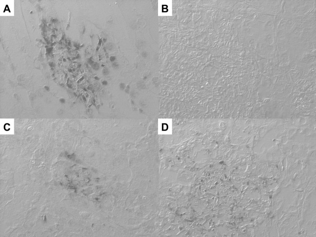
Specificity verification of the Trichosporon spp. PNA probe and assessments of rRNA retention and its hybridizability in experimentally infected mice. (A) ISH using the Trichosporon spp. PNA probe in renal tissue from mice infected with T. asahii. Strong positive signals against 28S rRNA of Trichosporon spp. were observed in the specimen. (B) ISH using the Trichosporon spp. PNA probe in renal tissue from mice infected with C. albicans. Positive signals were not observed in the specimen. (C) ISH result with the panfungal PNA probe in renal tissue from mice infected with T. asahii. Strong positive signals were observed in the specimen. (D) ISH result with the panfungal PNA probe in renal tissue from mice infected with C. albicans. Strong positive signals were observed in the specimen. Magnification, ×400.
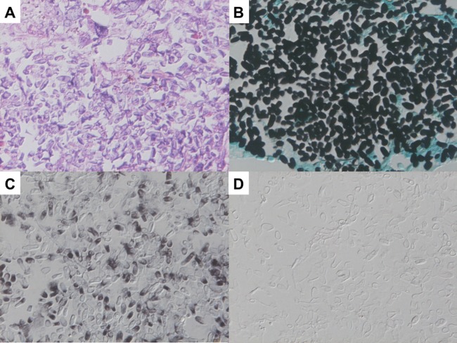
Results of ISH with a pulmonary lesion of disseminated trichosporonosis confirmed by DNA sequence analysis. (A) Pathological findings with hematoxylin and eosin stain. Histological examination revealed foci consisting of yeast formations of organisms. (B) Findings with Grocott's stain. Grocott's stain showed oval or square yeast-like elements within foci of infection. (C) Result of ISH with the Trichosporon spp. PNA probe. The PNA probe against Trichosporon spp. was strongly reactive with the yeast-like elements of Trichosporon spp. (D) ISH result with the C. albicans PNA probe. The PNA probe against C. albicans was not reactive with any Trichosporon spp. organisms.
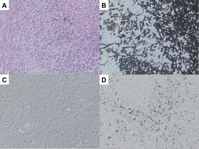
Results of ISH for pulmonary lesions in the case of culture-proven C. albicans. (A) Pathological findings with hematoxylin and eosin stain. Histological examination revealed foci consisting of pseudohyphal and yeast formations of organisms. (B) Results with Grocott's stain. Grocott's stain showed oval yeast-like and pseudohyphal elements within the foci of infection. (C) ISH result with the Trichosporon spp. PNA probe. The PNA probe against Trichosporon spp. was not reactive with any organisms of C. albicans. (D) ISH result with the C. albicans PNA probe. The PNA probe against C. albicans was strongly reactive with pseudohyphal and yeast-like elements of C. albicans.
Similar articles
-
Histopathological study on the prevalence of trichosporonosis in formalin-fixed and paraffin-embedded tissue autopsy sections by in situ hybridization with peptide nucleic acid probe.Med Mycol. 2020 Jun 1;58(4):460-468. doi: 10.1093/mmy/myz096. Med Mycol. 2020. PMID: 31535126 Free PMC article.
-
Identification of Fusarium species in formalin-fixed and paraffin-embedded sections by in situ hybridization using peptide nucleic acid probes.J Clin Microbiol. 2011 Mar;49(3):808-13. doi: 10.1128/JCM.01149-10. Epub 2010 Nov 24. J Clin Microbiol. 2011. PMID: 21106796 Free PMC article.
-
[Histopathological Diagnosis of Invasive Fungal Infections in Formalin-Fixed and Paraffin-Embedded Tissues in Conjunction with Molecular Methods].Med Mycol J. 2018;59(1):E7-E18. doi: 10.3314/mmj.17-00016. Med Mycol J. 2018. PMID: 29491339 Japanese.
-
[Development and evaluation of nucleic acid-based techniques for an auxiliary diagnosis of invasive fungal infections in formalin-fixed and paraffin-embedded (FFPE) tissues].Med Mycol J. 2012;53(4):241-5. doi: 10.3314/mmj.53.241. Med Mycol J. 2012. PMID: 23257724 Review. Japanese.
-
Technical Aspects and Applications for Developing in situ Hybridization Procedures for Formalin-Fixed and Paraffin-Embedded (FFPE) Tissues for Diagnosis of Fungal Infections.Med Mycol J. 2017;58(1):E33-E37. doi: 10.3314/mmj.16-00025. Med Mycol J. 2017. PMID: 28250362 Review.
Cited by
-
Differentiation of the emerging human pathogens Trichosporon asahii and Trichosporon asteroides from other pathogenic yeasts and moulds by using species-specific monoclonal antibodies.PLoS One. 2014 Jan 2;9(1):e84789. doi: 10.1371/journal.pone.0084789. eCollection 2014. PLoS One. 2014. PMID: 24392156 Free PMC article.
-
Histopathological study on the prevalence of trichosporonosis in formalin-fixed and paraffin-embedded tissue autopsy sections by in situ hybridization with peptide nucleic acid probe.Med Mycol. 2020 Jun 1;58(4):460-468. doi: 10.1093/mmy/myz096. Med Mycol. 2020. PMID: 31535126 Free PMC article.
-
Pathophysiological implication of reversed CT halo sign in invasive pulmonary mucormycosis: a rare case report.Diagn Pathol. 2013 May 17;8:82. doi: 10.1186/1746-1596-8-82. Diagn Pathol. 2013. PMID: 23683872 Free PMC article.
-
Rare fungal infectious agents: a lurking enemy.F1000Res. 2017 Oct 31;6:1917. doi: 10.12688/f1000research.11124.1. eCollection 2017. F1000Res. 2017. PMID: 29152230 Free PMC article. Review.
References
-
- Nahass GT, Rosenberg SP, Leonardi CL, Penneys NS. 1993. Disseminated infection with Trichosporon beigelii. Report of a case and review of the cutaneous and histologic manifestations. Arch. Dermatol. 129:1020–1023 - PubMed
-
- Walsh TJ. 1989. Trichosporonosis. Infect. Dis. Clin. North Am. 3:43–52 - PubMed
-
- Walsh TJ, Groll A, Hiemenz J, Fleming R, Roilides E, Anaissie E. 2004. Infections due to emerging and uncommon medically important fungal pathogens. Clin. Microbiol. Infect. 10(Suppl. 1):48–66 - PubMed
-
- Kume H, Yamazaki T, Togano T, Abe M, Tanuma H, Kawana S, Okudaira M. 2011. Epidemiology of visceral mycoses in autopsy cases in Japan: comparison of the data from 1989, 1993, 1997, 2001, 2005 and 2007 in the Annual of Pathological Autopsy Cases in Japan. Med. Mycol. J. 52:117–127 - PubMed
-
- Shimodaira K, Okubo Y, Nakayama H, Wakayama M, Shinozaki M, Ishiwatari T, Sasai D, Nemoto T, Takahashi K, Ishii T, Saji T, Shibuya K. 2012. Trends in the prevalence of invasive fungal infections from an analysis of annual records of autopsy cases of Toho University. Mycoses 55:435–443 - PubMed
Publication types
MeSH terms
Substances
LinkOut - more resources
Full Text Sources
