Interspecies protein substitution to investigate the role of the lyssavirus glycoprotein
- PMID: 23100360
- PMCID: PMC3709617
- DOI: 10.1099/vir.0.048827-0
Interspecies protein substitution to investigate the role of the lyssavirus glycoprotein
Abstract
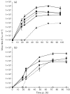
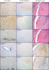
European bat lyssaviruses type 1 (EBLV-1) and type 2 (EBLV-2) circulate within bat populations throughout Europe and are capable of causing disease indistinguishable from that caused by classical rabies virus (RABV). However, the determinants of viral fitness and pathogenicity are poorly understood. Full-length genome clones based on the highly attenuated, non-neuroinvasive, RABV vaccine strain (SAD-B19) were constructed with the glycoprotein (G) of either SAD-B19 (SN), of EBLV-1 (SN-1) or EBLV-2 (SN-2). In vitro characterization of SN-1 and SN-2 in comparison to wild-type EBLVs demonstrated that the substitution of G affected the final virus titre and antigenicity. In vivo, following peripheral infection with a high viral dose (10(4) f.f.u.), animals infected with SN-1 had reduced survivorship relative to infection with SN, resulting in survivorship similar to animals infected with EBLV-1. The histopathological changes and antigen distribution observed for SN-1 were more representative of those observed with SN than with EBLV-1. EBLV-2 was unable to achieve a titre equivalent to that of the other viruses. Therefore, a reduced-dose experiment (10(3) f.f.u.) was undertaken in vivo to compare EBLV-2 and SN-2, which resulted in 100 % survivorship for all recombinant viruses (SN, SN-1 and SN-2) while clinical disease developed in mice infected with the EBLVs. These data indicate that interspecies replacement of G has an effect on virus titre in vitro, probably as a result of suboptimal G-matrix protein interactions, and influences the survival outcome following a peripheral challenge with a high virus titre in mice.
Figures


Similar articles
-
Lyssavirus infection: 'low dose, multiple exposure' in the mouse model.Virus Res. 2014 Mar 6;181:35-42. doi: 10.1016/j.virusres.2013.12.029. Epub 2013 Dec 29. Virus Res. 2014. PMID: 24380842
-
Chimeric rabies viruses for trans-species comparison of lyssavirus glycoprotein ectodomain functions in virus replication and pathogenesis.Berl Munch Tierarztl Wochenschr. 2012 May-Jun;125(5-6):219-27. Berl Munch Tierarztl Wochenschr. 2012. PMID: 22712419
-
Susceptibility of sheep to European bat lyssavirus type-1 and -2 infection: a clinical pathogenesis study.Vet Microbiol. 2007 Dec 15;125(3-4):210-23. doi: 10.1016/j.vetmic.2007.05.031. Epub 2007 Jun 15. Vet Microbiol. 2007. PMID: 17706900
-
Molecular Epidemiology and Evolution of European Bat Lyssavirus 2.Int J Mol Sci. 2018 Jan 5;19(1):156. doi: 10.3390/ijms19010156. Int J Mol Sci. 2018. PMID: 29303971 Free PMC article. Review.
-
European bat lyssaviruses: an emerging zoonosis.Epidemiol Infect. 2003 Dec;131(3):1029-39. doi: 10.1017/s0950268803001481. Epidemiol Infect. 2003. PMID: 14959767 Free PMC article. Review.
Cited by
-
Assessing Rabies Vaccine Protection against a Novel Lyssavirus, Kotalahti Bat Lyssavirus.Viruses. 2021 May 20;13(5):947. doi: 10.3390/v13050947. Viruses. 2021. PMID: 34065574 Free PMC article.
-
Characterisation of a Live-Attenuated Rabies Virus Expressing a Secreted scFv for the Treatment of Rabies.Viruses. 2023 Jul 31;15(8):1674. doi: 10.3390/v15081674. Viruses. 2023. PMID: 37632016 Free PMC article.
-
Pathogenicity and Immunogenicity of Recombinant Rabies Viruses Expressing the Lagos Bat Virus Matrix and Glycoprotein: Perspectives for a Pan-Lyssavirus Vaccine.Trop Med Infect Dis. 2017 Aug 9;2(3):37. doi: 10.3390/tropicalmed2030037. Trop Med Infect Dis. 2017. PMID: 30270894 Free PMC article.
-
Everything You Always Wanted to Know About Rabies Virus (But Were Afraid to Ask).Annu Rev Virol. 2015 Nov;2(1):451-71. doi: 10.1146/annurev-virology-100114-055157. Epub 2015 Jun 24. Annu Rev Virol. 2015. PMID: 26958924 Free PMC article. Review.
-
Taiwan Bat Lyssavirus: In Vitro and In Vivo Assessment of the Ability of Rabies Vaccine-Derived Antibodies to Neutralise a Novel Lyssavirus.Viruses. 2022 Dec 9;14(12):2750. doi: 10.3390/v14122750. Viruses. 2022. PMID: 36560754 Free PMC article.
References
-
- Amengual B., Whitby J. E., King A., Cobo J. S., Bourhy H. (1997). Evolution of European bat lyssaviruses. J Gen Virol 78, 2319–2328 - PubMed
-
- Blaney J. E., Wirblich C., Papaneri A. B., Johnson R. F., Myers C. J., Juelich T. L., Holbrook M. R., Freiberg A. N., Bernbaum J. G. & other authors (2011). Inactivated or live-attenuated bivalent vaccines that confer protection against rabies and Ebola viruses. J Virol 85, 10605–10616 10.1128/JVI.00558-11 - DOI - PMC - PubMed
-
- Cenna J., Tan G. S., Papaneri A. B., Dietzschold B., Schnell M. J., McGettigan J. P. (2008). Immune modulating effect by a phosphoprotein-deleted rabies virus vaccine vector expressing two copies of the rabies virus glycoprotein gene. Vaccine 26, 6405–6414 10.1016/j.vaccine.2008.08.069 - DOI - PMC - PubMed
Publication types
MeSH terms
Substances
LinkOut - more resources
Full Text Sources
Other Literature Sources

