. 2012 Dec 4;79(23):2241-8.
doi: 10.1212/WNL.0b013e31827689ad. Epub 2012 Oct 24.

Neurofascin as a target for autoantibodies in peripheral neuropathies

Affiliations

Neurofascin as a target for autoantibodies in peripheral neuropathies

Judy King Man Ng et al. Neurology. .

Abstract

Objectives: We asked whether autoantibodies against neurofascin (NF)186 or NF155, both localized at the nodes of Ranvier, are present in serum of patients with inflammatory neuropathy, and whether NF-specific monoclonal antibodies are pathogenic in vivo.

Methods: We cloned human NF155 and NF186, and developed an ELISA and cell-based assay to screen for antibodies to human NF in a total of 434 donors including 294 patients with Guillain-Barré syndrome variants acute inflammatory demyelinating polyneuropathy (AIDP), acute motor axonal neuropathy, and chronic inflammatory demyelinating polyneuropathy (CIDP). We characterized reactive samples by isotyping, tissue section staining, and epitope mapping. We also injected NF-specific monoclonal antibodies IV into rats with experimental autoimmune neuritis.

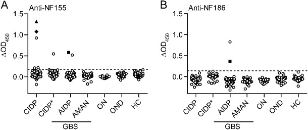
Results: We detected autoantibodies to NF by ELISA in 4% of patients with AIDP and CIDP, but not in controls. Most positive samples contained immunoglobulin G (IgG)1, IgG3, or IgG4 antibodies directed to only one isoform of NF. Two patients with CIDP showed particularly high (1:10,000 dilution) NF155-specific reactivity in both assays and stained paranodes. Two other patients with CIDP who benefited from plasma exchange exhibited antibodies to NF155 by ELISA, and upon affinity purification, antibodies to both isoforms were observed by both assays. Anti-NF monoclonal antibodies enhanced and prolonged induced neuritis in rats.

Conclusions: Autoantibodies to NF are detected in a very small proportion of patients with AIDP and patients with CIDP, but may nevertheless be pathogenic in these cases.


Figures

Figure 1
Figure 1. Autoantibodies to NF155 and NF186 in a very small proportion of patients with neuropathy
Serum samples from patients with chronic inflammatory demyelinating polyneuropathy (CIDP) (n = 117), acute inflammatory demyelinating polyneuropathy (AIDP) (n = 65), acute motor axonal neuropathy (AMAN) (n = 50), other neuropathies (ON) (n = 20), and controls with other neurologic diseases (OND) (n = 61) and healthy controls (HC) (n = 77) were tested for autoantibodies to neurofascin (NF)155 and NF186 by ELISA. Plasma exchange samples from a separate cohort of patients with CIDP (CIDP*; n = 40) were tested at 100 μg/mL immunoglobulin concentration. ELISA reactivities to NF155 (A) and NF186 (B) are shown as baseline-subtracted optical density reading at 450 nm (ΔOD450; response to NF minus bovine serum albumin reactivity). The cutoff (dashed line) represents the mean of OND group plus 4 SDs. Two CIDP samples marked with special symbols (▲, ♦) showed reactivity by both ELISA and flow cytometry to NF155 (more details in figures 3 and e-2, C–K). One AIDP sample marked with a special symbol (■) showed reactivity by ELISA to both isoforms of NF. GBS = Guillain-Barré syndrome.
Figure 2
Figure 2. ratNF155-NS0 is not a specific antigen to detect antibodies to human neurofascin (NF)
Serum screening by ELISA using ratNF155-NS0 (A) included samples from patients with chronic inflammatory demyelinating polyneuropathy (CIDP) (n = 82), acute inflammatory demyelinating polyneuropathy (AIDP) (n = 54), acute motor axonal neuropathy (AMAN) (n = 50), and controls with other neurologic diseases (OND) (n = 61) and healthy controls (HC) (n = 77). The response to ratNF155-NS0 minus bovine serum albumin reactivity (delta OD) is shown. Most of the patients and control donors show a response against this antigen. The mean for each group is shown. Closed circles represent positive samples identified by ELISA or flow cytometry using human NF155 and NF186. Antibodies purified over ratNF155-NS0 conjugated column were tested by (B) ELISA and (C) Western blot for reactivity against HEK293–derived NF155 and NF186, ratNF155-NS0, and unrelated NS0-derived antigens: contactin-2-NS0 and oligodendrocyte myelin glycoprotein (OMgp-NS0). The number above each lane in (C) corresponds to the antigen with the same number in (B). Western blot analysis procedure is detailed in appendix e-5. Anti-pan-NF mAb (A4/3.4) was also tested by ELISA as a positive control for reactivity against NF155, NF186, and ratNF155-NS0 (B). GBS = Guillain-Barré syndrome.
Figure 3
Figure 3. Features of antibodies to neurofascin (NF) in a patient with chronic inflammatory demyelinating polyneuropathy (CIDP)
Reactivity to NF155 was seen by flow cytometry (A–C) up to a dilution of 1:10,000 (B). (A) Reactivity to NF155 (blue line) was compared with reactivity to NF186 (orange line) and to control cells (black, filled). (B) The mean fluorescence intensity (MFI) ratio is plotted for level of reactivity above background. (C) The NF155 reactivity was mediated by immunoglobulin (Ig)G4 with minor contribution from IgG1. Reactivity to NF155 was also seen by ELISA (D–F) up to a dilution of 1:10,000 (E) by IgG4 and weakly by IgG1, IgG3, IgM, and IgA (F). Serum staining (G, left) colocalizes with Caspr staining (G, middle) on longitudinal rat spinal cord sections. The scale bar represents 10 μm. (H) NF186 differs from NF155 by substitution of Fn3-Fn4 with Fn4-Mucin-Fn5 (Ig = immunoglobulin-like domain; Fn = fibronectin type III domain). (I) A scheme of super green fluorescent protein (sGFP) fusion truncated NF variants is shown beside the corresponding serum reactivity by flow cytometry. Reactivities to truncated NF variants and to negative control cells are shown as sGFP intensity vs serum reactivity. The fragment recognized by both NF155-reactive serum samples is boxed.
Figure 4
Figure 4. Two different monoclonal antibodies to neurofascin enhance and prolong experimental autoimmune neuritis
Lewis rats immunized with P2 peptide were injected with either anti-pan-neurofascin monoclonal antibodies A12/18.1 mouse immunoglobulin (Ig)G2a (A) or A4/3.4 mouse IgM (B) (closed symbols) or their respective isotype controls (open symbols) at disease onset (day 14 after experimental autoimmune neuritis [EAN] induction, indicated by arrowhead). They were scored blinded daily for EAN disease severity. Values (mean ± SEM) represent EAN clinical scores from 6 rats per group, pooled from 2 independent experiments.

Comment in

References

    1. Lehmann HC, Meyer zu Horste G, Kieseier BC, Hartung HP. Review: pathogenesis and treatment of immune-mediated neuropathies. Ther Adv Neurol Disord 2009;2:261–281 - PMC - PubMed
    1. Hughes RAC, Cornblath DR. Guillain-Barré syndrome. Lancet 2005;366:1653–1666 - PubMed
    1. Willison HJ, Yuki N. Peripheral neuropathies and anti-glycolipid antibodies. Brain 2002;125:2591–2625 - PubMed
    1. Kaida K, Kusunoki S. Antibodies to gangliosides and ganglioside complexes in Guillain-Barré syndrome and Fisher syndrome: mini-review. J Neuroimmunol 2010;223:5–12 - PubMed
    1. Illa I, Ortiz N, Gallard E, et al. Acute axonal Guillain-Barré syndrome with IgG antibodies against motor axons following parenteral gangliosides. Ann Neurol 1995;38:218–224 - PubMed

Publication types

MeSH terms