. 2012 Oct 24;32(43):15076-85.
doi: 10.1523/JNEUROSCI.3128-12.2012.

Ventral tegmental area glutamate neurons: electrophysiological properties and projections

Affiliations

Ventral tegmental area glutamate neurons: electrophysiological properties and projections

Thomas S Hnasko et al. J Neurosci. .

Abstract

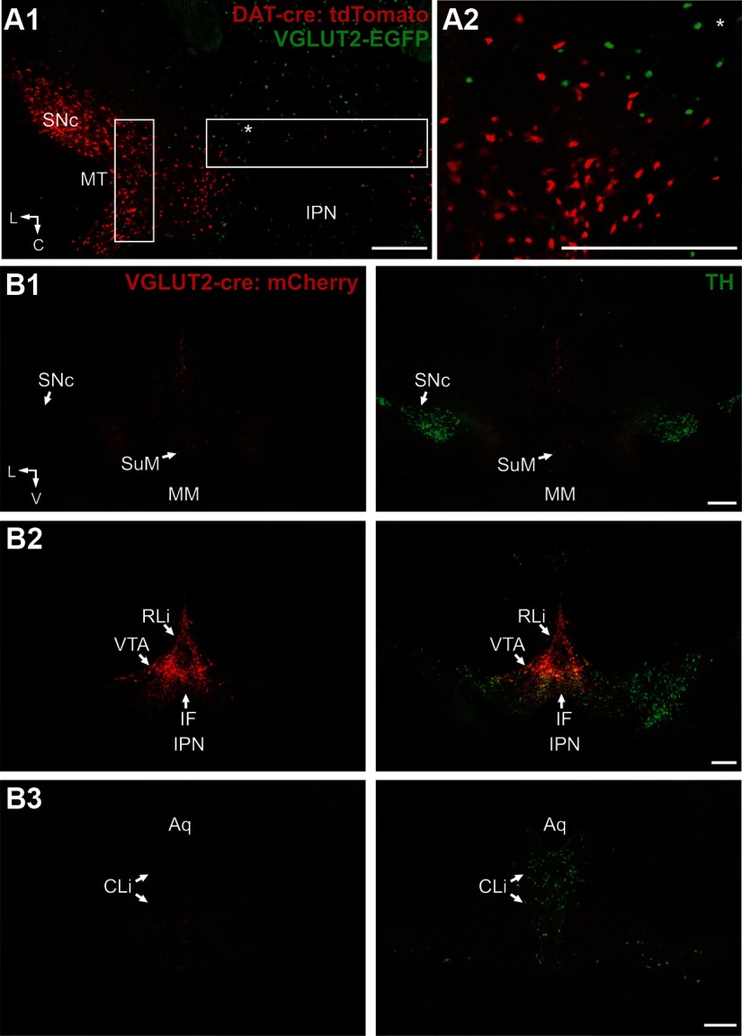
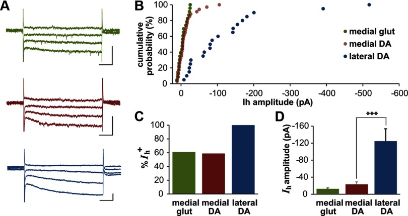
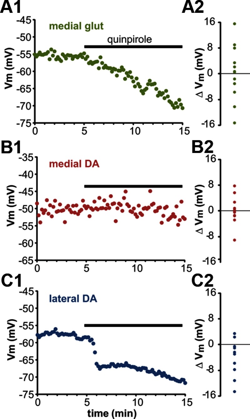
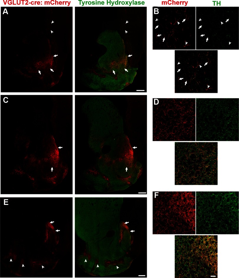
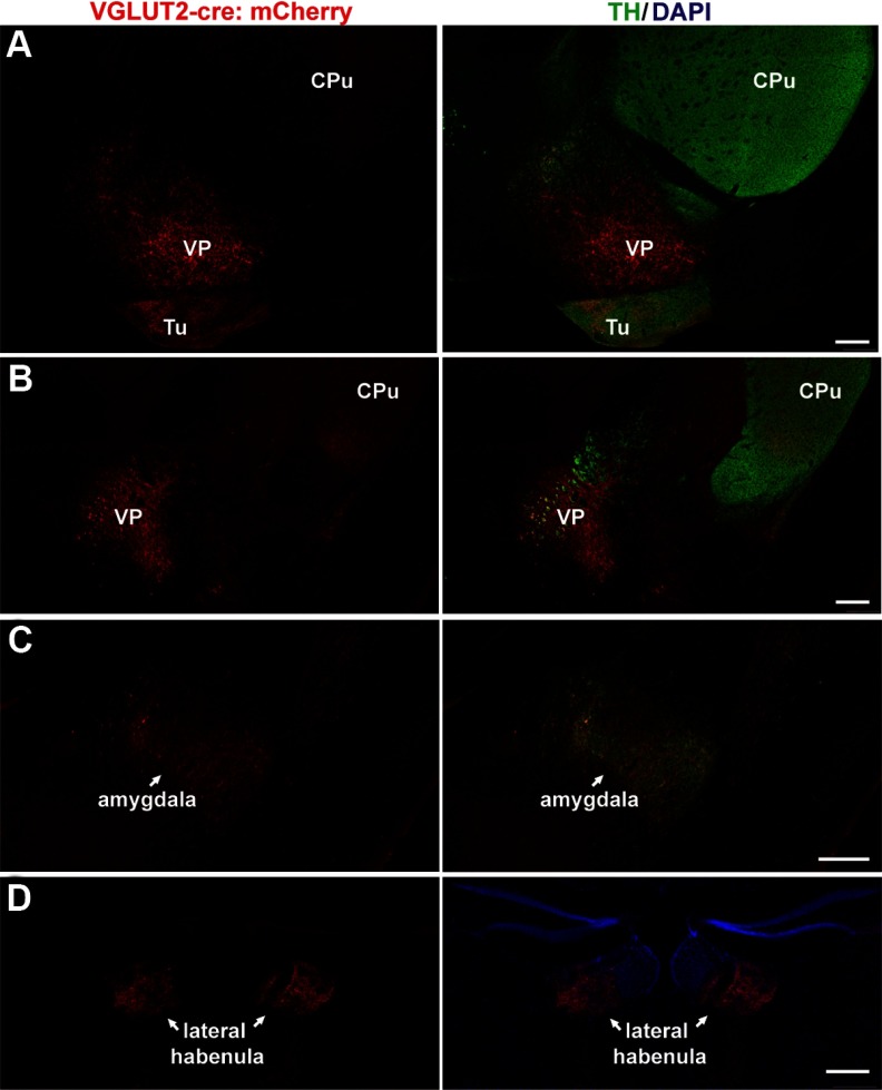
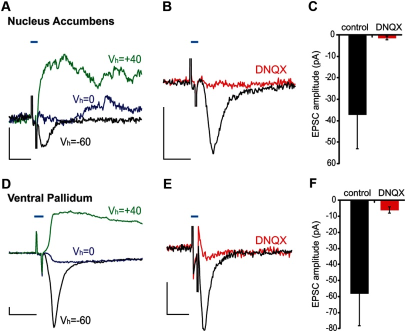
The ventral tegmental area (VTA) has a central role in the neural processes that underlie motivation and behavioral reinforcement. Although thought to contain only dopamine and GABA neurons, the VTA also includes a recently discovered population of glutamate neurons identified through the expression of the vesicular glutamate transporter VGLUT2. A subset of VGLUT2(+) VTA neurons corelease dopamine with glutamate at terminals in the NAc, but others do not express dopaminergic markers and remain poorly characterized. Using transgenic mice that express fluorescent proteins in distinct cell populations, we now find that both dopamine and glutamate neurons in the medial VTA exhibit a smaller hyperpolarization-activated current (I(h)) than more lateral dopamine neurons and less consistent inhibition by dopamine D(2) receptor agonists. In addition, VGLUT2(+) VTA neurons project to the nucleus accumbens (NAc), lateral habenula, ventral pallidum (VP), and amygdala. Optical stimulation of VGLUT2(+) projections expressing channelrhodopsin-2 further reveals functional excitatory synapses in the VP as well as the NAc. Thus, glutamate neurons form a physiologically and anatomically distinct subpopulation of VTA projection neurons.


Figures

Figure 1.
Figure 1.
Identification of VTA glutamate neurons. A1, Horizontal section through the VTA of a mouse expressing GFP under the control of VGLUT2 regulatory elements (VGLUT2-GFP), Cre recombinase under the control of DAT regulatory elements (DAT-Cre), and the Rosa26 flox-stop tdTomato reporter to identify glutamate and catecholamine neurons, respectively. For whole-cell recordings, GFP+ glutamate and tdTomato+ dopamine neurons were defined as medial (within the horizontal box rostral to the interpeduncular nucleus, IPN), or lateral (within the vertical box near MT, the medial terminal nucleus of the accessory optic tract). A2, Magnified image of medial VTA (reference asterisk (*) marks the same location in A1 and A2). B1, B2, B3, Conditional viral vector AAV-EF1α-DIO-ChR2-mCherry was injected into the VTA of VGLUT2-Cre mice, and >3 weeks later coronal sections from rostral (bregma, −2.9 mm) (B1), central (bregma, −3.7 mm) (B2), and caudal (bregma, −4.3 mm) (B3) midbrain were stained for mCherry (red) and TH (green). In this mouse, virus spread (indicated by mCherry expression) was limited to the VTA and supramammillary nucleus (SuM). Scale bars, 250 μm; MM, mammillary nucleus; IF, interfascicular nucleus; RLi, rostral linear nucleus; CLi, caudal linear nucleus; Aq, aqueduct; C, caudal; L, lateral; V, ventral.
Figure 2.
Figure 2.
Medial dopamine and glutamate neurons express less hyperpolarization-activated current, Ih, than lateral VTA dopamine neurons. A, Representative traces of Ih+ medial glutamate (green), medial dopamine (red), and lateral dopamine (blue) neurons in the VTA. Recording in voltage clamp, the cells were held at −60 mV and jumped sequentially to −50, −80, −100, and −120 mV. Scale bars, 100 pA and 100 ms. B, Cumulative frequency distribution of Ih amplitudes after a −60 to −120 mV step shows much larger Ih in lateral VTA dopamine neurons. C, The lateral VTA contains a larger fraction of cells that express detectable Ih. D, Average Ih amplitude of Ih+ cells. Data were analyzed by one-way ANOVA across groups (main effect of group; F(2,51) = 11.0, p < 0.001) followed by Tukey's post hoc test (*p < 0.001); n = 14 (medial glut), 20 (medial DA), 20 (lateral DA).
Figure 3.
Figure 3.
Response to D2 dopamine receptor (D2R) activation differs among medial glutamate, medial dopamine, and lateral dopamine VTA neurons. A1, A2, A VTA glutamate neuron hyperpolarizes in response to quinpirole (A1), but a scatter plot distribution of responses shows that VTA glutamate neurons respond heterogeneously to D2R agonist application (A2). B1, B2, A medial VTA dopamine neuron does not respond to quinpirole (B1), but the distribution shows similarly heterogeneous responsiveness to D2 stimulation by medial VTA dopamine neurons (B2). C1, C2, Lateral VTA dopamine neurons hyperpolarize in response to quinpirole as shown by the example time course (C1) and distribution of responses (C2). Several cells inhibited by quinpirole were firing at the time of drug application and so were not included in the scatter plot distributions of ΔVm.
Figure 4.
Figure 4.
VTA glutamate neurons project to nucleus accumbens and prefrontal cortex. A, B, More than 3 weeks after stereotactic injection of AAV-EF1α-DIO-ChR2-mCherry into the medial VTA (Fig. 1B), a coronal section through the striatum (A) shows strong labeling of glutamatergic projections (red) from the VTA to the medial and ventromedial shell of the nucleus accumbens (NAc) (arrows). Sparse labeling also occurs in the PFC (arrowheads). Sections were double stained for TH (green) to identify the projections from midbrain dopamine neurons. A confocal image of the PFC (B) shows mCherry+ glutamate fibers that colocalize with TH (arrows) and others which do not (arrowheads). C, D, In a coronal section through the central NAc (C), dense mCherry+ glutamatergic fibers project throughout the shell of the NAc, in particular medially (arrows). A confocal image in the NAc shell (D) demonstrates widespread colocalization of mCherry+ (glutamatergic) and dopaminergic fibers. E, F, A coronal section through the caudal NAc (E) shows mCherry+ VTA glutamate projections concentrated in the dorsal cone of the medial shell (arrow) and (F) colocalizing with TH by confocal microscopy. Glutamate fibers from the VTA are also observed in the rostral finger-like projections of the VP (arrowheads), and these label only sparsely for TH (see Fig. 5). Scale bars, A, C, E, 250 μm; B, D, F, 50 μm.
Figure 5.
Figure 5.
VTA glutamate neurons project to ventral pallidum, amygdala, and lateral habenula. More than 3 weeks after injection of AAV-EF1α-DIO-ChR2-mCherry (Fig. 1B), processes in rostral (A) and caudal (B) regions of the VP stain strongly for VGLUT2 (red, arrows) but only sparsely for TH (green). In contrast, fibers of the medial forebrain bundle and the caudal caudate–putamen (CPu) dorsal to the VP stain strongly for TH (A, B). Tu, Olfactory tubercle. C, Glutamate fibers from the VTA (arrows) also innervate the amygdala, along with TH+ dopamine terminals. D, The LHb receives dense bilateral innervation (arrows) by mCherry+ VTA neurons (red). No significant TH expression was observed in this region (data not shown). Scale bars, 250 μm.
Figure 6.
Figure 6.
VTA projections form functional synapses in both the nucleus accumbens and ventral pallidum. A, More than 3 weeks after stereotactic injection of AAV-EF1α-DIO-ChR2-mCherry into the medial VTA, striatal slices show light-evoked currents in NAc neurons. Representative traces from NAc neurons held at the potentials indicated show both AMPAR-mediated (black trace) and NMDAR-mediated (green trace) excitatory currents. B, C, The AMPAR antagonist DNQX (red trace) blocks the AMPAR-mediated currents observed at −60 mV (5.9% ± 3.9% residual current following DNQX, p < 0.001 by within-cell paired t test, n = 5) D–F, Neurons in the VP exhibit similar light-evoked synaptic currents (D) and those mediated by AMPAR are also DNQX-sensitive (E, F) (9.9% ± 3.9% residual current following DNQX, p < 0.001 by within-cell paired t test, n = 10). Black scale bars, 20 pA, 10 ms; blue bar represents the 2 ms blue light pulse.

Similar articles

Cited by

References

    1. Bäckman CM, Malik N, Zhang Y, Shan L, Grinberg A, Hoffer BJ, Westphal H, Tomac AC. Characterization of a mouse strain expressing Cre recombinase from the 3′ untranslated region of the dopamine transporter locus. Genesis. 2006;44:383–390. - PubMed
    1. Borgius L, Restrepo CE, Leao RN, Saleh N, Kiehn O. A transgenic mouse line for molecular genetic analysis of excitatory glutamatergic neurons. Mol Cell Neurosci. 2010;45:245–257. - PubMed
    1. Brischoux F, Chakraborty S, Brierley DI, Ungless MA. Phasic excitation of dopamine neurons in ventral VTA by noxious stimuli. Proc Natl Acad Sci U S A. 2009;106:4894–4899. - PMC - PubMed
    1. Cameron DL, Wessendorf MW, Williams JT. A subset of ventral tegmental area neurons is inhibited by dopamine, 5-hydroxytryptamine and opioids. Neuroscience. 1997;77:155–166. - PubMed
    1. Chuhma N, Zhang H, Masson J, Zhuang X, Sulzer D, Hen R, Rayport S. Dopamine neurons mediate a fast excitatory signal via their glutamatergic synapses. J Neurosci. 2004;24:972–981. - PMC - PubMed

Publication types

MeSH terms