. 2012 Oct 24;32(43):15112-23.
doi: 10.1523/JNEUROSCI.1729-12.2012.

Loss of interleukin receptor-associated kinase 4 signaling suppresses amyloid pathology and alters microglial phenotype in a mouse model of Alzheimer's disease

Affiliations

Loss of interleukin receptor-associated kinase 4 signaling suppresses amyloid pathology and alters microglial phenotype in a mouse model of Alzheimer's disease

Brent Cameron et al. J Neurosci. .

Abstract

Alzheimer's disease (AD) is typified by the deposition of amyloid in the brain, which elicits a robust microglial-mediated inflammatory response that is associated with disease exacerbation and accelerated progression. Microglia are the principal immune effector cells in the brain and interact with fibrillar forms of Aβ (fAβ) through a receptor complex that includes Toll-like receptors (TLR) 2/4/6 and their coreceptors. Interleukin receptor-associated kinases (IRAKs) are essential intracellular signaling molecules for transduction of TLR signals. Studies of mouse models of AD in which the individual TLRs are knocked out have produced conflicting results on roles of TLR signaling in amyloid homeostasis. Therefore, we disrupted a common downstream TLR signaling element, IRAK4. We report that microglial IRAK4 is necessary in vitro for fAβ to activate the canonical pro-inflammatory signaling pathways leading to activation of p38, JNK, and ERK MAP kinases and to generate reactive oxygen species. In vivo the loss of IRAK4 function results in decreased Aβ levels in a murine model of AD. This was associated with diminished microgliosis and astrogliosis in aged mice. Analysis of microglia isolated from the adult mouse brain revealed an altered pattern of gene expression associated with changes in microglial phenotype that were associated with expression of IRF transcription factors that govern microglial phenotype. Further, loss of IRAK4 function also promoted amyloid clearance mechanisms, including elevated expression of insulin-degrading enzyme. Finally, blocking IRAK function restored olfactory behavior. These data demonstrate that IRAK4 activation acts normally to regulate microglial activation status and influence amyloid homeostasis in the brain.


Figures

Figure 1.
Figure 1.
IRAK4 is required for fAβ stimulated pro-inflammatory signaling and ROS generation. A, Primary microglia were isolated from WT C57BL/6J or IRAK4 KI mice (P0–P2) and were treated with 5 μm fAβ1–42 for the indicated times. Cell lysates were collected and evaluated by Western blot for the indicated phosphorylated proteins. Blots were stripped and reprobed for nonphosphorylated proteins as loading controls. B, Microglia were treated with 100 ng/ml LPS, 2.5 μm fAβ1–42, or 390 nm PMA in the presence of NBT. Cells (>300 from 3 separate fields) were counted and the fraction blue formazan-positive cells determined. Data represent mean ± SEM for three independent experiments. ***p ≤ 0.05, one-way ANOVA with Tukey's post hoc analysis.
Figure 2.
Figure 2.
Loss of IRAK4 kinase activity reduces Aβ levels in vivo. Serial extraction of soluble (DEA extract) (A, B) and insoluble (formic acid extract) Aβ (C, D) was performed on brain homogenates of 4-month-old (A, C) or 8-month-old (B, D) APPPS1 or APPPS1;IRAK4KI/KI. Aβx-40 and Aβx-42 were detected using sandwich ELISA. At 4 months mean total Aβ (ng Aβ/mg total protein) for APPPS1 mice were DEA soluble Aβ40 = 3.05 and Aβ42 = 46.26 and formic acid (FA) soluble Aβ40 = 67.67 and Aβ42 = 637.39. At 8 months mean total Aβ values for APPPS1 mice were DEA soluble Aβ40 = 3.22 and Aβ42 = 44.95 and FA soluble Aβ40 = 88.96 and Aβ42 = 1500.00. These data represent normalized mean ± SEM. N ≥ 5 animals/genotype/age. *p ≤ 0.05, Student's two-tailed t test.
Figure 3.
Figure 3.
Loss of IRAK4 kinase activity reduces dense core plaques. APPPS1 (A, C, E, G) or APPPS1;IRAK4KI/KI (B, D, F, H) mice were killed at the indicated age and coronal sections were stained with thioflavin S. Cortical (I) and hippocampal (L) thioS-positive deposits from matching sections were counted and averaged. Plaque area in the cortex (J) and hippocampus (Hpc) (M) was quantified using ImagePro software. Plaque diameter in the cortex (K) and in the hippocampus (N) was quantified using ImagePro software. Graphs represent mean ± SEM at each age. N ≥ 5 animals/genotype/age. **p ≤ 0.01, Student's two-tailed t test for APPPS1 vs. APPPS1;IRAK4 KI.
Figure 4.
Figure 4.
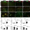
IRAK4 kinase activity is necessary for reactive gliosis. APPPS1 or APPPS1;IRAK4KI/KI mice were killed at 4 months (A–D) or 8 months (E–L) and coronal sections were stained for microglia (Iba1, green, A, B, E, F, I, J) or astrocytes (GFAP, green; C, D, G, H, K, L) and total amyloid (6e10, red; all images). A–H, 10× representative cortical images are shown. High-magnification representative images at 8 months of age are shown (I–L). Cortical area occupied by microglia (Iba1, M), astrocytes (GFAP, N), and Aβ (6E10, O) was quantified. 6E10-positive plaque diameter (P) was calculated. Cortical area occupied by microglia (Iba1, Q) or astrocytes (GFAP, R) was normalized to the corresponding Aβ plaque area (6E10). All image analysis (M–R) was performed with ImagePro software. Graphs represent mean ± SEM at each age. N ≥ 5 animals/genotype/age. *p ≤ 0.05, ***p ≤ 0.001, Student's two-tailed t test for APPPS1 vs. APPPS1;IRAK4 KI.
Figure 5.
Figure 5.
Loss of IRAK4 activity alters the inflammatory response of microglia. Microglia from the brain of 4-month-old (A, B) or 8-month-old (C, D) C57BL/6J, IRAK4KI/KI, APPPS1, or APPPS1;IRAK4KI/KI were isolated. mRNA was isolated, converted to cDNA, and analyzed by quantitative PCR. The data are expressed as RQ values with 95% confidence interval indicated. Representative M1 cytokines and markers (A, C) and M2 cytokines and markers (B, D) were assessed. All genotypes were normalized to expression of the individual genes in C57BL/6J (dotted line) using the ΔΔCt method. All statistics were done at the ΔCt level using the ΔCt ± SEM as indicated by Yuan et al. (2006). Samples were compared using one-way ANOVA with Tukey's post hoc test; n ≥ 8 animals/genotype/age. *p ≤ 0.05, **p ≤ 0.01, ***p ≤ 0.001, for APPPS1 vs. APPPS1;IRAK4KI/KI or Student's t test, #p ≤ 0.05 for APPPS1 vs. APPPS1–21;IRAK4KI/KI.
Figure 6.
Figure 6.
IRF transcriptional machinery shifts to an M2 state with loss of IRAK4 activity. Whole-brain homogenates were analyzed by Western blots for IRF4 and IRF5 at 4 months (A) and 8 months (B). Densitometric quantification of IRF4 (C) and IRF5 (D) of Western blots, normalized to β-actin, was performed. Graphs represent mean ± SEM; n ≥ 6 animals/genotype/age. Samples were compared with Student's t test. *p ≤ 0.05, **p ≤ 0.01, ***p ≤ 0.001. Microglia from the brain of 4-month-old (E) or 8-month-old (F) C57BL/6J, IRAK4KI/KI, APPPS1, or APPPS1;IRAK4KI/KI were isolated. mRNA was isolated, converted to cDNA, and analyzed via quantitative PCR. The data are expressed as RQ values with 95% confidence interval indicated. All genotypes were normalized to expression of the individual genes in C57BL/6J (dotted line) using the ΔΔCt method. All statistics were done at the ΔCt level using the ΔCt ± SEM as indicated by Yuan et al. (2006). Samples were compared using one-way ANOVA with Tukey's post hoc test; n ≥ 8 animals/genotype/age. *p ≤ 0.05, **p ≤ 0.01, and ***p ≤ 0.001, for APPPS1 vs. APPPS1;IRAK4KI/KI or Student's t test, and ##p ≤ 0.01 for APPPS1 vs. APPPS1–21;IRAK4KI/KI.
Figure 7.
Figure 7.
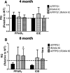
Loss of IRAK4 activity preserves amyloid clearance pathways. Microglia from the brain of 4-month-old (A) or 8-month-old (B) C57BL/6J, IRAK4KI/KI, APPPS1, or APPPS1;IRAK4KI/KI were isolated. mRNA was isolated, converted to cDNA, and analyzed via quantitative PCR. Graphs represent RQ values with 95% confidence interval indicated. All genotypes were normalized to expression of the individual genes in C57BL/6J (dotted line) using the ΔΔCt. All statistics were done at the ΔCt level using the ΔCt ± SEM as indicated by Yuan et al. (2006). Samples were compared using one-way ANOVA with Tukey's post hoc test; n ≥ 8 animals/genotype/age. *p ≤ 0.05, or ##p ≤ 0.01 (via Student's t test) for APPPS1 vs. APPPS1;IRAK4KI/KI.
Figure 8.
Figure 8.
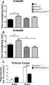
Loss of IRAK4 activity restores novel odor investigation. C57BL/6J, APPPS1;IRAK4KI/KI, and APPPS1;IRAK4KI/KI animals were subjected to a novel odor habituation protocol. Investigation times for Trial 1 odors are presented for 4-month-old mice (A) and 8-month-old mice (B). Four different odor presentations for Trial 1 for each animal were averaged such that each animal only contributed a single data point. Data represent mean ± SEM; n ≥ 5 animals/genotype/age. *p ≤ 0.05, one-way ANOVA with Tukey's post hoc test. APPPS1 or APPPS1;IRAK4KI/KI mice were killed at 4 or 8 months and coronal sections were stained with thioflavin S. Piriform cortex thioS-positive deposits from matching sections were counted and averaged (C). Data represent mean ± SEM; n ≥ 5 animals/genotype/age. **p ≤ 0.01, Student's t test within each age group.

References

    1. Ahyi AN, Chang HC, Dent AL, Nutt SL, Kaplan MH. IFN regulatory factor 4 regulates the expression of a subset of Th2 cytokines. J Immunol. 2009;183:1598–1606. - PMC - PubMed
    1. Akiyama H, et al. Inflammation and Alzheimer's disease. Neurobiol Aging. 2000;21:383–421. - PMC - PubMed
    1. An H, Hou J, Zhou J, Zhao W, Xu H, Zheng Y, Yu Y, Liu S, Cao X. Phosphatase SHP-1 promotes TLR- and RIG-I-activated production of type I interferon by inhibiting the kinase IRAK1. Nat Immunol. 2008;9:542–550. - PubMed
    1. Anderson CF, Mosser DM. A novel phenotype for an activated macrophage: the type 2 activated macrophage. J Leukoc Biol. 2002;72:101–106. - PubMed
    1. Bamberger ME, Harris ME, McDonald DR, Husemann J, Landreth GE. A cell surface receptor complex for fibrillar beta-amyloid mediates microglial activation. J Neurosci. 2003;23:2665–2674. - PMC - PubMed

Publication types

MeSH terms