. 2012 Nov 13;109(46):18944-9.
doi: 10.1073/pnas.1203306109. Epub 2012 Oct 25.

RAB-5 and RAB-10 cooperate to regulate neuropeptide release in Caenorhabditis elegans

Affiliations

RAB-5 and RAB-10 cooperate to regulate neuropeptide release in Caenorhabditis elegans

Nikhil Sasidharan et al. Proc Natl Acad Sci U S A. .

Abstract

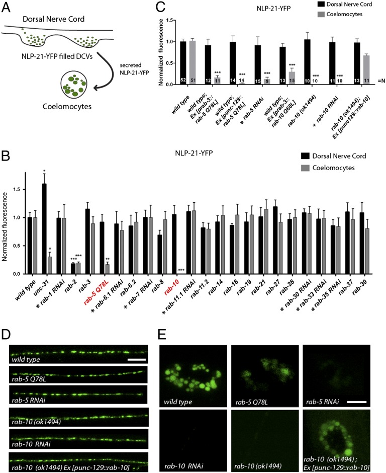
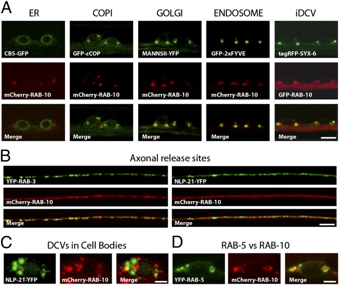
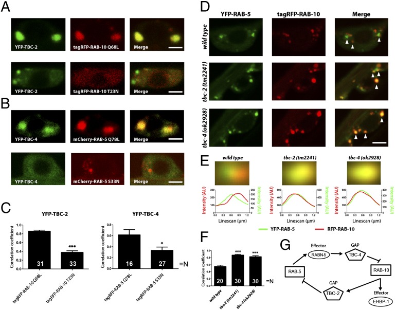
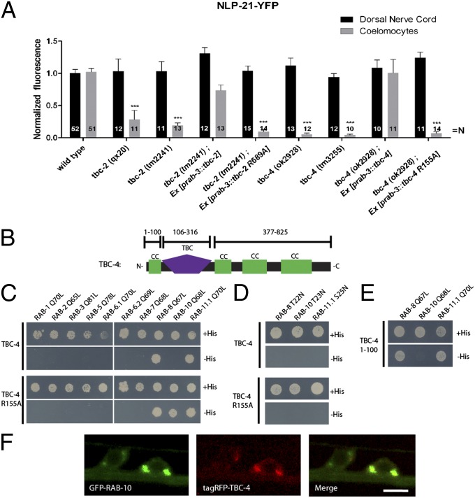
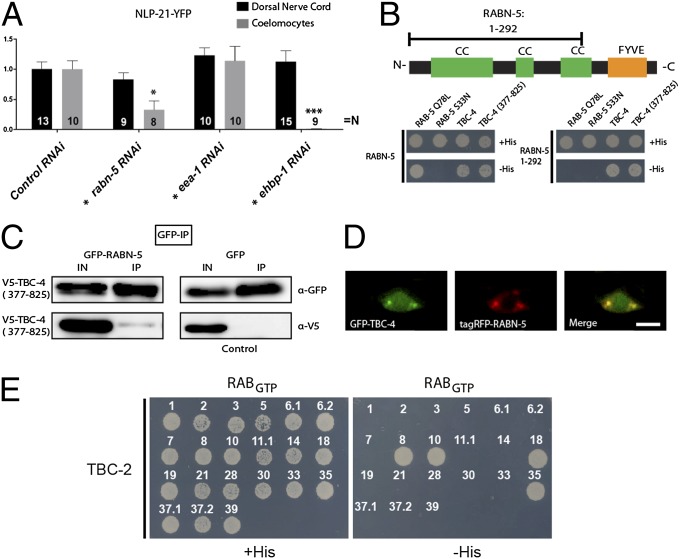
Neurons secrete neuropeptides from dense core vesicles (DCVs) to modulate neuronal activity. Little is known about how neurons manage to differentially regulate the release of synaptic vesicles (SVs) and DCVs. To analyze this, we screened all Caenorhabditis elegans Rab GTPases and Tre2/Bub2/Cdc16 (TBC) domain containing GTPase-activating proteins (GAPs) for defects in DCV release from C. elegans motoneurons. rab-5 and rab-10 mutants show severe defects in DCV secretion, whereas SV exocytosis is unaffected. We identified TBC-2 and TBC-4 as putative GAPs for RAB-5 and RAB-10, respectively. Multiple Rabs and RabGAPs are typically organized in cascades that confer directionality to membrane-trafficking processes. We show here that the formation of release-competent DCVs requires a reciprocal exclusion cascade coupling RAB-5 and RAB-10, in which each of the two Rabs recruits the other's GAP molecule. This contributes to a separation of RAB-5 and RAB-10 domains at the Golgi-endosomal interface, which is lost when either of the two GAPs is inactivated. Taken together, our data suggest that RAB-5 and RAB-10 cooperate to locally exclude each other at an essential stage during DCV sorting.


Conflict of interest statement

The authors declare no conflict of interest.

Figures

Fig. 1.
Fig. 1.
RAB-5 and RAB-10 are regulators of DCV secretion. (A) Schematic describing the NLP-21-YFP assay used for analyzing DCV secretion in C. elegans. (B) All rab GTPase mutants were systematically tested for defects in NLP-21-YFP secretion. For Rabs where no mutant was available, tissue-specific knockdown was conducted in DA and DB cholinergic motoneurons (indicated by *) or a dominant-active variant was expressed and analyzed. Note: RNAi experiments were normalized to knockdown of mock vector (L4440). Error bars indicate SEM. ***P < 0.001; **P < 0.01; *P < 0.05 (one-way ANOVA with Bonferroni post test). Representative pictures are shown in Fig. S1. (C) Secretion of NLP-21-YFP is impaired in rab-5 and rab-10 mutants Error bars indicate SEM. ***P < 0.001; (one-way ANOVA with Bonferroni post test). (D and E) NLP-21-YFP fluorescence levels in the DNC are unaffected (D), whereas fluorescence levels in the coelomocytes are decreased (E). Tissue-specific knockdown of rab-5 and rab-10 in neurons showed similar defects in secretion. [Scale bars: 6 μm (in DNC); 4 μm (in coelomocytes).]
Fig. 2.
Fig. 2.
Colocalization analysis of RAB-10. (A) Fine-mapping of the subcellular localization of RAB-10 in VNC neurons revealed no overlap with markers for the ER (CB5-GFP) and early Golgi/COPI vesicle (GFP-ɛCOP) and partial colocalization with markers for the medial Golgi (MannsII-YFP), endosomes (GFP-2xFYVE), and iDCVs (tagRFP-Syx-6). (Scale bar: 4 μm.) (B) RAB-10 is also enriched in axons at DNC synapses and showed colocalization with the SV marker (YFP-RAB-3) and DCV marker (NLP-21-YFP). (Scale bar: 5 μm.) (C) RAB-10 also displayed partial colocalization with the DCV marker (NLP-21-YFP) in neuronal cell bodies. (Scale bar: 1.5 μm.) (D) RAB-10 and RAB-5 localize to adjacent domains that rarely colocalize. (Scale bar: 1.5 μm.) Note: GFP-RAB-10 and tagRFP-SYX-6 are false-colored to enable simpler viewing.
Fig. 5.
Fig. 5.
GAP recruitment and domain formation by RAB-5 and RAB-10. (A) Typical images indicating the colocalization of dominant-active (Q68L) or -inactive (T23N) RAB-10 (fused to tagRFP, red), with the RAB-5 GAP, TBC-2 (fused to YFP, green). (B) Similar experiment performed for RAB-5 (fused to mCherry, red) and TBC-4 (fused to YFP, green). (Scale bar: 2 μm.) (C) Pearson’s correlation coefficients were calculated for the different genotypes. Note that the active Rab variants correlate with each other’s GAPs to significantly higher levels than inactive variants. ***P < 0.001; *P < 0.05 (Student's t test). (D) Colocalization between YFP-RAB-5 (green) and tagRFP-RAB-10 (red) in the presence or absence of the two GAPs. Arrowheads denote individual organelles. (Scale bar: 2.5 μm.) Note the increase in colocalization upon deletion of TBC-2 (middle images) or TBC-4 (bottom images). (E) The green and red images of YFP-RAB-5–containing organelles were averaged to determine organelle size and the distance between the positions of the RAB-5 and RAB-10 domains (see ref. and SI Materials and Methods for details). The organelle colors indicate that the overlap is much higher in absence of TBC-2 or TBC-4. To obtain numeric information, we performed line scans in the two color channels (lower graphs). The separation between the peaks of the line scans indicates the distance between the green and red fluorescence signals (45); Gaussian fits to the individual scans indicate the organelle (domain) sizes. Note that whereas the two Rabs colocalize perfectly in the absence of TBC-2 or TBC-4, their signals are shifted by ∼210 nm in the wild type (a value much lower than the organelle diameter, ∼570 nm; 75–99 organelles were averaged for each genotype). (F) Pearson’s correlation coefficients, calculated as above, give an additional indication that RAB-5 and RAB-10 colocalize better in the absence of TBC-2 or TBC-4. ***P < 0.001 (Student’s t test). (G) Scheme describing the interplay between RAB-5 and RAB-10 (see Discussion for details).
Fig. 3.
Fig. 3.
Two Rab GAPs display impairments in DCV release similar to rab-5 and rab-10 mutants. (A) The Rab GAP mutants, tbc-2 and tbc-4, have severe defects in DCV release. Error bars indicate SEM. ***P < 0.001 (one-way ANOVA with Bonferroni post test). (B) Schematic representation of the domain structure of TBC-4. CC, CC domain; TBC, tre-2/cdc16/Bub2 domain. (C and D) Full-length TBC-4 interacts with RAB-8 and RAB-11.1 (C) in a GTP-dependent manner (D). A catalytically inactive mutant of TBC-4 (R155A) specifically interacts with RAB-10 in a GTP-dependent manner. (E) RAB-8 and RAB-11.1 interact with an N-terminal CC domain of TBC-4 (1–100 aa). (F) TBC-4 and RAB-10 localize to the same compartments in VNC neurons. (Scale bar: 4 μm.)
Fig. 4.
Fig. 4.
Two Rab effectors also display impairments in DCV release. (A) Depletion of the RAB-5 and RAB-10 effectors, RABN-5 and EHBP-1, respectively, show defects in DCV release. Depletion of another RAB-5 effector, EEA-1, did not reveal any defect. Tissue-specific RNAi was conducted (as indicated by *). Error bars indicate SEM. ***P < 0.001; *P < 0.05 (one-way ANOVA with Bonferroni post test). (B) Schematic representation of the domain structure of RABN-5. Y2H analysis of RABN-5 and TBC-4 showed that the C-terminal CC domains of TBC-4 (377–825 aa) interacts with the N-terminal CC domains of RABN-5 (1–292 aa). CC, CC domains; FYVE, FYVE domain. (C) Coimmunoprecipitation of V5-TBC-4 (377–825 aa) and GFP-RABN-5 demonstrated that they also interact when expressed in COS7 cells. (D) TBC-4 and RABN-5 also localize to similar compartments in VNC neurons. (Scale bar: 4 μm.) (E) Y2H interaction analysis of full-length TBC-2 against C. elegans Rabs. TBC-2 interacts with GTP-bound RAB-8, RAB-10, RAB-18, and RAB-35.

Similar articles

Cited by

References

    1. Hu Z, Pym EC, Babu K, Vashlishan Murray AB, Kaplan JM. A neuropeptide-mediated stretch response links muscle contraction to changes in neurotransmitter release. Neuron. 2011;71:92–102. - PMC - PubMed
    1. Chalasani SH, et al. Neuropeptide feedback modifies odor-evoked dynamics in Caenorhabditis elegans olfactory neurons. Nat Neurosci. 2010;13:615–621. - PMC - PubMed
    1. Takamori S, et al. Molecular anatomy of a trafficking organelle. Cell. 2006;127:831–846. - PubMed
    1. Zhao B, et al. Transport of receptors, receptor signaling complexes and ion channels via neuropeptide-secretory vesicles. Cell Res. 2011;21:741–753. - PMC - PubMed
    1. Tooze SA, Martens GJ, Huttner WB. Secretory granule biogenesis: Rafting to the SNARE. Trends Cell Biol. 2001;11:116–122. - PubMed

Publication types

MeSH terms

LinkOut - more resources