. 2012 Oct 26;151(3):590-602.
doi: 10.1016/j.cell.2012.08.042.

A mouse model of Salmonella typhi infection

Affiliations

A mouse model of Salmonella typhi infection

Ramkumar Mathur et al. Cell. .

Abstract

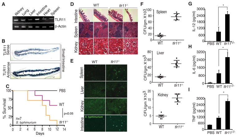
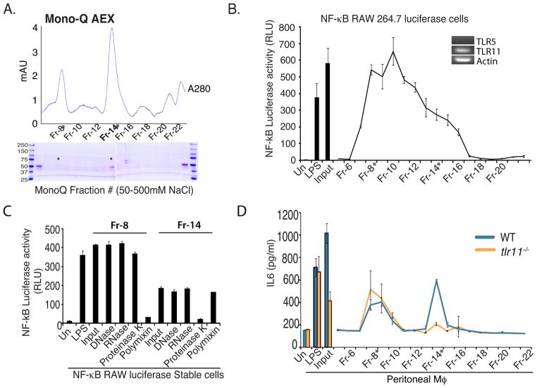
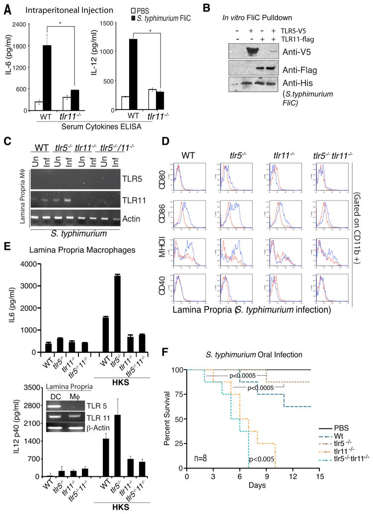
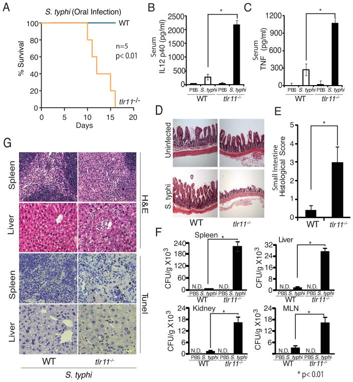
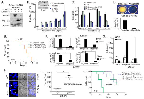
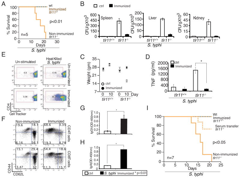
Salmonella spp. are gram-negative flagellated bacteria that can cause food- and waterborne gastroenteritis and typhoid fever in humans. We now report that flagellin from Salmonella spp. is recognized in mouse intestine by Toll-like receptor 11 (TLR11). Absence of TLR11 renders mice more susceptible to infection by S. Typhimurium, with increased dissemination of the bacteria and enhanced lethality. Unlike S. Typhimurium, S. Typhi, a human obligatory pathogen that causes typhoid fever, is normally unable to infect mice. TLR11 is expressed in mice, but not in humans, and remarkably, we find that tlr11(-/-) mice are efficiently infected with orally administered S. Typhi. We also find that tlr11(-/-) mice can be immunized against S. Typhi. Therefore, tlr11(-/-) mice represent a small-animal model for the study of the immune response to S. Typhi and for the development of vaccines against this important human pathogen.


Figures

Figure 1
Figure 1. TLR11 is expressed in intestine and protects against S. typhimurium
(A) RT PCR analysis of TLR11 in various organs. (B) In situ hybridization staining (antisense) for TLR11 in small-intestine tissue section. (C) Survival of wt and tlr11−/− mice (n=7) orally infected with S. typhimurium (108 CFU). (D) Tissue damage during S. typhimurium infection shown by H&E staining in small intestine, spleen and kidney on day 5 post infection. (E) Intestinal invasion of S. typhimurium expressing GFP. (F) Dissemination of GFP S. typhimurium in orally infected wt or tlr11−/− mice was detected on day 5 post infection by measuring CFU per gram of spleen, liver and kidney. (G-I) Day 5 serum cytokines from wt or tlr11−/− mice orally infected with S. typhimurium measured by ELISA. * p<0.001
Figure 2
Figure 2. TLR11 recognizes S. typhimurium flagellin
(A) The TLR11 ligand was purified by mechanical disruption of S. typhimurium followed by sequential DEAE-sephacel and CMSephacel chromatography. Fractions with stimulatory activity in Raw-κB-luc cells were further subjected to MonoQ FPLC with a linear NaCl gradient. Shown is absorbance at 280nm over the course of the elution. The fractions indicated were resolved by SDS PAGE and proteins were visualized by Coomassie staining. (B) Stimulatory activity of MonoQ fractions was assessed in Raw-κB-luc cells. (C) Fractions #8 and #14 were subjected to DNase, RNase, Proteinase K and Polymixin B treatment and assessed for stimulatory activity in Raw-κB-luc cells. (D) Peritoneal macrophages from WT or tlr11−/− mice were stimulated with Mono-Q fractions and IL-6 production was analyzed by ELISA.
Figure 3
Figure 3. Recognition of S. typhimurium flagellin by TLR5 and TLR11
(A) WT and tlr11−/− mice (n=5) were injected intraperitoneally with recombinant S. typhimurium flagellin and 2 hours later serum IL-6 and IL-12 were measured by ELSIA. * p<0.01. (B) TLR5 and TLR11 were expressed in HEK293 cells for 48 hours and TLR/flagellin complexes were formed by incubating cell lysates with His-tagged recombinant S. typhimurium flagellin (1μg/ml) overnight at 40C. Binding was assessed by Ni-sepharose pulldown followed by Western blotting with Anti-Flag, Anti-V5, and Anti-His antibodies. (C) Wild type, tlr11−/− and tlr5−/−/ tlr11−/− mice were challenged with S. typhimurium and 5-days later mice were sacrificed to obtained lamina propria macrophages by sorting (CD11CCD11b+ MHC-II+). RNA was prepared and analyzed for TLR5 and TLR11 expression by RT-PCR. (D) Lamina propria cells from above were analyzed for macrophage activation markers (CD80, CD86, MHC-II, CD40) by FACS. The blue traces are from infected mice. (E) Lamina propria macrophages and DCs were sorted from wild type mice and analyzed for TLR5 and TLR11 expression by RT-PCR (inset). Cells from wild type, tlr11−/− and tlr5− /−/ tlr11−/− mice were stimulated in vitro with heat killed S. typhimurium and IL-6 (upper graph) and IL-12 (lower graph) production was measured by ELISA. (F) Wild type, tlr11−/− and tlr5−/−/ tlr11−/− mice (n=8) were challenged with S. typhimurium.
Figure 4
Figure 4. Tlr11/ mice are susceptible to oral infection with Salmonella typhi
(A) Survival of wild type and tlr11−/− mice (n=5 mice) orally infected with 109 CFU S. typhi (S. typhi-Ty2, ATCC 700931). (B) Day 10 serum IL-12 p40 from mice orally infected with 109 CFU S. typhi per mouse. (C) ELISA analysis of serum TNF from mice orally infected with S. typhi as in (B). * p<0.01 (D) Histology of orally infected wild type and tlr11−/− mice was assessed on day 10 post infection using small intestine sections and (E) histological score plotted. (F) S. typhi dissemination detected by Colony forming units (CFU) per gram of tissue on LB plates from spleen, MLN, liver and kidney. (G) H&E staining and tunel staining of tissues from day 10 post infection with S. typhi. The arrows indicate tunel positive cells.
Figure 5
Figure 5. S. typhi infection of tlr11−/− mice requires flagellin
(A) TLR/flagellin complexes were formed by incubating cells expressing TLR11 or TLR5 with His-tagged recombinant S. typhi flagellin. Binding was assessed by Ni-sepharose pulldown followed by Western blotting with Anti-Flag, Anti-V5, and Anti-His antibodies. (B) S. typhimurium and S. typhi flagellin were cloned in pET-15b vector and flagellin was purified by affinity chromatography (inset). RAW NF-kB luciferase cells were stimulated with increasing doses of flagellin and assayed for luciferase activity. (C) Wild type, tlr11−/− and tlr5−/−/ tlr11−/− peritoneal macrophages were stimulated with Flagellin (500ng/ml) and supernatant IL6 was measured by ELISA. (D) Aflagellate S. typhi was obtained from the Salmonella Genome Stock center (SGSC; Calgary, Canada) and examined for motility on soft agar plate, grown in a humid chamber overnight at 370C, bacterial growth were measured from center and plotted. (E) WT and tlr11−/− mice (n=5) were orally infected with flagellate or aflagellate Salmonella typhi (5×10⁸ CFU). (F) Mice were sacrificed at day 10 and tissue CFU following dissemination into spleen, kidney, liver and lung were measured using LBagar plates. * p<0.01 (G) On day-10 serum cytokines were analyzed by ELISA. (H) Wild type and tlr11−/− mouse peritoneal macrophages were infected with S. typhi at an MOI of 10 for 6 hours and stained with DAPI (indicated by arrows). (I) Macrophages infected for 2 hours were washed three times, placed in media with 100 μg/ml gentamycin and incubated for four hours. Macrophages lysates prepared with Triton-X100 containing media were plated on the LB agar plates to determine CFU. (J) Wild type and tlr11−/− mice (n=8) were orally inoculated with flagellate and aflagellate Salmonella typhimurium (CFU-108/ mice).
Figure 6
Figure 6. Generation of protective immunity in tlr11−/− mice by immunization
(A) WT and tlr11−/− mice immunized with or without 108 CFU equivalents of heat killed S. typhi (HKS) at day - 30, -26, -22, -18, and -14 were challenged by oral administration of 5X10⁸ CFU live S. typhi on day 0 (n=5 per group). (B) Day 10 post infection CFU from spleen, liver, and kidney homogenates measured by plating on LB agar plates. (C) Weight of WT and tlr11−/− mice injected with or without heat killed S. typhi, and subsequently orally challenged with live S. typhi as in (A) (n=5). (D) TNF ELISA performed from serum samples collected at day 10 post infections of HKS immunized versus control, non-immunized mice (as in A). * p<0.01 (E) Proliferation of Cell Tracker labeled CD4 T cells cultured in vitro for 48 hours with antigen presenting cells simulated with HKS was analyzed by FACS. (F) Memory phenotype of splenic T cells from tlr11−/− mice, either non-immunized or HKS immunized, was assessed by analysis of CD44 and CD62L by FACS. (G,H). Serum IgG1 and IgA measured by ELISA from uninfected animals immunized as in (A). (I) Survival of WT and tlr11−/− mice infected with S. typhi. The tlr11−/− mice were either nonimmunized or HKS immunized or were injected with serum (100μl) from immunized WT mice, before challenge with live, orally administered S. typhi (CFU 5×10⁸).

Comment in

References

    1. Arricau N, Hermant D, Waxin H, Ecobichon C, Duffey PS, Popoff MY. The RcsB-RcsC regulatory system of Salmonella typhi differentially modulates the expression of invasion proteins, flagellin and Vi antigen in response to osmolarity. Mol Microbiol. 1998;29:835–850. - PubMed
    1. Carvalho FA, Aitken JD, Gewirtz AT, Vijay-Kumar M. TLR5 activation induces secretory interleukin-1 receptor antagonist (sIL-1Ra) and reduces inflammasome-associated tissue damage. Mucosal Immunol. 2011;4:102–111. - PMC - PubMed
    1. Collins FM, Carter PB. Growth of salmonellae in orally infected germfree mice. Infection and immunity. 1978;21:41–47. - PMC - PubMed
    1. Crump JA, Luby SP, Mintz ED. The global burden of typhoid fever. Bull World Health Organ. 2004;82:346–353. - PMC - PubMed
    1. Denning TL, Wang YC, Patel SR, Williams IR, Pulendran B. Lamina propria macrophages and dendritic cells differentially induce regulatory and interleukin 17-producing T cell responses. Nat Immunol. 2007;8:1086–1094. - PubMed

Publication types