Review
. 2012 Nov;28(3):483-513.
doi: 10.1016/j.cvfa.2012.07.009.

Gross lesions of alimentary disease in adult cattle

Affiliations
Review

Gross lesions of alimentary disease in adult cattle

Bradley L Njaa et al. Vet Clin North Am Food Anim Pract. 2012 Nov.

Abstract

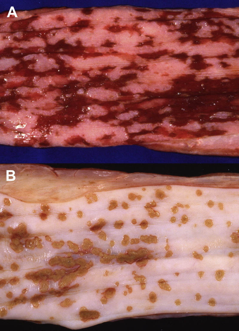
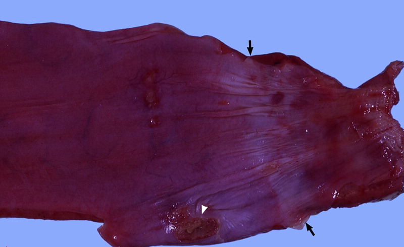
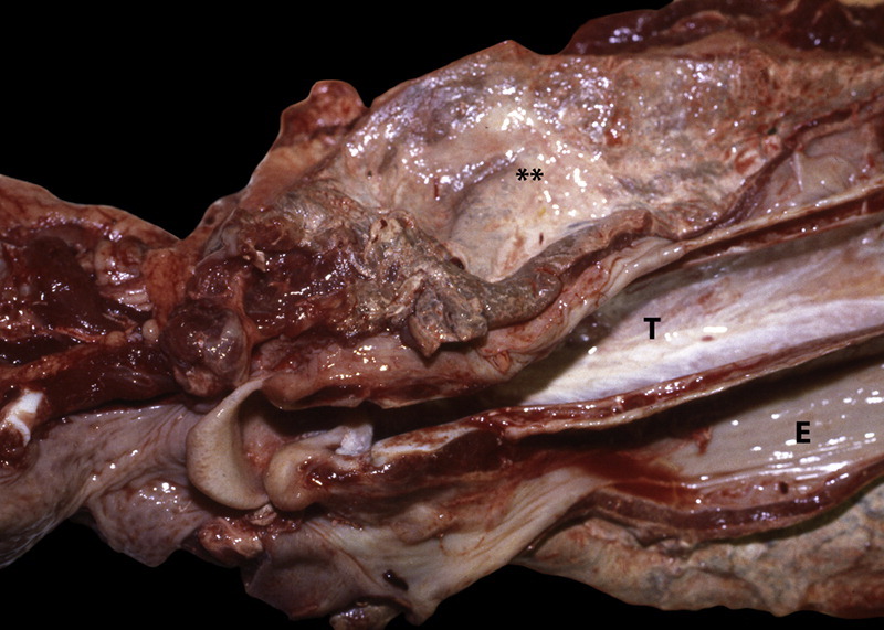
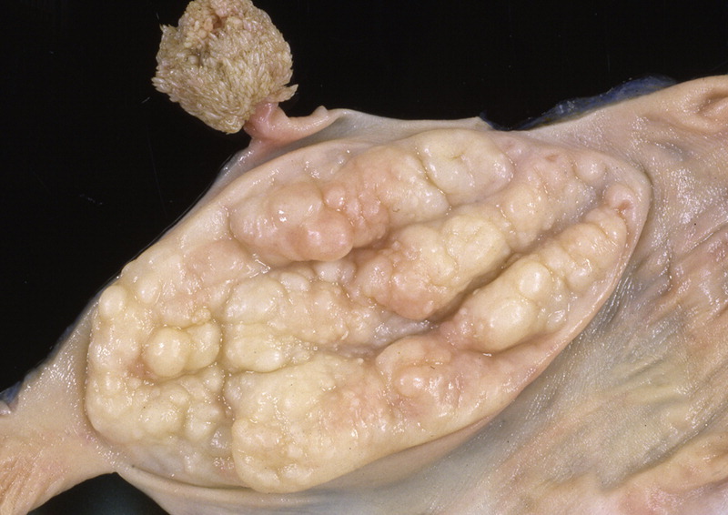
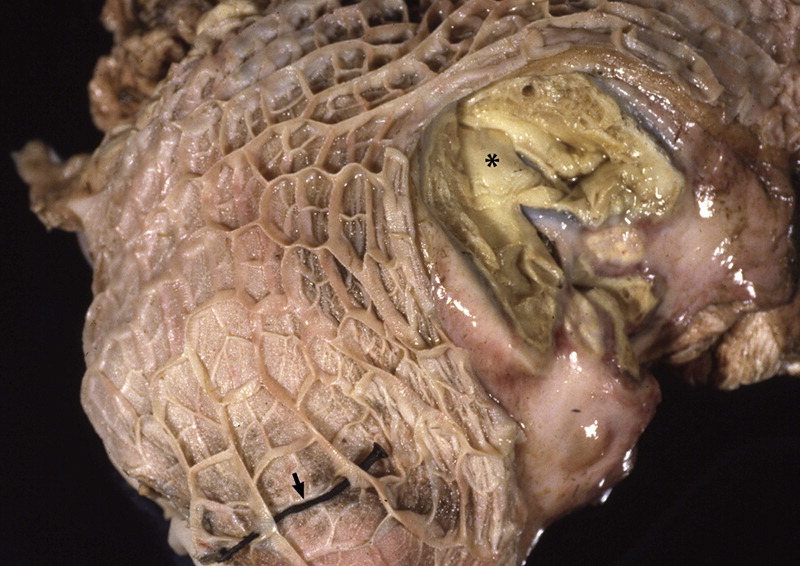
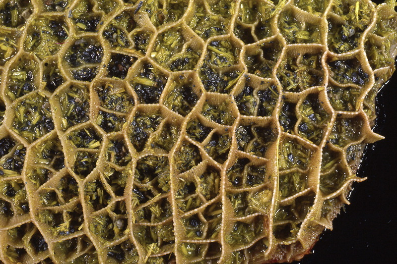
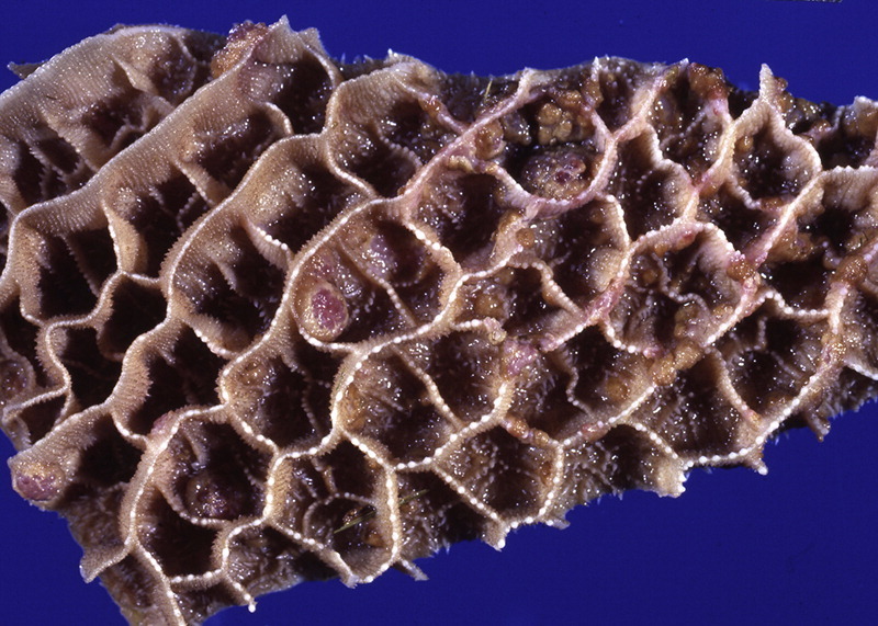
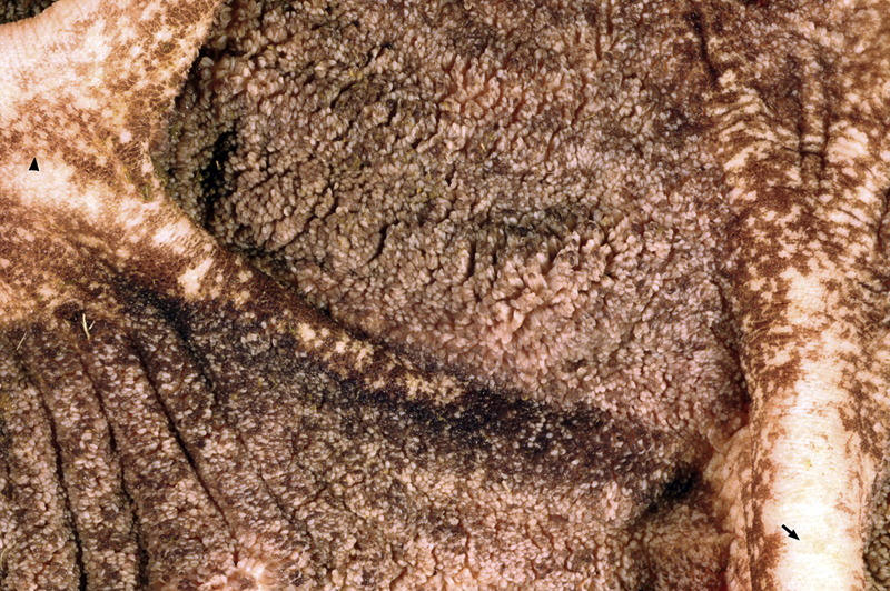
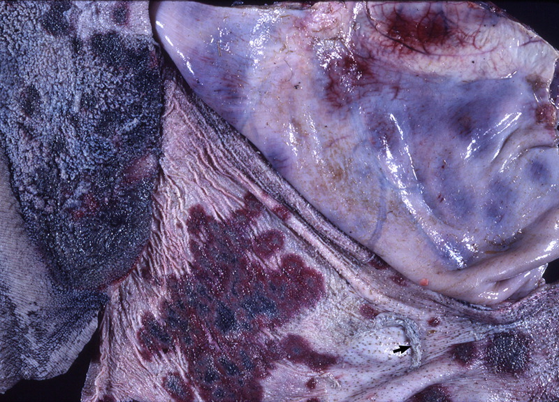
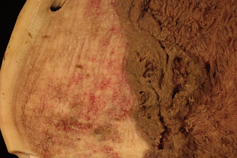
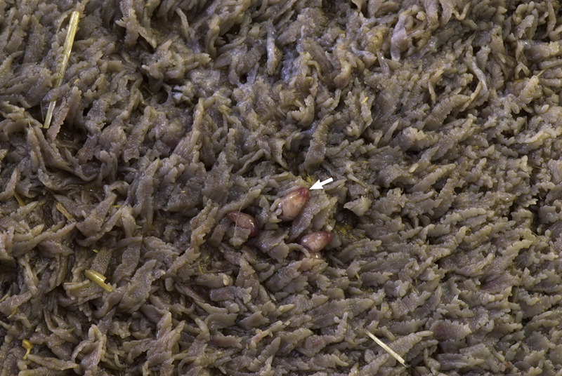
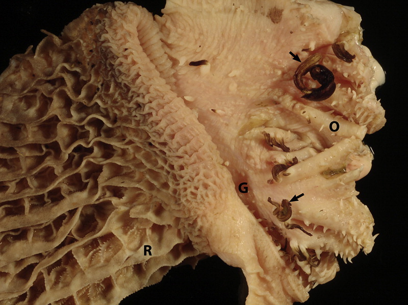
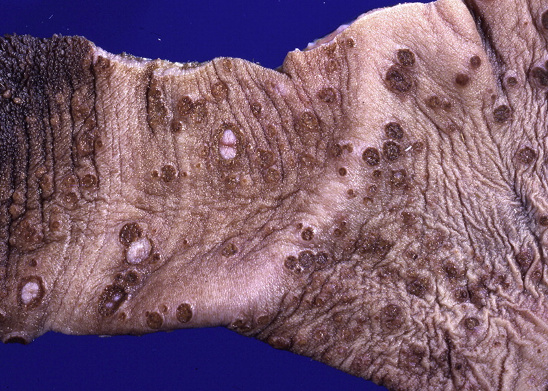
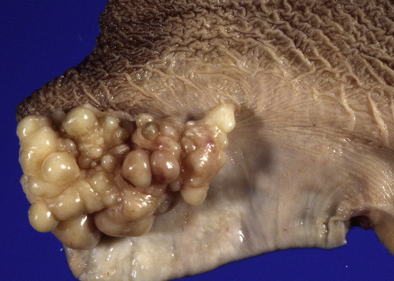
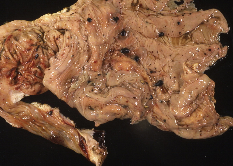
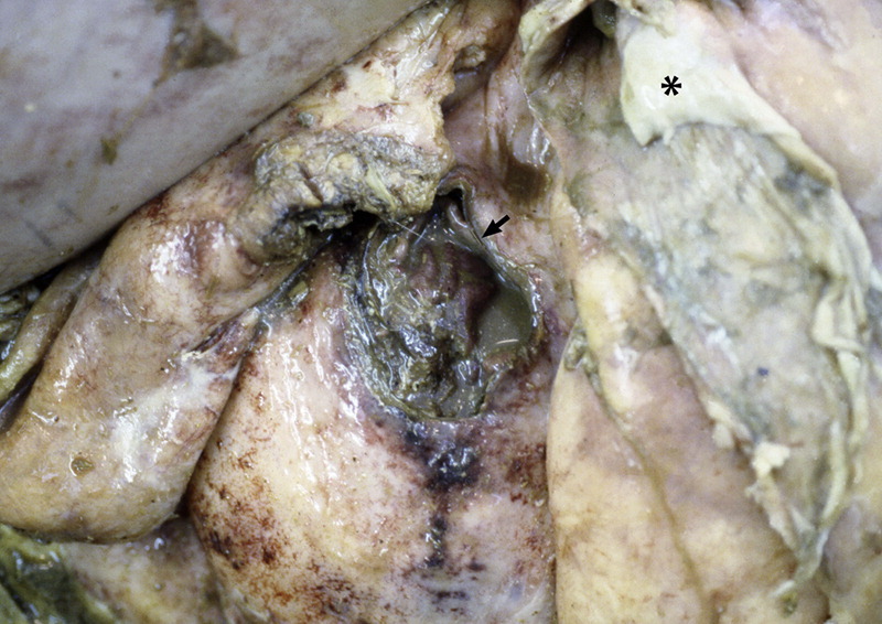
The purpose of the gross necropsy examination of the gastrointestinal tract is to recognize the presence of lesions, thus requiring a basic understanding of its normal appearance and anatomy. This article highlights gross changes to the gastrointestinal tract of adult cattle that help place the disease processes into broad categories. Although few gross lesions reach the zenith of pathognomonic, there are numerous lesions that, when considered in aggregate with history (eg, number of animals affected, environment, duration of signs, time of onset relative to management changes, previous management) and clinical signs, can help narrow the spectrum of causes, provide a basis for a strong presumptive diagnosis, and focus diagnostic test selection.


Figures

Fig. 1
Fig. 1
Abdominal viscera. The abdomen is opened, exposing the viscera. The forestomachs occupy most of the abdominal space. Typically, the abomasum (asterisk) is a darker reddish purple compared with the forestomachs.
Fig. 2
Fig. 2
Congested jejunum. The mucosa is intensely hyperemic, which can be normal. This is not considered enteritis because there is a lack of blotchy hemorrhage, the mucosa has a prominent, velvety sheen, and there is no evidence of fibrinous exudation. The serosal surfaces look normal.
Fig. 3
Fig. 3
Focal small intestinal obstruction. An intraluminal obstruction (arrow) is present, resulting in a markedly dilated and congested proximal bowel and mostly empty distal bowel.
Fig. 4
Fig. 4
Bovine papular stomatitis involving the oropharynx. Numerous circular (arrow) lesions are scattered throughout the surface of the hard and soft palate. Several of the lesions are less circular and have a more proliferative superficial exudation (arrowhead).
Fig. 5
Fig. 5
Periodontal disease caused by fluorosis. Enamel erosion and hypoplasia and black discoloration are caused by excess fluoride ingestion. The white material on the surface of the teeth is what remains of the enamel.
Fig. 6
Fig. 6
Periodontal enamel defect caused by BVDV. This bilaterally symmetric enamel defect in deciduous incisor teeth is cause by a previous in utero infection with BVDV.
Fig. 7
Fig. 7
Methemoglobinemia. The blood on the left is muddy, red brown, collected from an animal exposed to excess nitrates. The blood on the right is from a normal animal.
Fig. 8
Fig. 8
Hard and soft palate erosions. The oral mucosa of the hard and soft palate is multifocally eroded with areas (arrowheads) that lack the normal glistening sheen and are typically darker than the surrounding unaffected mucosa. BVDV was confirmed as the cause in this case based on enzyme-linked immunosorbent immunoassay and immunohistochemistry techniques.
Fig. 9
Fig. 9
Lingual erosions. There are shallow (arrowhead) and deeper erosions (arrows) affecting the mucosal surface of the tongue. The lingual papillae covering the surface of the torus linguae (T) are blunted. This is the tongue from the same animal in Fig. 8 that was confirmed positive for BVDV.
Fig. 10
Fig. 10
Ruptured vesicles in the lingual mucosa. The lingual mucosa is focally ulcerated (arrows) in areas where vesicles have ruptured. This animal was a confirmed case of foot-and-mouth disease during an outbreak in Bolivia in 2007.
Fig. 11
Fig. 11
Necroulcerative lingual gingivitis. At the rostral base of the torus linguae (T), in the lingual fossa is an area of dark red ulcerative inflammation within which are embedded grass awns mixed with other feed material.
Fig. 12
Fig. 12
Lumpy jaw. (A) Lateral view of marked swelling with superficial cutaneous ulceration and exudation. Most of the swelling is caused by boney proliferation associated with osteomyelitis. Most commonly, Actinomyces bovis is cultured from these lesions. (B) Rostral view of severe mandibular osteomyelitis with marked distortion of the mandibular incisors.
Fig. 13
Fig. 13
Actinobacillosis or “wooden” tongue. (A) The tongue is markedly enlarge with several areas of chronic ulceration along its lateral edge. (B) Cross section through a case of wooden tongue with numerous pyogranulomas (arrow) scattered throughout the tongue. This is typically caused by Actinobacillus lignieresii.
Fig. 14
Fig. 14
Lingual and pharyngeal squamous cell carcinoma. A large area of ulceration is present at the base of the tongue. This was associated with chronic ingestion of bracken fern.
Fig. 15
Fig. 15
Bloat line. Cranial to the level of the thoracic inlet (arrow), the esophagus is markedly congested, whereas caudal to this the esophagus is pale. This is an indication that elevated intraruminal pressure could not be alleviated resulting in severe vascular compromise and formation of a “bloat line.”
Fig. 16
Fig. 16
Esophageal Gongylonema pulchrum infestation. Serpentine mucosal or submucosal parasites in the esophagus (arrow) are typical of G pulchrum infestation. When found in the rumen, they are Gongylonema verrucosum.
Fig. 17
Fig. 17
Bovine papular stomatitis, esophagus. Numerous round to oval papules are present. The largest one is centrally ulcerated. Bovine papular stomatitis virus, often affecting cattle that may have impaired immunity, causes these lesions.
Fig. 18
Fig. 18
Multifocal linear esophageal erosions. Numerous mucosal erosions are scattered throughout this esophagus. With lesions this numerous and shallow, BVD must be strongly considered. This animal was confirmed positive for BVDV.
Fig. 19
Fig. 19
Esophageal ulceration. (A) An acute, hemorrhagic, ulcerative lesion affecting the esophageal mucosa. (B) A subacute ulcerative esophagitis with superficial crusting.
Fig. 20
Fig. 20
Esophageal stricture. A focal esophageal foreign body has resulted in areas of ulceration (white arrowhead) and a circumferential area of pallor and constriction (arrows).
Fig. 21
Fig. 21
Pharyngeal and periesophageal cellulitis or fasciitis. Both the trachea (T) and esophagus (E) have been displaced by a regional area of necrosis and inflammation (**). Any type of forceful injury to the pharynx can result in perforation, edema, necrosis, and inflammation. In this case, it was caused by improperly administered medicinal boluses by a mechanical dispensing device (ie, balling gun).
Fig. 22
Fig. 22
Esophageal papillomatosis. A regional area of mucosal proliferation is present in the distal esophagus and a focal, pedunculated mass. Both are papillomas.
Fig. 23
Fig. 23
Reticuloperitonitis. The reticulum is firmly adherent to the diaphragm with localized area of peritonitis (asterisk). A nail is embedded in the wall of the reticulum (arrow). It is most likely that this nail or another sharp, slender piece of metal penetrated through the wall of the reticulum to cause a localized peritonitis. If this inflammation tracks to the pericardial sac, it is called traumatic reticulopericarditis.
Fig. 24
Fig. 24
Lead intoxication. Variably sized, sliver-gray, metallic particles are present within the mucosal compartments of the reticulum. These represent lead shavings obtained from a source of lead in a field, such as used batteries.
Fig. 25
Fig. 25
Bovine papular stomatitis, reticulum.
Fig. 26
Fig. 26
Mucosal erosion, rumen. Early indication of shallow mucosal erosions is the loss of the normal greenish black coloration of the mucosa (arrowhead). Closer inspection reveals numerous shallow erosions in the mucosa (arrow). This case was confirmed BVDV positive.
Fig. 27
Fig. 27
Multifocal visceral hemorrhages. Forestomachs, abomasum, and intestines have mural hemorrhages. In this particular case, the cause was the thrombocytopenic form of BVD.
Fig. 28
Fig. 28
Rumenitis. Arborizing islands of adherent mucosa cover small portions of the underlying submucosa (asterisk). The edges of these attached portions of epithelium are thickened and lack rumen papillae. These areas represent areas of rumenitis and epithelial injury. The remaining portions that lack epithelium represent normal postmortem sloughing of mucosa (**). P, rumen pillar.
Fig. 29
Fig. 29
Necrobacillosis. This focal area of necrosis is surrounded by a red rim of congestion. This represents a local response to proliferation of Fusobacterium necrophorum.
Fig. 30
Fig. 30
Mucosal hyperplasia caused by superficial mycotic rumentitis, from Candida albicans proliferation.
Fig. 31
Fig. 31
Mycotic rumenitis. There are multifocal to coalescing areas of transmural hemorrhage affecting the rumen. These represent areas of fungal proliferation, on the surface squamous epithelium and in the walls of blood vessels. In this case, an initial ruminal acidosis caused by excess grain consumption caused significant mucosal injury allowing invasion and proliferation of fungi (ie, Mucor spp, or Aspergillus spp). In unaffected portions of the mucosa, the mucosal epithelium is lifting off in sheets (arrow).
Fig. 32
Fig. 32
Rumen mucosal epithelium normally sloughs easily from the submucosa several hours after death. If the rumen mucosal epithelium is firmly adherent, suspect a previous rumenitis (see Fig. 28).
Fig. 33
Fig. 33
Rumen flukes. Attached to the rumen surface and partially camouflaged by the rumen papillae are three rumen flukes (arrow). In this case, these are presumptively Paramphistomum spp.
Fig. 34
Fig. 34
Unguiculliform papillae. These papillae (arrows) may become quite prominent and gnarly near the reticular groove (G) along the junction of omasum (O) and reticulum (R) but are normal.
Fig. 35
Fig. 35
Bovine papular stomatitis, rumen.
Fig. 36
Fig. 36
Rumen papilloma. This focally proliferative, papillary growth is typical of a rumen papilloma.
Fig. 37
Fig. 37
Abomasal hemorrhage. Multifocally throughout the abomasal mucosa are variably sized dark red areas of hemorrhage. Any type of bleeding diathesis can result in this lesion. This particular example was a case of thrombocytopenic BVD.
Fig. 38
Fig. 38
Perforated abomasal ulcer with peritonitis. This is a serosal view of a perforated abomasal ulcer (arrow) that has resulted in a severe fibrinous peritonitis (asterisk). This is more commonly seen in young calves but less frequently can affect adult cattle.
Fig. 39
Fig. 39
Right displacement and volvulus of the abomasum. (A) The dark red to black viscus is the abomasum. Displacement and volvulus has resulted in severe vascular compromise and tissue death. (B) The mucosal surface of a similarly affected abomasum is extremely dark red and edematous caused by severe congestion and hemorrhage and necrosis.
Fig. 40
Fig. 40
Proliferative abomasitis. This is most commonly the result of Ostertagia spp infestation and associated inflammation. (A) Marked proliferation of all abomasal rugae. (B) Higher magnification of another example with focal areas of inflammation.
Fig. 41
Fig. 41
Abomasal lymphoma. (A) Multifocal areas of mural thickening is one manifestation of abomasal lymphoma. (B) Most rugal folds in this abomasum are discolored pale tan and diffusely thickened because of infiltration by neoplastic lymphocytes.
Fig. 42
Fig. 42
Enteritis. The mucosal surface of this small intestinal section lacks the normal velvety, glistening sheen and instead is dull, partially covered by fibrin mixed with feed material, and has multifocal areas of mucosal hemorrhage.
Fig. 43
Fig. 43
Hemorrhagic serositis. There are multifocal red areas over the serosa representing areas of mural hemorrhage and necrosis. This same injury could be observed from the mucosal surface indicating evidence for enteritis.
Fig. 44
Fig. 44
Hemorrhagic enteritis. The intestinal mucosa is red and congested with large mats of blood clots mixed with fibrin strands and feed material. This lesion has several potential causes but in this case is an example of coccidiosis.
Fig. 45
Fig. 45
Fibrinonecrotizing enteritis. The intestinal wall is mottled red and tan. The mucosa has lost is typical velvety sheen and is overlayed by a mat of fibrin. There is a large luminal clot of fibrin (asterisk). This lesion was caused by Clostridium perfringens.
Fig. 46
Fig. 46
Peyer's patch necrosis caused by BVD. (A) Along the antimesenteric edge of this section of small intestine is an area of necrosis and hemorrhage that corresponds to Peyer's patches. (B) Mucosal surface of small intestine depicting necrosis of Peyer's patches (arrow) with adherent fibrin and necrotic debris. This lesion is typical for BVDV infections.
Fig. 47
Fig. 47
Chronic button ulcers, colon. Numerous nodules are present with raised, rounded edges and central regions of necrosis. These lesions are a mucosal response to chronic Salmonella spp infection, which was confirmed in this case.
Fig. 48
Fig. 48
Granulomatous ileitis. (A) An opened section of small intestine with a proliferative, corrugated appearance. This lesions is virtually pathognomonic for Johne's disease in cattle. (B) Partially opened section of small intestine with a similarly proliferative mucosa. Additionally, there are serosal lymphatics that are prominent because of a granulomatous lymphangitis (arrows), a more subtle lesion also typical for Johne's disease.
Fig. 49
Fig. 49
Jejunal hemorrhagic syndrome. (A) Unopened loops of small intestine, focally hemorrhagic and mildly dilated because of a luminal blood clot. These luminal blood clots tend to lodge and obstruct. (B) Opened segment of small intestine with luminal clots of blood but a mucosal surface that appears grossly normal.
Fig. 50
Fig. 50
Massive omental fat necrosis. This is a lesion that occurs most commonly in Channel Island cattle. Lesions vary from clinically incidental to obstructive. The necrotic fat is typically saponified and palpably hard.

Similar articles

Cited by

  • Bovine trichomoniasis: A hidden threat to reproductive efficiency.
    Hermadi HA, Khairullah AR, Damayanti Y, Safitri E, Tyasningsih W, Warsito SH, Fauzia KA, Wardhani BWK, Ekawasti F, Wibowo S, Fauziah I, Moses IB, Kurniasih DAA, Kusala MKJ, Julaeha J. Hermadi HA, et al. Open Vet J. 2024 Nov;14(11):2722-2730. doi: 10.5455/OVJ.2024.v14.i11.1. Epub 2024 Nov 30. Open Vet J. 2024. PMID: 39737020 Free PMC article. Review.

References

    1. Schummer A., Nickel R., Sack W.O. The viscera of the domestic mammals. 2nd revised edition. Springer-Verlag; New York: 1979. The alimentary canal of the ruminants; p. 148. 168.
    1. Dyce K.M., Sack W.O., Wensing C.J. Textbook of veterinary anatomy. 3rd edition. Saunders; Philadelphia: 2002. The head and ventral neck of the ruminants; pp. 636–637.
    1. Helman R.G. Interpretation of basic gross pathologic changes of the digestive tract. Vet Clin North Am Food Anim Pract. 2000;16:1–22. - PubMed
    1. Thompson K. Bones and joints. In: Maxie M.G., editor. 5th edition. vol. 1. Elsevier; 2007. pp. 98–99. (Jubb, Kennedy, and Palmer’s Pathology of domestic animals).
    1. Brown C.C., Baker D.C., Barker I.K. Alimentary System. In: Maxie M.G., editor. 5th edition. vol. 2. Elsevier; 2007. pp. 20–21. (Jubb, Kennedy, and Palmer’s Pathology of domestic animals).

MeSH terms